Photoluminescence enhancement of MoS2/CdSe quantum rod heterostructures induced by energy transfer and exciton–exciton annihilation suppression†
Yang
Luo
a,
Hangyong
Shan
a,
Xiaoqing
Gao
 b,
Pengfei
Qi
 a,
Yu
Li
a,
Bowen
Li
a,
Xin
Rong
a,
Bo
Shen
a,
Han
Zhang
a,
Feng
Lin
a,
Zhiyong
Tang
a,
Yu
Li
a,
Bowen
Li
a,
Xin
Rong
a,
Bo
Shen
a,
Han
Zhang
a,
Feng
Lin
a,
Zhiyong
Tang
 c and
Zheyu
Fang
 *a
*a
aSchool of Physics, State Key Laboratory for Mesoscopic Physics, Academy for Advanced Interdisciplinary Studies, Collaborative Innovation Center of Quantum Matter, and Nano-optoelectronics Frontier Center of Ministry of Education, Peking University, Beijing 100871, P. R. China. E-mail: zhyfang@pku.edu.cn
bCollege of Physics and Optoelectronic Engineering, Shenzhen University, Guangdong 518060, P. R. China
cCAS Key Laboratory of Nanosystem and Hierarchical Fabrication, CAS Center for Excellence in Nanoscience, National Center for Nanoscience and Technology, Beijing 100190, P. R. China
First published on 31st March 2020
Abstract
Energy transfer in heterostructures is an essential interface interaction for extraordinary energy conversion properties, which promote promising applications in light-emitting and photovoltaic devices. However, when atomic-layered transition metal dichalcogenides (TMDCs) act as the energy acceptor because of strong Coulomb interactions, the transferred energy can be consumed by nonradiative exciton annihilations, which hampers the development of light-emitting devices. Hence, revealing the mechanism of energy transfer and the related relaxation processes from the aspect of the acceptor in the heterostructure is key to reducing nonradiative loss and optimizing luminescence. Here, we study the exciton dynamics from the standpoint of the acceptor in MoS2/CdSe quantum rod (QR) heterostructures and realize efficiently enhanced photoluminescence (PL). Through femtosecond pump–probe measurements, it is directly observed that energy transfer from CdSe QRs largely raises the exciton population of the acceptor, MoS2, providing a larger emission “source”. In addition, the dielectric environment introduced by CdSe QRs efficiently enhances the PL by suppressing exciton–exciton annihilation (EEA). This study provides new insights for on-chip applications such as light-emitting diodes and optical conversion devices based on low dimensional semiconductor heterostructures.
New ConceptsWe reveal the unique mechanism of PL enhancement in MoS2/CdSe quantum rod (QR) heterostructures from the standpoint of the acceptor. The mechanism involves energy transfer and suppression of exciton–exciton annihilation. The physical mechanism of energy transfer has been proposed for several decades. However, how the transferred energy influences the exciton decay of the acceptor has not been addressed. In monolayered two-dimensional transition metal dichalcogenides (TMDCs), the nonradiative exciton annihilation is more intense compared to other semiconductors due to the strong reduced dimension, and thus the transferred energy can dissipate in a nonradiative way, which seriously hinders terminal luminescence efficiency. Using ultrafast pump–probe measurements, we demonstrate that the transferred energy can “re-excite” excitons in the MoS2 acceptor and the nonradiative annihilation process can be effectively suppressed by the CdSe QRs. Additionally, the contribution of energy transfer and exciton–exciton annihilation to exciton relaxation can be modulated to improve the final photoluminescence. Our work advances the basic comprehension of the exciton dynamics of the energy acceptor and paves the way for developments in new high-performance optical conversion, on-chip light emission, and light-sensing devices. |
Introduction
With the development of nanotechnology, low dimensional heterostructures have attracted vast attention due to novel effects such as energy transfer,1,2 charge transfer,3–5 and Moiré superlattice properties,6 yielding potential applications in light-harvesting and photoelectron devices.7,8 Nonradiative energy transfer in heterostructures is a process that transfers energy from a “donor” to an “acceptor” through near-field interactions to achieve energy conversion.9,10 In particular, with atomic transition metal dichalcogenide (TMDC)/quantum confined semiconductor (quantum rods,11 quantum dots,12 and quantum nanoplatelets13) heterostructures, energy transfer can help atomic TMDCs break their light emission limit by providing extra energy from quantum rods (QRs), which can lead to high quantum yields of monolayered TMDC-based devices. In recent years, approaches such as inducing an electrical field,14 altering the number of TMDC layers,15 tuning the donor size of the heterostructures16 as well as changing the polarization between the donor and acceptor17 have been employed to modulate energy transfer in heterostructures effectively.However, most of the above-mentioned studies are focused on the energy transfer efficiency and exciton relaxation properties from the perspective of the donor. The acceptor usually acts as the final output terminal in applications, where the transferred energy can decay by both radiative and nonradiative channels. When the thickness of the acceptor in TMDCs is reduced to the monolayer scale, nonradiative relaxation such as exciton–exciton annihilation (EEA) and high order exciton–exciton annihilation (HOEEA) become more active, which accelerates exciton consumption and the exciton lifetime decreases to tens of picoseconds.18 Thus, a large portion of the transferred energy in the acceptor is consumed in a nonradiative way, which strongly attenuates the PL emission. In order to achieve applications in high quantum yield devices, it is urgent to study the exciton relaxation process from the perspective of acceptor and explore ways to suppress the nonradiative decay of excitons.
In this study, we investigate the exciton relaxation dynamics of MoS2/CdSe QR heterostructures from the standpoint of the energy acceptor to reveal its unique mechanism of PL enhancement. The femtosecond pump–probe technique was employed to investigate how the transferred energy affects the exciton relaxation of MoS2 and the effective suppression of the nonradiative decay. This study provides a new physical understanding of the exciton dynamics of low dimensional heterostructures and lays the foundation for highly efficient solar cells, ultralow-threshold lasers, light-emitting diodes, and sensitive optical detectors.
Results and discussion
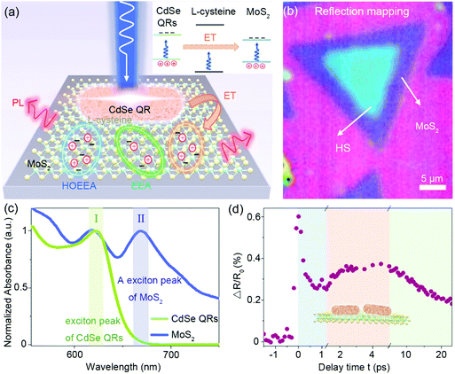
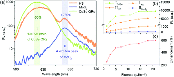
In the heterostructure, CdSe QRs with L-cysteine ligands are distributed on a MoS2 monolayer (Fig. 1(a)). Excitons of MoS2 and CdSe QRs are pumped using 400 nm laser excitation. The reduced Coulomb screening of the MoS2 monolayer enables multi-exciton interactions such as HOEEA and EEA occur in a very short time. The energy band structures of the MoS2 monolayer, CdSe QRs, and L-cysteine molecules are shown in the inset, and the corresponding band values have been reported in previous studies.19–21 As the energy of the excitation photon is around 3.05 eV, which is lower than the excitation energy of L-cysteine, only excitons in CdSe and MoS2 are excited and a high energy barrier is formed by the L-cysteine molecules. In addition, L-cysteine is an amino acid with high resistance that can suppress interfacial charge transfer even in a single layer.22 Thus, the effects of charge transfer between CdSe QRs and MoS2 are very weak, and the dominating interaction between CdSe QRs and the MoS2 monolayer is energy transfer through near-field coupling. Processes such as energy transfer, EEA, and HOEEA affect the final light emission of the MoS2 monolayer. To obtain the morphology of the heterostructure, reflection mapping was performed (Fig. 1(b)). The homogeneous blue area indicates that CdSe QRs are distributed almost uniformly on the top of the MoS2 monolayer, which indicates that optical effects induced by the aggregation of QRs are trivial (AFM mapping is shown in Fig. S13, ESI†). The corresponding absorption spectra of CdSe QRs and MoS2 monolayer are shown in Fig. 1(c). The signals of the CdSe QRs and MoS2 monolayer are normalized by the intensity of peak I and II, respectively, to highlight the peak positions. Peak I, located at 620 nm, is the exciton peak of CdSe QRs and peak II, at 670 nm, is the A exciton of the MoS2 monolayer, which provides wavelength information for pump–probe measurements. In the time-resolved differential reflection measurement (Fig. 1(d)), the signal of MoS2 in the heterostructure displays three different decay processes corresponding to the exciton relaxation by HOEEA, energy transfer, and EEA, as seen in Fig. 1(a). The unique exciton decay of the MoS2 acceptor in the heterostructure contributes to PL enhancement, which will be discussed later and is shown in Fig. 3.PL measurements were employed to study the light-emitting properties of the heterostructure, with the PL spectra of the pristine MoS2 monolayer and CdSe QRs for comparison. As shown in Fig. 2(a), peak I and II are located at 624 nm and 672 nm and are attributed to the PL peaks of the MoS2 monolayer and CdSe QRs, respectively. After forming the MoS2/CdSe QR heterostructure, the PL peak I has approximately 50% attenuation and the attenuation of peak II increases to about 230%. In the presence of the CdSe layer, the amount of excitation light that reaches MoS2 is reduced and the PL of MoS2 should be weakened, which is contrary to the experimental results. This implies that the different amount of excitation light is not the reason for the enhanced PL of MoS2. The typical PL spectrum of energy transfer is shown in Fig. 2(a) with the donor signal decreasing and the acceptor signal increasing. This is the direct evidence for energy transfer from the CdSe QRs to the MoS2 monolayer.23 If the charge transfer dominates, the PL signals of the donor and acceptor should both decrease.24,25 Thus, it is evident that energy transfer is the dominating interaction in the heterostructure. Since the PL intensity of CdSe QRs is much larger than that of MoS2, the 50% decrease in the energy of CdSe QRs is large enough to result in a 230% energy enhancement of MoS2. In order to further explore the light-emitting properties of the heterostructure, the excitation fluence was increased from 1.4 μJ cm−2 to 21.6 μJ cm−2 (Fig. S1, ESI†). On one hand, the PL peaks I and II of the heterostructure grow with similar trend, as shown in Fig. 2(b), which suggests that the energy transfer from CdSe to MoS2 has modulated the light emission of the heterostructure. On the other hand, the MoS2 in the heterostructure is further enhanced as the excitation fluence increases, and it reaches 500% when the pump fluence increases to 21.6 μJ cm−2 (Fig. 2(c)). Since the rate of the nonradiative energy transfer is related to distance, spectral overlap, and the relative polarization between the donor and acceptor, the energy transfer induced enhancement of MoS2 in the heterostructure should not be altered by a change in the pump fluence, implying that there is another mechanism that improves the radiative emission of MoS2.
PL emission is decided by the exciton population (N) and the radiative and nonradiative channels of the exciton relaxation process. In order to explore the mechanism of enhanced PL, theoretical analysis of the exciton relaxation dynamics of the heterostructure from the aspect of MoS2 is employed.
In a pristine MoS2 monolayer, the relaxation process, including exciton recombination, EEA, and HOEEA, can be expressed as26–28
 | (1) |
When energy transfer plays a role in exciton relaxation, the relaxation equation should be modified. In the energy transfer process, the exciton population of the donor (ND) can be simplified to ND = ND1e−kETt, where ND1 is a constant, and kET represents the energy transfer rate. When the donor consumes ΔND(ET) in the energy transfer process, the acceptor acquires excitons ΔNA = ΔND(ET). Thus, the acquired exciton population of the acceptor (NA) can be written as  , indicating that for the acceptor, there is an “exciton re-excitation” process (Section S7, ESI†). Based on the above analysis, the exciton population of the acceptor can be expressed as:
 | (2) |
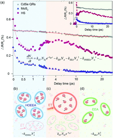
The exciton relaxation dynamics of the pristine MoS2 monolayer and heterostructure were analyzed, as shown in Fig. 3(a). It should be noted that in this study the effects of coherent phonons were excluded (Section S7, ESI†). For the pristine MoS2 monolayer, it can be clearly observed that the exciton relaxation consists of two different processes, as displayed in eqn (1). In the first 1 ps, high order interactions among excitons, HOEEA, dominates the decay process and is expressed as  . As time goes by, the interactions of HOEEA diminish and are followed by EEA, with the kinetic equation:
  . The value of kEEA is obtained as kEEA = 0.417 ± 0.050 cm2 s−1, which is in agreement with that obtained in previous studies,29,30 verifying the reliability of these experimental methods and the analytical approaches used in this work.
As for the heterostructure, the exciton relaxation can be described by eqn (2). However, the modified model in eqn (2) has four free parameters, which hinder the analysis of the experimental data. Luckily, the four relaxation processes dominate at different time scales. Thus, it is reasonable to divide the whole relaxation process into four parts, with each part having one free parameter. In the beginning, excitons quickly decay to the band edge level after they are pumped to high energy levels. Under this condition, the “crowded” excitons tend to undergo high order interactions, HOEEA, contributing to the rapid descent of NA (Fig. 3(b)). About 1 ps later, the effect of HOEEA is rapidly weakened, and the role of energy transfer emerges, leading to an overall enhancement of NA, as shown in Fig. 3(c). The corresponding dominant relaxation equation is expressed as  , with the energy transfer rate as kET = 17.41 ± 0.13 ns−1. When t > 5 ps, the effect of energy transfer is gradually diminished, and EEA starts to dominate the relaxation process and NA decreases again (Fig. 3(d)).
Compared to the signal of the pristine MoS2 monolayer, the most obvious difference is the increase in NA after the HOEEA process, which is the direct evidence of energy transfer. It should be mentioned that this is the first direct observation of the increase in NA induced by energy transfer, benefitting from the ultra-short probe time step length of 100 fs. In the heterostructure, energy is transferred from donor CdSe QRs to acceptor MoS2 in the form of additional excitons. These excitons are equivalent to an extra “source” for PL, which lays the foundation for the enhanced PL of the heterostructure. However, if the following nonradiative interaction (EEA) in the MoS2 acceptor is active, there still would be a large portion of excitons decayed in a nonradiative way, impeding luminescence of the material. In this study, the EEA of the heterostructure is effectively suppressed, with kEEA(HS)/kEEA(MoS2) ≈ 0.091. The existence of CdSe QRs affects the dielectric environment with a larger refractive index than air. Additionally, the Coulomb interactions in MoS2 can be reduced by leaking into the surrounding environment,31 resulting in a smaller kEEA(HS) compared to kEEA(MoS2) (Fig. S7, ESI†). Alternatively, surface modification by CdSe QRs can effectively suppress the EEA process to reduce the nonradiative loss. The existence of CdSe QR-induced energy transfer optimizes the dielectric environment, contributing to the enhanced PL of the heterostructure.
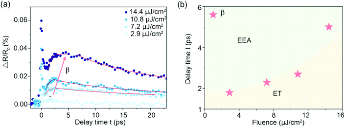
In order to further optimize the PL emission of the heterostructure, the contribution of energy transfer and EEA to the exciton relaxation process was investigated. When the contribution of energy transfer becomes larger, more excitons are provided for emission. When the contribution of EEA gets higher, more excitons decay through a nonradiative pathway. Through tuning the fluence of the pump laser, the effect of energy transfer and EEA on exciton relaxation can be efficiently modulated. In Fig. 4(a), the modulation is reflected by the emerging time of inflection point β between the two processes. At point β, the effect of energy transfer diminishes and EEA starts to dominate the exciton relaxation dynamics. By increasing the pump fluence from 2.9 μJ cm−2 to 14.4 μJ cm−2, the appearance time of point β is increased, where the effect of energy transfer on exciton relaxation is stronger in comparison to that of EEA, as shown in Fig. 4(b). As energy transfer is independent of the excitation fluence, the variation in point β indicates a change to EEA. When the pump fluence is relatively low, EEA can be mediated by some defect trapping processes.32 In this situation, kEEA keeps reducing until all of the defect states become occupied when the pump fluence is increasing. Thus, in the heterostructure, EEA shows a decreasing trend as the pump fluence increases (Fig. S11, ESI†), resulting in an increase in exciton due to the effects of energy transfer compared to EEA, ultimately improving PL under high fluence excitation.
Conclusions
In conclusion, the mechanism of PL enhancement from the standpoint of the acceptor in the MoS2/CdSe QR heterostructure was studied using the femtosecond pump–probe technique. Energy transfer induced “extra excitons” and the CdSe QRs induced the suppression of nonradiative EEA, which resulted in the PL enhancement of the heterostructure. It is the first direct observation of an increase in NA induced by energy transfer using the time-resolved differential reflection signal. An energy transfer term was added to modify the exciton relaxation dynamics equation to systematically investigate the exciton relaxation dynamics from the standpoint of the acceptor, which has far-reaching significance for more sophisticated multi-physical processes. Our work advances the basic comprehension of the exciton dynamics of the energy acceptor and paves the way for developments of new high-performance optical conversion, on-chip light emission, and light-sensing devices.Methods
Sample fabrication
The MoS2 monolayer was grown using the chemical vapor deposition (CVD) method at 850 °C, with MoO3 and pristine sulphur as the reaction materials. The silicon substrate was covered by a 540 nm SiO2 film.33 CdSe QRs with a diameter of 3.75 ± 0.47 nm and an aspect ratio of 4.82 ± 0.81 were synthesized in trioctylphosphine oxide, following the thermal injection technique (Fig. S12, ESI†). Subsequent ligand exchange allowed QRs to be coated with cysteine without geometric destruction.34,35 To avoid bubbles caused by the strain effect, the heterostructure was fabricated by spin-coating a CdSe QR solution onto the MoS2 monolayer, followed by water evaporation in a nitrogen cabinet at room temperature.Optical measurements
Reflection mapping and absorption spectra were measured using a hyperspectral imaging system (Cytoviva HIS V3) and detected using a spectrometer (Horiba IHR550) with a 100× objective lens (Olympus MPlanFL N, NA = 0.9). The absorption spectra were recorded from the relative reflection change of sample compared with the substrate. Raman spectra were recorded using a home-built optical system and detected using a spectrometer (Horiba IHR550) with a 50× objective lens (Olympus LUCPlanFL N, NA = 0.5) and a 2400 g mm−1 grating for high resolution.Pump–probe and PL measurements were conducted using a home-build optical system. A detailed description can be referred to in previous study.36 Femtosecond pulse with a repetition rate of 80 MHz and pulse duration of ∼84 fs was produced using a mode-locked oscillator (Tsunami 3941C-25XP). The output 800 nm pulse was split into two parts. One was focused on a BBO crystal to generate the 400 nm pump light, which was modulated using a chopper with 1500 MHz and the other one passed through a photonic fiber crystal (Newport SCG-800) to produce supercontinuum white light. The time delay between the pump and probe pulse was controlled by a steeper linear stage (Newport M-ILS150PP) and limited by a mode-locked oscillator. Only two pump wavelengths were available: 400 nm and 800 nm. The pump and probe lights were focused by a 40× objective lens (Olympus LUCPlanFL N, NA = 0.6). The reflected probe signal was focused on a high-sensitivity photomultiplier (Thorlabs PMM02). The PL spectra were measured using a spectrometer (Princeton Action SP2500) with a 600 mm−1 grating. As the PL and pump–probe collection corresponded to the coupling of optical elements (lens, reflection mirror, filter, fibre, etc.) and the spectrograph, all of the measurement parameters remain unchanged to avoid the collection efficiently changing.
Author contributions
Z. F. and Y. L. conceived the idea and designed the research. Y. L. and H. S. performed the pump–probe and PL measurements. Y. L. and X. G. fabricated the samples. Y. L. and B. L. performed the reflection measurement. Y. L., P. Q., and H. Z. performed the theoretical simulation. Y. L., Z. F., P. Q., and H. Z. revised the manuscript. All authors discussed the results and commented on the manuscript.Conflicts of interest
There are no conflicts to declare.Acknowledgements
This work is supported by the National Key Research and Development Program of China (grant no. 2019YFA0210203, 2017YFA0205700 and 2017YFA0206000), National Science Foundation of China (grant no. 11674012, 61521004, 21790364, 61422501 and 11374023), Beijing Natural Science Foundation (grant no. Z180011 and L140007), and Foundation for the Author of National Excellent Doctoral Dissertation of China (grant no. 201420), National Program for Support of Top-notch Young Professionals (grant no. W02070003).References
- J. Shi, M. H. Lin, I. T. Chen, N. Mohammadi Estakhri, X. Q. Zhang, Y. Wang, H. Y. Chen, C. A. Chen, C. K. Shih, A. Alu, X. Li, Y. H. Lee and S. Gwo, Nat. Commun., 2017, 8, 35 CrossRef PubMed.
- X. Liu and J. Qiu, Chem. Soc. Rev., 2015, 44, 8714–8746 RSC.
- A. Henning, V. K. Sangwan, H. Bergeron, I. Balla, Z. Sun, M. C. Hersam and L. J. Lauhon, ACS Appl. Mater. Interfaces, 2018, 10, 16760–16767 CrossRef CAS PubMed.
- Y. Tan, L. Ma, Z. Gao, M. Chen and F. Chen, Nano Lett., 2017, 17, 2621–2626 CrossRef CAS PubMed.
- X. Shi, K. Ueno, T. Oshikiri, Q. Sun, K. Sasaki and H. Misawa, Nat. Nanotechnol., 2018, 13, 953–958 CrossRef CAS PubMed.
- B. Hunt, J. Sanchez-Yamagishi, A. Young, M. Yankowitz, B. J. LeRoy, K. Watanabe, T. Taniguchi, P. Moon, M. Koshino and P. Jarillo-Herrero, Science, 2013, 340, 1427–1430 CrossRef CAS PubMed.
- L. Pi, L. Li, K. Liu, Q. Zhang, H. Li and T. Zhai, Adv. Funct. Mater., 2019, 1904932, DOI:10.1002/adfm.201904932.
- N. R. Glavin, R. Rao, V. Varshney, E. Bianco, A. Apte, A. Roy, E. Ringe and P. M. Ajayan, Adv. Mater., 2019, e1904302, DOI:10.1002/adma.201904302.
- A. Raja, A. Montoya Castillo, J. Zultak, X. X. Zhang, Z. Ye, C. Roquelet, D. A. Chenet, A. M. van der Zande, P. Huang, S. Jockusch, J. Hone, D. R. Reichman, L. E. Brus and T. F. Heinz, Nano Lett., 2016, 16, 2328–2333 CrossRef CAS PubMed.
- L. Wu, Y. Chen, H. Zhou and H. Zhu, ACS Nano, 2019, 13, 2341–2348 CAS.
- D. Katz, T. Wizansky, O. Millo, E. Rothenberg, T. Mokari and U. Banin, Phys. Rev. Lett., 2002, 89, 086801 CrossRef PubMed.
- B. Peng, Z. Li, E. Mutlugun, P. L. Hernandez Martinez, D. Li, Q. Zhang, Y. Gao, H. V. Demir and Q. Xiong, Nanoscale, 2014, 6, 5592–5598 RSC.
- D. O. Sigle, L. Zhang, S. Ithurria, B. Dubertret and J. J. Baumberg, J. Phys. Chem. Lett., 2015, 6, 1099–1103 CrossRef CAS PubMed.
- D. Prasai, A. R. Klots, A. K. Newaz, J. S. Niezgoda, N. J. Orfield, C. A. Escobar, A. Wynn, A. Efimov, G. K. Jennings, S. J. Rosenthal and K. I. Bolotin, Nano Lett., 2015, 15, 4374–4380 CrossRef CAS PubMed.
- F. Prins, A. J. Goodman and W. A. Tisdale, Nano Lett., 2014, 14, 6087–6091 CrossRef CAS PubMed.
- S. Sampat, T. Guo, K. Zhang, J. A. Robinson, Y. Ghosh, K. P. Acharya, H. Htoon, J. A. Hollingsworth, Y. N. Gartstein and A. V. Malko, ACS Photonics, 2016, 3, 708–715 CrossRef CAS.
- N. Taghipour, P. L. Hernandez Martinez, A. Ozden, M. Olutas, D. Dede, K. Gungor, O. Erdem, N. K. Perkgoz and H. V. Demir, ACS Nano, 2018, 12, 8547–8554 CrossRef CAS PubMed.
- N. Kumar, Q. Cui, F. Ceballos, D. He, Y. Wang and H. Zhao, Phys. Rev. B: Condens. Matter Mater. Phys., 2014, 89, 125427 CrossRef.
- M. Beerbom, R. Gargagliano and R. Schlaf, Langmuir, 2005, 21, 3551–3558 CrossRef CAS PubMed.
- H. Shan, Y. Yu, X. Wang, Y. Luo, S. Zu, B. Du, T. Han, B. Li, Y. Li, J. Wu, F. Lin, K. Shi, B. K. Tay, Z. Liu, X. Zhu and Z. Fang, Light: Sci. Appl., 2019, 8, 9 CrossRef PubMed.
- J. Hu, L. Wang, L.-S. Li, W. Yang and A. P. Alivisatos, J. Phys. Chem. B, 2002, 106, 2447–2452 CrossRef CAS.
- X. Xu, S. Giménez, I. Mora-Seró, A. Abate, J. Bisquert and G. Xu, Mater. Chem. Phys., 2010, 124, 709–712 CrossRef CAS.
- J. Gu, X. Liu, E.-C. Lin, Y.-H. Lee, S. R. Forrest and V. M. Menon, ACS Photonics, 2017, 5, 100–104 CrossRef.
- X. Hong, J. Kim, S. F. Shi, Y. Zhang, C. Jin, Y. Sun, S. Tongay, J. Wu, Y. Zhang and F. Wang, Nat. Nanotechnol., 2014, 9, 682 CrossRef CAS PubMed.
- S. Kaniyankandy, S. Rawalekar and H. N. Ghosh, J. Phys. Chem. C, 2012, 116, 16271–16275 CrossRef CAS.
- D. Sun, Y. Rao, G. A. Reider, G. Chen, Y. You, L. Brezin, A. R. Harutyunyan and T. F. Heinz, Nano Lett., 2014, 14, 5625–5629 CrossRef CAS PubMed.
- H. Shan, Y. Yu, R. Zhang, R. Cheng, D. Zhang, Y. Luo, X. Wang, B. Li, S. Zu, F. Lin, Z. Liu, K. Chang and Z. Fang, Mater. Today, 2019, 24, 10–16 CrossRef CAS.
- S. Hao, M. Z. Bellus, D. He, Y. Wang and H. Zhao, Nanoscale Horiz., 2020, 5, 139–143 RSC.
- Y. Yu, Y. Yu, C. Xu, A. Barrette, K. Gundogdu and L. Cao, Phys. Rev. B: Condens. Matter Mater. Phys., 2016, 93, 201111 CrossRef.
- S. Mouri, Y. Miyauchi, M. Toh, W. Zhao, G. Eda and K. Matsuda, Phys. Rev. B: Condens. Matter Mater. Phys., 2014, 90, 155449 CrossRef.
- D. Jena and A. Konar, Phys. Rev. Lett., 2007, 98, 136805 CrossRef PubMed.
- B. Liu, Y. Meng, X. Ruan, F. Wang, W. Liu, F. Song, X. Wang, J. Wu, L. He, R. Zhang and Y. Xu, Nanoscale, 2017, 9, 18546–18551 RSC.
- S. Najmaei, Z. Liu, W. Zhou, X. Zou, G. Shi, S. Lei, B. I. Yakobson, J. C. Idrobo, P. M. Ajayan and J. Lou, Nat. Mater., 2013, 12, 754–759 CrossRef CAS PubMed.
- Z. A. Peng and X. Peng, J. Am. Chem. Soc., 2001, 123, 1389–1395 CrossRef CAS.
- X. Gao, X. Zhang, K. Deng, B. Han, L. Zhao, M. Wu, L. Shi, J. Lv and Z. Tang, J. Am. Chem. Soc., 2017, 139, 8734–8739 CrossRef CAS PubMed.
- Y. Yu, Z. Ji, S. Zu, B. Du, Y. Kang, Z. Li, Z. Zhou, K. Shi and Z. Fang, Adv. Funct. Mater., 2016, 26, 6394–6401 CrossRef CAS.
Footnote |
| † Electronic supplementary information (ESI) available. See DOI: 10.1039/c9nh00802k |
| This journal is © The Royal Society of Chemistry 2020 |


