Themed collection Optical Diagnosis (2014)

Optical diagnostics – spectropathology for the next generation
Malgorzata Baranska and Hugh J. Byrne, co-chairs of SPEC 2014, welcome you to this themed issue on the topic of Optical Diagnosis which focuses on recent developments of in-vivo, ex-vivo and in-vitro clinical applications of vibrational spectroscopy.

Analyst, 2015,140, 2064-2065
https://doi.org/10.1039/C5AN90024G
Spectropathology for the next generation: Quo vadis?
Vibrational spectroscopy for biomedical applications has shown great promise although its translation into clinical practice has, as yet, been relatively slow. This Editorial assesses the challenges facing the field and the potential way forward.
Analyst, 2015,140, 2066-2073
https://doi.org/10.1039/C4AN02036G
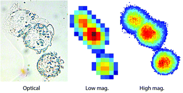
Enhanced FTIR bench-top imaging of single biological cells
A new optical system has recently been developed that enables infrared images to be obtained with a pixel resolution of 1 micron on a bench-top instrument using a thermal source.
Analyst, 2015,140, 2080-2085
https://doi.org/10.1039/C4AN02053G
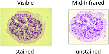
Rapid identification of goblet cells in unstained colon thin sections by means of quantum cascade laser-based infrared microspectroscopy
Mucin density is rapidly visualized in unstained, paraffin-embedded mouse colon tissue by means of mid-infrared spectroscopy using quantum cascade lasers.
Analyst, 2015,140, 2086-2092
https://doi.org/10.1039/C4AN02001D
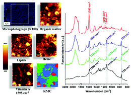
Raman spectroscopic studies of vitamin A content in the liver: a biomarker of healthy liver
Confocal Raman microspectroscopy was used in this study to identify hepatic stellate cells (HSCs) from healthy mice and mice with untreated and treated liver steatosis.
Analyst, 2015,140, 2074-2079
https://doi.org/10.1039/C4AN01878H
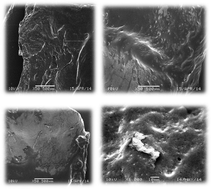
Inhibitors of thermally induced burn incidents – characterization by microbiological procedure, electrophoresis, SEM, DSC and IR spectroscopy
(DSC) and (TGA) investigations, acetate electrophoresis (CAE), infrared spectrometry (FTIR), scanning electron microscopy (SEM) and microbiological procedures were all carried out after heating the samples to a temperature sufficient for simulating a burn incident.
Analyst, 2015,140, 4599-4607
https://doi.org/10.1039/C5AN00329F
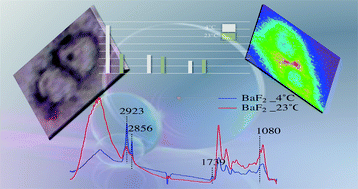
Cold shock induces apoptosis of dorsal root ganglion neurons plated on infrared windows
The effect of sample preparation and substrate choice in the apoptosis of dorsal root ganglion neurons using FTIR widefield microscopy.
Analyst, 2015,140, 4046-4056
https://doi.org/10.1039/C5AN00729A
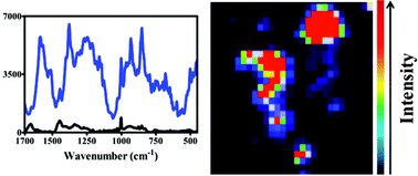
Gold nanoparticles as a substrate in bio-analytical near-infrared surface-enhanced Raman spectroscopy
“Large” nanoparticles potentially are a good starting point in order to derive informative NIR/IR SERS analysis of biological samples.
Analyst, 2015,140, 3090-3097
https://doi.org/10.1039/C4AN01899K
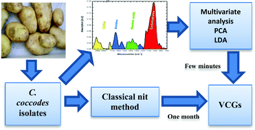
Assignment of Colletotrichum coccodes isolates into vegetative compatibility groups using infrared spectroscopy: a step towards practical application
FTIR spectroscopy may provide a specific, rapid, and inexpensive method for the successful classification of Colletotrichum coccodes isolates into vegetative compatibility groups.
Analyst, 2015,140, 3098-3106
https://doi.org/10.1039/C5AN00213C
Simultaneous intracellular redox potential and pH measurements in live cells using SERS nanosensors
In this paper we have presented the first example of multiplexing pH and redox responsive SERS nanosensors for intracellular live single cell measurement on a cell by cell basis.

Analyst, 2015,140, 2330-2335
https://doi.org/10.1039/C4AN02365J
The role of lipid droplets and adipocytes in cancer. Raman imaging of cell cultures: MCF10A, MCF7, and MDA-MB-231 compared to adipocytes in cancerous human breast tissue
We discussed the potential of lipid droplets in nonmalignant and malignant human breast epithelial cell lines as a prognostic marker in breast cancer.

Analyst, 2015,140, 2224-2235
https://doi.org/10.1039/C4AN01875C
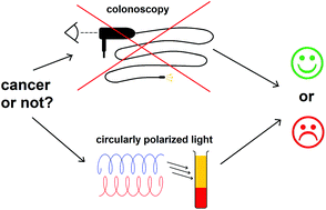
The potential of chiroptical and vibrational spectroscopy of blood plasma for the discrimination between colon cancer patients and the control group
Chiroptical spectroscopy is able to discriminate colon cancer patients from healthy controls.
Analyst, 2015,140, 2287-2293
https://doi.org/10.1039/C4AN01880J
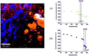
Raman microspectroscopy of noncancerous and cancerous human breast tissues. Identification and phase transitions of linoleic and oleic acids by Raman low-temperature studies
We present the results of Raman studies in the temperature range of 293–77 K on vibrational properties of linoleic and oleic acids and Raman microspectroscopy of human breast tissues at room temperature.
Analyst, 2015,140, 2134-2143
https://doi.org/10.1039/C4AN01877J
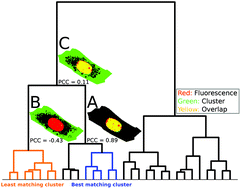
Colocalization of fluorescence and Raman microscopic images for the identification of subcellular compartments: a validation study
This paper introduces algorithms for identifying overlapping observations between Raman and fluorescence microscopic images of one and the same sample.
Analyst, 2015,140, 2360-2368
https://doi.org/10.1039/C4AN02153C
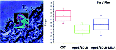
Identification of a biochemical marker for endothelial dysfunction using Raman spectroscopy
We provide evidence that phenylalanine/tyrosine (Phe/Tyr) ratio analysis by Raman spectroscopy discriminate endothelial dysfunction in ApoE/LDLR−/− mice as compared to control animals.
Analyst, 2015,140, 2185-2189
https://doi.org/10.1039/C4AN01998A
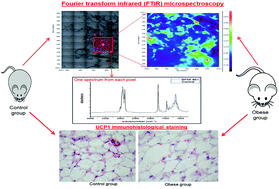
FTIR imaging of structural changes in visceral and subcutaneous adiposity and brown to white adipocyte transdifferentiation
FTIR microspectroscopy coupled with UCP1 immunohistological staining enables the detection of obesity-related molecular alterations and transdifferentiations in visceral and subcutaneous adipose tissues in spontaneously obese mice lines.
Analyst, 2015,140, 2205-2214
https://doi.org/10.1039/C4AN02008A
Evaluation of different tissue de-paraffinization procedures for infrared spectral imaging
Differential distribution of paraffin in a normal colon tissue section after various de-Waxing procedures in comparison to a paraffinized tissue.

Analyst, 2015,140, 2369-2375
https://doi.org/10.1039/C4AN02122C
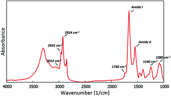
The biochemical changes in hippocampal formation occurring in normal and seizure experiencing rats as a result of a ketogenic diet
In this study, ketogenic diet-induced biochemical changes occurring in normal and epileptic hippocampal formations were compared.
Analyst, 2015,140, 2190-2204
https://doi.org/10.1039/C4AN01857E
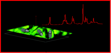
Raman spectroscopy and the material study of nanocomposite membranes from poly(ε-caprolactone) with biocompatibility testing in osteoblast-like cells
Modern medical treatment can be improved by nanotechnology methods for preparing nanocomposites with novel physical, chemical and biological properties.
Analyst, 2015,140, 2311-2320
https://doi.org/10.1039/C4AN02284J
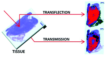
Comparison of transflection and transmission FTIR imaging measurements performed on differentially fixed tissue sections
FTIR microscopy of adjacent sections of tissue measured by transmission and transflection shows comparable images after UHCA.
Analyst, 2015,140, 2376-2382
https://doi.org/10.1039/C4AN02034K
Tissue phantoms to compare spatial and temporal offset modes of deep Raman spectroscopy
Tissue phantoms were created with embedded biomineral-simulating inclusions of varying size and depth, and formed of different mixtures of CaCO3 and hydroxyapatite, for comparison of deep Raman spectroscopy techniques.

Analyst, 2015,140, 2504-2512
https://doi.org/10.1039/C4AN01889C
Comparison of transmission and transflectance mode FTIR imaging of biological tissue
FTIR imaging from samples using translation rather than transmission mode leads to increased variance in the spectra. Whether this matters for spectral pathology is still a matter of debate.

Analyst, 2015,140, 2383-2392
https://doi.org/10.1039/C4AN01975J
A method for the comparison of multi-platform spectral histopathology (SHP) data sets
Results of a study comparing infrared imaging data sets collected on different instruments or instrument platforms are reported, along with detailed methods developed to permit such comparisons.
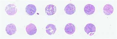
Analyst, 2015,140, 2465-2472
https://doi.org/10.1039/C4AN01879F
Statistical analysis of a lung cancer spectral histopathology (SHP) data set
We report results on a statistical analysis of an infrared spectral dataset comprising a total of 388 lung biopsies from 374 patients.
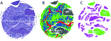
Analyst, 2015,140, 2449-2464
https://doi.org/10.1039/C4AN01832J
Rapid screening of classic galactosemia patients: a proof-of-concept study using high-throughput FTIR analysis of plasma
FTIR as a new approach to screen a rare disease.
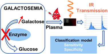
Analyst, 2015,140, 2280-2286
https://doi.org/10.1039/C4AN01942C
An attenuated total reflection (ATR) and Raman spectroscopic investigation into the effects of chloroquine on Plasmodium falciparum-infected red blood cells
Attenuated Total Reflection Fourier Transform Infrared (ATR-FTIR) and Raman spectroscopy were used to compare chloroquine (CQ)-treated and untreated cultured Plasmodium falciparum-infected human red blood cells (iRBCs).
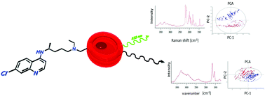
Analyst, 2015,140, 2236-2246
https://doi.org/10.1039/C4AN01904K
Label-free Raman imaging of the macrophage response to the malaria pigment hemozoin
Raman spectroscopy highlights biochemical changes that are spectrally or spatially related to the presence of the malaria pigment, hemozoin, inside macrophage cells, during the initial stages of exposure.
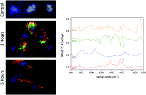
Analyst, 2015,140, 2350-2359
https://doi.org/10.1039/C4AN01850H
Development of a hierarchical double application of crisp cluster validity indices: a proof-of-concept study for automated FTIR spectral histology
The hierarchical double application of crisp cluster validity indices for automated spectral histology of a normal human colon.
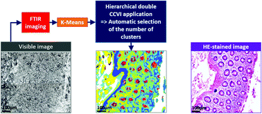
Analyst, 2015,140, 2439-2448
https://doi.org/10.1039/C4AN01937G
Clostridium difficile the hospital plague
Clostridium difficile infection (CDI) has become one of the major public health threats in the last two decades.
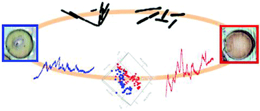
Analyst, 2015,140, 2513-2522
https://doi.org/10.1039/C4AN01947D
Analysis of the developing neural system using an in vitro model by Raman spectroscopy
We developed an in vitro model of early neural cell development.
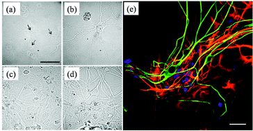
Analyst, 2015,140, 2344-2349
https://doi.org/10.1039/C4AN01961J
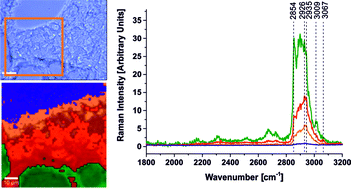
The lipid-reactive oxygen species phenotype of breast cancer. Raman spectroscopy and mapping, PCA and PLSDA for invasive ductal carcinoma and invasive lobular carcinoma. Molecular tumorigenic mechanisms beyond Warburg effect
The paper demonstrates that Raman imaging has reached a clinically relevant level in regard to breast cancer diagnosis applications.
Analyst, 2015,140, 2121-2133
https://doi.org/10.1039/C4AN01876A
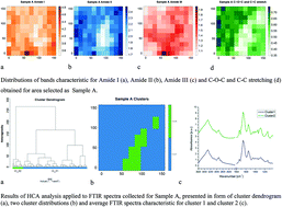
The pituitary gland under infrared light – in search of a representative spectrum for homogeneous regions
This work focuses on obtaining unique representative FTIR spectrum characteristic for one type of cells architecture. Presented idea is based on using of HCA for data evaluation to search for uniform patterns within samples from the perspective of FTIR spectra.
Analyst, 2015,140, 2156-2163
https://doi.org/10.1039/C4AN01985G
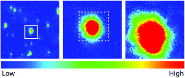
Rapid biodiagnostic ex vivo imaging at 1 μm pixel resolution with thermal source FTIR FPA
Novel high spatial resolution (1 × 1 μm pixel) FTIR imaging with commercial benchtop instrument yields data comparable to that from synchrotron sources.
Analyst, 2015,140, 2493-2503
https://doi.org/10.1039/C4AN01982B
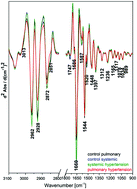
Plasma biomarkers of pulmonary hypertension identified by Fourier transform infrared spectroscopy and principal component analysis
In this work FTIR studies on blood plasma in the rat models of hypertension are reported.
Analyst, 2015,140, 2273-2279
https://doi.org/10.1039/C4AN01864H
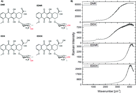
Nuclear accumulation of anthracyclines in the endothelium studied by bimodal imaging: fluorescence and Raman microscopy
Anthracycline antibiotics display genotoxic activity towards cancer cells but their clinical utility is limited by their cardiac and vascular toxicity.
Analyst, 2015,140, 2302-2310
https://doi.org/10.1039/C4AN01882F
Identification of spectral biomarkers for type 1 diabetes mellitus using the combination of chiroptical and vibrational spectroscopy
Chiroptical spectroscopy is able to detect conformational changes of plasmatic biomolecules during type 1 diabetes mellitus.

Analyst, 2015,140, 2266-2272
https://doi.org/10.1039/C4AN01874E
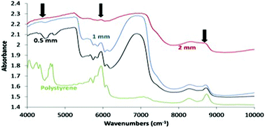
Wavelength-dependent penetration depth of near infrared radiation into cartilage
In this study, we established how the depth of penetration into cartilage varies throughout the NIR frequency range (4000–10 000 cm−1).
Analyst, 2015,140, 2093-2100
https://doi.org/10.1039/C4AN01987C
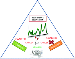
Recurrence prediction in oral cancers: a serum Raman spectroscopy study
Serum Raman spectroscopy was explored for prediction of oral cancer recurrence in before surgery and after surgery blood samples. Findings suggest RS of post-surgery samples may help in prediction of recurrence.
Analyst, 2015,140, 2294-2301
https://doi.org/10.1039/C4AN01860E
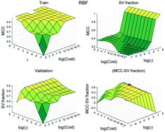
Competitive evaluation of data mining algorithms for use in classification of leukocyte subtypes with Raman microspectroscopy
In this study Raman spectral data from peripheral blood mononuclear cells (PBMCs) is used for the competitive evaluation of three data-mining models in discriminating a highly pure population of T-cell lymphocytes from other myeloid cells within the PBMCs fraction.
Analyst, 2015,140, 2473-2481
https://doi.org/10.1039/C4AN01887G
Infrared micro-spectroscopy for cyto-pathological classification of esophageal cells
We report results from a study utilizing infrared spectral cytopathology (SCP) to detect abnormalities in exfoliated esophageal cells.
Analyst, 2015,140, 2215-2223
https://doi.org/10.1039/C4AN01884B
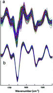
High throughput absorbance spectra of cancerous cells: a microscopic investigation of spectral artifacts
Infrared spectra of cell smears change in shape with cell density.

Analyst, 2015,140, 2393-2401
https://doi.org/10.1039/C4AN01834F
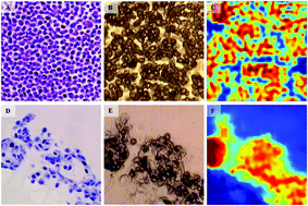
Infrared imaging of MDA-MB-231 breast cancer cell line phenotypes in 2D and 3D cultures
Breast cancer cell lines in 2D (top) and 3D (bottom) culture: H&H, unstained bright field, and IR images.
Analyst, 2015,140, 2336-2343
https://doi.org/10.1039/C4AN01833H
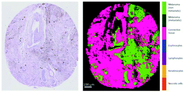
Infrared imaging of primary melanomas reveals hints of regional and distant metastases
FTIR imaging can identify the main cell types of melanoma tumors and can help identify primary melanomas with the highest risk of metastases.
Analyst, 2015,140, 2144-2155
https://doi.org/10.1039/C4AN01831A
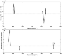
Multivariate statistical methodologies applied in biomedical Raman spectroscopy: assessing the validity of partial least squares regression using simulated model datasets
In the drive towards biomedical applications of Raman spectroscopy, it is critically important to validate the data analysis tools.
Analyst, 2015,140, 2482-2492
https://doi.org/10.1039/C4AN02167C
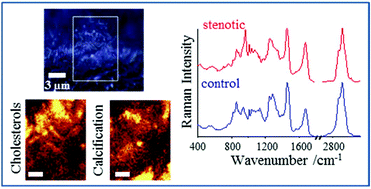
Raman microspectroscopy of human aortic valves: investigation of the local and global biochemical changes associated with calcification in aortic stenosis
Raman microspectroscopy was applied to characterize the local and global biochemical changes associated with calcification in human stenotic aortic valves.
Analyst, 2015,140, 2164-2170
https://doi.org/10.1039/C4AN01856G
An infrared spectral signature of human lymphocyte subpopulations from peripheral blood
Peripheral blood cytotoxic T cells (CD8+), helper T cells (CD4+) and regulatory T cells (T reg) have unique spectral signatures in the mid-infrared.

Analyst, 2015,140, 2257-2265
https://doi.org/10.1039/C4AN02247E
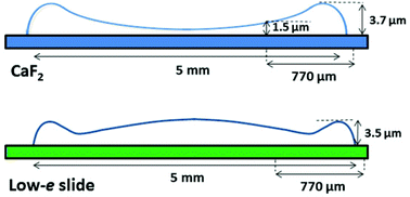
Transmission versus transflection mode in FTIR analysis of blood plasma: is the electric field standing wave effect the only reason for observed spectral distortions?
Fourier transform infrared (FTIR) microspectroscopy is assessed in terms of two techniques (i.e., transmission and transflection) as a method for rapid measurements of blood plasma.
Analyst, 2015,140, 2412-2421
https://doi.org/10.1039/C4AN01842G
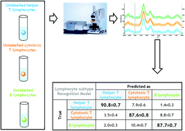
Label-free phenotyping of peripheral blood lymphocytes by infrared imaging
FTIR imaging enables to effectively discriminate lymphocyte subpopulations without antibody labelling.
Analyst, 2015,140, 2247-2256
https://doi.org/10.1039/C4AN01855A
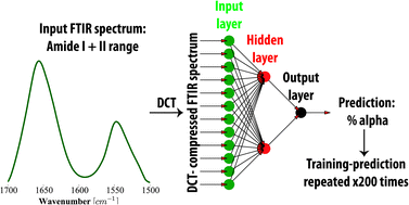
The combination of artificial neural networks and synchrotron radiation-based infrared micro-spectroscopy for a study on the protein composition of human glial tumors
Protein-related changes associated with the development of human brain gliomas are of increasing interest in modern neuro-oncology.
Analyst, 2015,140, 2428-2438
https://doi.org/10.1039/C4AN01867B
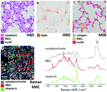
Raman microimaging of murine lungs: insight into the vitamin A content
The composition of mice lung tissue was investigated using Raman confocal microscopy at 532 nm excitation wavelength supported with different experimental staining techniques as well as DFT calculations.
Analyst, 2015,140, 2171-2177
https://doi.org/10.1039/C4AN01881H
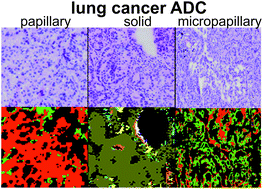
Marker-free automated histopathological annotation of lung tumour subtypes by FTIR imaging
Automated detection of lung cancer adenocarcinoma subtypes by FTIR imaging is presented in this study for the first time.
Analyst, 2015,140, 2114-2120
https://doi.org/10.1039/C4AN01978D
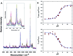
SERS-based monitoring of the intracellular pH in endothelial cells: the influence of the extracellular environment and tumour necrosis factor-α
The intracellular pH plays an important role in various cellular processes.
Analyst, 2015,140, 2321-2329
https://doi.org/10.1039/C4AN01988A
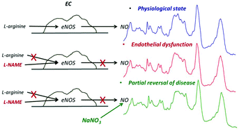
Biochemical changes of the endothelium in the murine model of NO-deficient hypertension
Alterations in the α-helix and β-sheet content and the lipid-to-protein ratio are the most striking features of hypertension development in the vascular endothelium.
Analyst, 2015,140, 2178-2184
https://doi.org/10.1039/C4AN01870B
Comparison of FTIR transmission and transfection substrates for canine liver cancer detection
FTIR spectroscopy is a widely used technique that provides insights into disease processes at the molecular level.
Analyst, 2015,140, 2402-2411
https://doi.org/10.1039/C4AN01901F
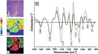
Biomolecular characterization of adrenal gland tumors by means of SR-FTIR
Preliminary FTIR measurements of adrenal gland biocomposition as the first step to find a spectral biomarker of malignancy phenotype.
Analyst, 2015,140, 2101-2106
https://doi.org/10.1039/C4AN01891E
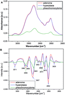
Label-free determination of lipid composition and secondary protein structure of human salivary noncancerous and cancerous tissues by Raman microspectroscopy
The applications of optical spectroscopic methods in cancer detection open new possibilities in oncological diagnostics.
Analyst, 2015,140, 2107-2113
https://doi.org/10.1039/C4AN01394H
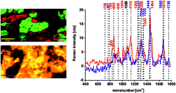
Assessment of the statistical significance of classifications in infrared spectroscopy based diagnostic models
Permutation testing in the evaluation of the statistical significance for infrared based classification of biological samples.
Analyst, 2015,140, 2422-2427
https://doi.org/10.1039/C4AN01783H
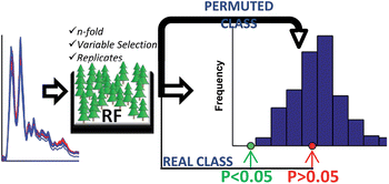
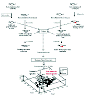
A preliminary Raman spectroscopic study of urine: diagnosis of breast cancer in animal models
Analysis of urine by Raman spectroscopy (RS) as an alternative screening and diagnostic tool for breast cancer..
Analyst, 2015,140, 456-466
https://doi.org/10.1039/C4AN01703J
About this collection
This collection of papers highlights work from the SPEC 2014 conference on Optical Diagnosis held in Krakow, Poland, 17-22 August 2014. Guest Edited by Malgosia Baranska and Hugh J. Byrne. New articles will be added to this collection as they are published.