
 Open Access Article
 This Open Access Article is licensed under a Creative Commons Attribution-Non Commercial 3.0 Unported Licence
Recent advances in biomimetic taste-based biosensors and their applications
Jialu
Kang
a,
Jiejing
Liu
a,
Yufei
Geng
a,
Yuxuan
Yuan
a,
Shuge
Liu
a,
Yushuo
Tan
a,
Liping
Du
*ab and
Chunsheng
Wu
 *ab
*ab
aInstitute of Medical Engineering, Department of Biophysics, School of Basic Medical Sciences, Health Science Center, Xi'an Jiaotong University, Xi'an, Shaanxi 710061, China. E-mail: duliping@xjtu.edu.cn; wuchunsheng@xjtu.edu.cn
bKey Laboratory of Environment and Genes Related to Diseases (Xi'an Jiaotong University), Ministry of Education of China, Xi'an, China
First published on 22nd October 2024
Abstract
The biological taste sensing system has a sensitive perception ability for taste substances (tastants) and is considered as one of the most efficient chemical sensing systems in nature. With the rapid development of human society, biomimetic taste-based biosensors have become increasingly important to improve human life quality and ensure human health, and have been widely applied in many fields such as food safety, biomedicine, and public health. In recent years, researchers have been devoted to developing a new type of chemical sensing system. Among them, biomimetic olfactory-based biosensors have shown promising prospects and potential applications compared to traditional chemical sensors due to the utilization of well-developed natural molecular recognition mechanisms. Biomimetic taste-based biosensors usually employ biologically originated taste cells, taste receptors, taste buds, taste organoids and lipid membranes as sensitive elements, combined with secondary transducers to achieve specific and sensitive detection of tastants in order to obtain comparable detection performance to that of the biological taste system. This review summarizes the most recent advances in biomimetic taste-based biosensors based on biological taste sensing elements. First, the basic principle of biomimetic taste-based biosensors is briefly introduced. Then, the system composition and development of biomimetic taste-based biosensors are outlined and discussed in detail, with a focus on the preparation technology of sensitive elements and their coupling with transducers. In addition, the performance of biomimetic taste-based biosensors and their applications in food quality testing and basic and clinical research are summarized. Finally, the current challenges and development trends of biomimetic taste-based biosensors are proposed and discussed.
Introduction
Taste is a critical physiological sensation, typically categorized into five basic tastes: sour, sweet, bitter, salty, and umami.1–3 It largely determines the selection of food by organisms, allowing them to promptly replenish nutrients essential for survival based on their needs.4,5 Taste plays an essential role in the regulation of feeding, bodily nutrition, and metabolic control. Clinically, certain diseases (such as tongue cancer) and treatment plans (such as radiotherapy for head and neck tumours) can lead to taste disorders or even loss of taste.6–8 However, research on taste has significantly lagged behind studies on vision, hearing, touch, and smell, leading to a lack of timely interventions and treatments for taste abnormalities in clinical settings. Additionally, with the advancement of society and civilization, the demand for higher precision and accuracy in detecting liquid components has increased—something that traditional taste receptors struggle to meet.9,10The human taste organs are taste buds, each of which contains 50 to 100 taste cells.11–13 These taste cells send specific signals related to different taste substances to the brain, which then processes the signals and, through experiences or neural networks, classifies or judges the taste substances, ultimately forming the perception of taste. Biomimetic taste-based biosensors are a novel class of biosensors designed to mimic the signal transduction mechanisms of human taste and the transmission and processing of taste signals by neuronal systems, thereby developing instruments for the detection and analysis of taste substances. Traditional liquid-phase detection technologies, such as electronic tongues, have achieved certain success but are mostly developed based on electrochemical principles, which makes it difficult for them to replicate the biological characteristics of the human tongue and, consequently, to identify the taste profiles of complex mixtures.14 On the other hand, biomimetic taste-based biosensors, which are based on biological sensitive elements, offer a new solution to this challenge. Biomimetic taste-based biosensors combine the high information capture capability of traditional liquid phase detection technologies (such as electronic tongues) with the high sensitivity and specificity of biosensors, thereby improving sensitivity, response time and specificity. This greatly expands the application scope of traditional liquid phase detection methods, overcomes the challenges of mimicking human biological characteristics, and provides a new method for quickly, simply and accurately collecting taste information. It shows broad application potential in many fields such as food detection, fundamental research, clinical research, and pharmaceutical research.15,16
In recent decades, biomimetic taste-based biosensors have attracted more and more attention and significant progress has been achieved. However, there is very limited literature on recent advances in this field. This review provides a comprehensive summary of the most recent advances in biomimetic taste-based biosensors based on biological taste sensing elements. First, the basic principle of biomimetic taste-based biosensors is briefly introduced. Then, the system composition and development of biomimetic taste-based biosensors are outlined and discussed in detail, with a focus on the preparation technology of sensitive elements and their coupling with transducers. In addition, the performance of biomimetic taste-based biosensors and their applications in food quality testing and basic and clinical research are summarized. Finally, the current challenges and development trends of biomimetic taste-based biosensors are proposed and discussed.
Recognition mechanism of the mammalian taste sensing system
The generation of taste sensation in mammals originates from the detection of chemical stimuli presented by the tastants in the oral cavity through taste buds, which are clusters of specialized taste cells responsible for sensing taste signals. There are various types of papillae distributed on the different regions of the tongue surface. At present, it is known that there are one to hundreds of taste buds located in one papillae. These taste buds send the detected taste signals to the brain via afferent cranial nerves.17 There are ∼50–100 taste cells located in a taste bud, which can be classified into four categories based on their ultrastructural features (i.e. type I (dark), type II (light), type III (intermediate), and type IV cells). At present, it is known that type II taste cells are responsible for the detection of sweet, bitter, and umami taste substances, while type III cells are responsible for the detection of sour and salty.6 Each taste responsive cell contains membrane proteins known as taste receptors (Fig. 1). These taste receptors could interact with chemical substances in the oral cavity. There are five types of basic taste, each corresponding to the detection of one of the five basic tastes: sour, sweet, bitter, salty, and umami. | ||
| Fig. 1 Schematic diagram showing the structure of a mammalian tongue. (A) The bumps on the surface of the tongue are called papillae. (B) Taste buds are hidden beneath the surface of the papillae and barely poke out. (C) Each taste bud is made up of a cluster of cells, which are packed together like segments of an orange. (D) The cells making up taste buds store special taste receptors at their tips, which respond to tastants presented by foods.18 | ||
Once the taste receptors detect chemical stimuli, the taste bud cells transduce the stimulus into electrical signals or the release of neuro transmitters through intracellular signal transduction pathways. These taste responsive signals are received by afferent nerve fibers distributed across the tongue and transmitted from the back of the oral cavity to the relevant areas in the cerebral cortex responsible for taste perception (Fig. 2), where the brain processes and makes judgments regarding the taste.18
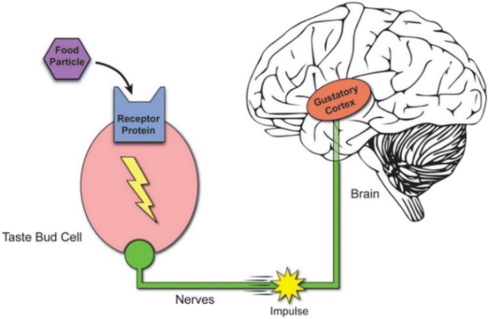 | ||
| Fig. 2 Schematic diagram showing taste signal transduction and transmission pathways. Taste signals begin when food tastants are sensed by taste receptors on the taste bud cells. When the receptors sense different kinds of tastants, the taste bud cells could transduce the corresponding taste signals and send them to the nervous system, which relays the impulse to the brain. This diagram is simplified and shows a taste bud cell with one receptor. In reality, each taste bud cell could have millions of receptors.18 | ||
The study of the molecular basis of taste perception is a relatively young and interesting field. Here, we present an overview of the recent advances and what is currently understood to be the primary molecular mechanisms of taste perception. Signal transduction mechanisms for salty taste perception: the low-sodium-sensing cell is depolarized by the influx of Na+via the amiloride-sensitive epithelial sodium channel (ENaC). Additional influx of Na through the voltage-gated sodium channel (VGNaC) creates an action potential that ultimately leads to the release of ATP through CALHM1/3, without the involvement of intracellular calcium (Ca2+).2,3 The mechanistic overview of signal transduction in sweet, bitter, and umami taste perception: tastants bind to cell surface G protein-coupled receptors (GPCRs) and initiate a signaling cascade through phospholipase Cβ2 (PLCβ2) and inositol 1,4,5-triphosphate (IP3) that mobilizes Ca2+ from the endoplasmic reticulum (ER) by activating IP3 receptor type 3 (IP3R3), thus increasing intracellular Ca2+ concentration. The spike in intracellular calcium activates transient receptor potential cation channel subfamily M member 5 (TRPM5) channels that depolarize the plasma membrane and create action potentials via voltage-gated Na+ channels (VGNa+ C). These changes in the membrane potential and the increased presence of intracellular Ca2+ trigger the release of ATP into the channel synapse through CALHM1/3 s. This stimulates the gustatory neuron, thus completing transduction of the signal from the taste cell to the afferent.2,4,5
Basic principle of biomimetic taste-based biosensors
Biomimetic taste-based biosensors can measure taste signals without distinguishing between individual chemical substances, employing a taste-sensing system that mimics the response mechanism of the mammalian taste system. These biosensors can quantify taste characteristics. Biomimetic taste-based biosensors mainly consist of two components: the primary sensitive element, which is a bioactive material that binds to specific taste substances and generates a specific response, and the secondary transducer, which is a micro/nano sensor or device that is able to convert the responsive signals of sensitive elements into physical signals, such as optical or electrical signals, which are easier to process.9,15 The basic principle of these biosensors is based on the utilization of sensitive elements to detect chemical signals presented by food, which can subsequently be transduced by the secondary transducer into electrical or optical signals. As a result, sensitive elements play a crucial role in determining the performance of these biosensors. Biomimetic taste-based biosensors usually utilize bioactive materials from biological taste systems as sensitive elements, which include taste buds, taste responsive cells, taste receptors, and taste organoids. Thus, to some extent, these biomimetic taste-based biosensing systems inherit some of the advantages of biological taste systems, such as rapid response, high sensitivity, and good specificity. On the other hand, the rapid advancement in microfabrication processes also greatly facilitates the development of various micro/nano sensors or devices for the purpose of transducing responsive signals from sensitive elements with high efficiency, which could thus improve the performance of the whole biomimetic taste-based biosensing system.Preparation of sensitive elements and their coupling with transducers
For the design and construction of biomimetic taste-based biosensors, one of the key steps is to prepare sufficient functional bioactive materials that could respond to certain taste signals. At present, a wide range of sensitive elements can be utilized in biomimetic taste-based biosensors. Basically, any biological materials that exhibit taste-responsive characteristics can serve as sensitive elements in these biosensors, including taste cells, taste receptors, taste buds, taste bud organoids, and sensitive membranes. On the other hand, it is also very important to couple the sensitive elements with transducers with high efficiency. Various strategies have been employed to improve the coupling efficiency of bioactive materials with transducers. By the following, we will introduce different types of biomimetic taste-based biosensors in detail based on the different sensitive elements.In addition, since taste receptors can also be expressed in other parts of the mammalian body, such as the digestive tract, respiratory system, brain, and heart, cells beyond taste epithelial cells, such as cardiomyocytes, can also be used to construct biomimetic taste-based biosensors. For example, rat cardiomyocytes have been used as taste sensing elements, coupling a MEA as a transducer.21 The specific detection of two bitter substances (benzydamine and diphenidol) and one umami compound (MSG) was achieved, with a detection limit of 10−6 M. Similarly, a coupled sensor array integrating taste and olfactory sensing has been developed based on HL-1 cardiomyocytes with a MEA, which was used for drug screening and evaluation for treating tachycardia.22 An in vitro tachycardia model was established using isoproterenol as the stimulant. The proposed sensor array facilitated the screening of potential drugs for treating tachycardia, such as salicin, artemisinin, bufalin, and azelaic acid, all of which activated specific receptors on HL-1 cells. This research not only paved a new path for building integrated taste and olfactory sensors, but also provided a powerful tool for quantitatively evaluating potential tachycardia treatments. Furthermore, chemesthetic cells from the intestinal secretin tumor cell line (STC-1) have been applied to taste recognition.23 Multivariate data processing through partial least squares discriminant analysis (PLS-DA) was employed to decode the taste-specific cellular responses from ion chromatography fingerprints, enabling the distinction between bitter, sweet, and umami tastes. The PLS-DA model achieved nearly perfect identification of cell response ion chromatogram fingerprints, improving classification accuracy from 80.0% to 98.9% (Fig. 3 and 4). More recently, a bio-hybrid tongue has been proposed based on a hypothalamic neuronal network (HNN) chip coupled with a MEA.24 This HNN-on-a-chip bio-hybrid tongue serves as a valuable in vitro tool for studying hypothalamic neurons, elucidating glucose-sensing mechanisms, and understanding the function of hypothalamic neurons.
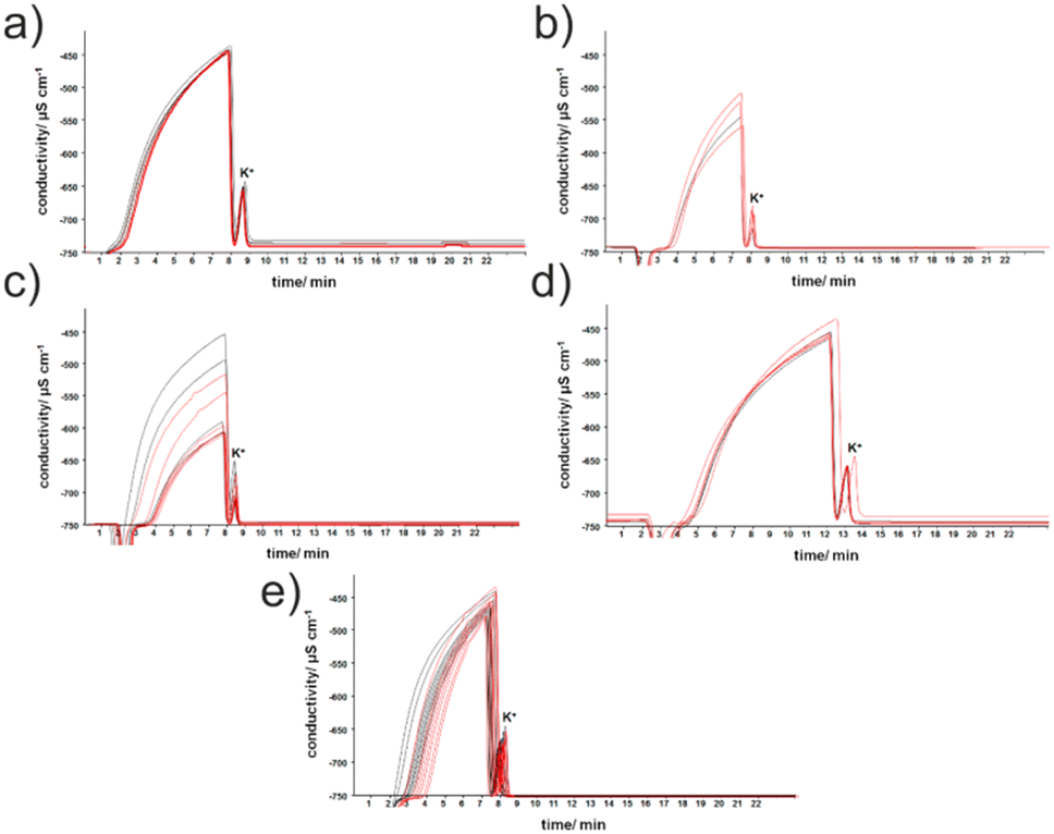 | ||
| Fig. 3 Examples of chromatograms (red) of Dulbecco-modified phosphate buffered saline (DPBS) added to intestinal secretin tumor cell line (STC-1) cells after treatment with 1 mM solutions of: (a) aspartame, (b) citric acid, (c) caffeine citrate, (d) sodium L-glutamate, and (e) sodium chloride. Black denotes control groups. Controls were carried out in the absence of taste substances.23 | ||
 | ||
| Fig. 4 Partial least squares discriminant analysis (PLS-DA) classification of cellular responses towards five basic tastants: (A) 3D PLS-DA score plot of chromatographic fingerprints of the STC-1 cellular response; (B) and (C) confusion matrices for basic taste recognition.23 | ||
![[double bond, length as m-dash]](https://www.rsc.org/images/entities/char_e001.gif) S groups. On the other hand, a taste-based biosensor has been developed by integrating the ligand-binding domain of the umami receptor type 1 member 1 venus flytrap (T1R1 VFT) with a graphene-based field-effect transistor (FET) transducer, enabling real-time detection of monosodium L-glutamate (MSG) with high sensitivity (∼1 nM) and specificity.29
S groups. On the other hand, a taste-based biosensor has been developed by integrating the ligand-binding domain of the umami receptor type 1 member 1 venus flytrap (T1R1 VFT) with a graphene-based field-effect transistor (FET) transducer, enabling real-time detection of monosodium L-glutamate (MSG) with high sensitivity (∼1 nM) and specificity.29
A biomimetic taste-based biosensor has been developed by expressing the ligand-binding domain of the human taste receptor T1R1-VFT and coupling it with gold nanoparticles (AuNPs) as the recognition elements.30 By combining the properties of semiconductor and noble metal materials, a bimetallic composite nanomaterial was synthesized as the substrate, creating a biosensor to detect the synergistic effects of umami substances. More recently, Escherichia coli (E. coli) bacteria have also been used to express the human bitter taste receptor, hT2R4, which makes it possible to detect bitter substances.31 The study innovatively employed a simple and low-cost electrochemical device based on an indium tin oxide (ITO)-based electrolyte-semiconductor (ES) structure as a transducer, allowing for real-time evaluation of receptor-activated bacterial metabolic responses. On the other hand, a biosensor has been proposed based on the ligand-binding domain of the human sweet taste receptor protein (T1R2 VFT).32 This biosensor demonstrated biomimetic characteristics and initiated the application of immune recognition in taste-based biosensors, allowing for the precise and sensitive differentiation of sweet substances from other related taste compounds, with a detection limit of 5.1 pM, far lower than that of taste biosensors without immune amplification. This biomimetic electrochemical taste biosensor serves as a novel screening platform for taste development and sheds light on the mechanisms of sweet taste perception. In addition, a nanobionic taste biosensor was constructed based on the human taste receptor protein hTAS2R38, carboxylated polypyrrole nanotubes (CPNT), and a FET, which exhibited high sensitivity and selectivity.33 This biosensor was able to detect taste in mixtures and food with human-like taste performance. However, since a single receptor cannot respond to multiple taste sensations simultaneously, it is challenging for receptor-based biomimetic taste sensors to detect multiple taste types at the same time.25
Meanwhile, a taste biosensor has been developed by the deposition of tyrosinase and glucose oxidase onto polypyrrole (Ppy) or Ppy/AuNP composites for detecting phenols and sugars, which are two key quality indicators for grapes and wine.35 The results indicated that both enzyme materials played a significant role in the sensor's response process. Similarly, a taste biosensor has been developed using bovine serum albumin (BSA), ovalbumin (OVA), and β-lactoglobulin (BLG), along with modified gold nanoparticles (AuNPs).36 These biosensors, based on the differential affinity between biorecognition receptors and various analytes, were able to detect and differentiate acrylamide and six of its analogs (such as acetamide, nicotinamide, methacrylamide, N-tert-butylacrylamide, diacetone acrylamide, and N-(hydroxymethyl)acrylamide). This has significant implications for food safety monitoring in the food industry.
The tissue-derived method involves directly digesting taste bud tissues to obtain taste buds, which are then cultured into taste bud organoids.42 Specifically, the tongues of mice or rats are extracted, followed by collagenase injection to acquire the tongue epithelium and the isolation of taste papillae. The obtained taste papillae tissues are further digested using trypsin (Fig. 5) and mixed with Matrigel, after which they are seeded onto culture plates, allowing for the growth of organoids. Subsequently, their physiological characteristics are characterized through the detection of taste lineage marker mRNA and protein expression levels, along with immunofluorescence techniques.
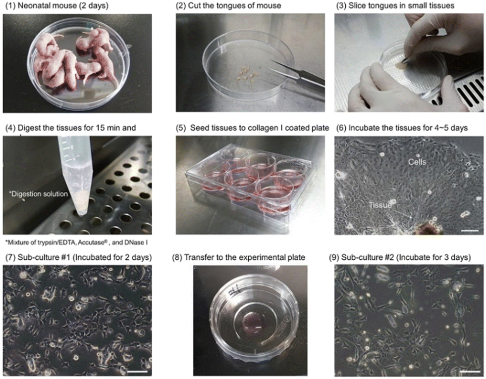 | ||
| Fig. 5 Tissue-derived taste bud organoid culture procedures.42 | ||
The stem/progenitor cell-derived method involves pre-labelling the taste stem/progenitor cells obtained from tissues, followed by fluorescence-activated cell sorting (FACS) to enable single-cell culture of taste stem/progenitor cells.43 These cells are then induced to divide and differentiate, forming taste bud organoids that respond similarly to the original tissue (Fig. 6). Once the taste bud organoids are obtained, they can be coupled with MEA electrodes to effectively simulate the biological taste sensing system, allowing for substance detection.
 | ||
| Fig. 6 Lgr5+ cell-derived taste bud organoid culture. (A) Lgr5+ cells in the taste papilla. (B) Results of FACS sorting of Lgr5+ cells. (C) Single Lgr5+ cell (green). (D) Representative images of taste bud organoids on different days in culture.43 | ||
A coupling method is presented here in detail; taste organoids were harvested on day 14 and put onto microelectrodes of the MEA chip overnight in the medium. After that, the medium was withdrawn and a volume of 3 μL Matrigel was then gently pipetted from the top to where the organoids were located, followed by a 20 min curing process in an incubator at 37 °C. For the construction of the detection system, 1 mL medium was added to the taste organoids-on-a-chip and the chip was put on the MEA2100-System which was connected to the computer. The cell culture incubator was used to maintain an essential environment (37 °C, 5% CO2) for taste organoids-on-a-chip maintenance. For recognition of the tastants, the medium in the chip was replaced with a medium containing the taste stimulus and the extracellular changes were recorded.6
Performances of biomimetic taste-based biosensors
Biomimetic taste-based biosensors are analytical tools capable of recognizing and detecting specific taste qualities. Their standout features include a low detection limit, high selectivity and sensitivity, improved stability and reproducibility, cost-effectiveness, and particularly, high-throughput detection capabilities. Factors that can enhance the detection performance of biomimetic taste-based biosensors include:1 improving components that facilitate the binding between biomolecules and taste substances,2 employing biomolecules as sensitive elements, and (3) optimizing methods for signal detection.The development of intelligent portable devices has been central to sensor research and is the ultimate goal for the development of various types of biosensors. These devices, characterized with miniaturization, intelligence, and excellent detection capabilities (such as high specificity, sensitivity, reproducibility, and robustness), enable rapid, accurate, and on-site detection, which are the hallmarks of high-quality sensors. In recent years, the rapid advancement of microfluidic integrated devices has provided a potential platform for the smart and portable design of biomimetic taste-based biosensors, with applications ranging from the construction of in vitro clinical diagnostic models53,54 to drug screening and transport studies.55,56 Their advantages, including miniaturization, integration, automation, and high throughput, offer promising directions for the complex, high-throughput detection of taste substances. Moreover, the use of 3D biomimetic micro-structures and biomaterials (such as biocompatible hydrogels) allows for the cultivation of organs and tissues on microfluidic platforms,57 overcoming the limitations of traditional 2D cell cultures and animal experiments. These 3D culture systems enable a more physiologically relevant environment for in vitro tissues and organs, providing a reliable platform for cultivating and functionally validating taste organoids.
Applications of biomimetic taste-based biosensors
After years of development, biomimetic taste-based biosensors have found widespread applications in industrial fields such as food quality testing, as well as in biomedical fields. Although the new generation of ex vivo molecular and cellular taste biosensors is still in its early stages, their taste detection performance has been repeatedly validated in laboratory settings. These biosensors are now gradually entering the application testing phase, with four main areas of application.Food detection
Food detection is essential for ensuring food safety and quality. Biomimetic taste-based biosensors, using various biological sensitive materials (such as the advanced taste sensing systems of insects), enable the detection of specific molecules and play a crucial role in protecting consumers' rights and promoting the healthy development of the domestic food market.Various biomimetic taste-based biosensors have been developed for the applications of food quality detection. For example, a differential colorimetric nano-biosensor platform has been developed using three biological receptor units (bovine serum albumin, ovalbumin, and β-lactoglobulin) to distinguish and quantify multiple foodborne carcinogens, including acrylamide and six of its analogs.36 This platform could accurately distinguish foodborne amides based on their amine subgroups, IARC (International Agency for Research on Cancer) carcinogen classifications, and food additive types, even at ultra-low concentrations (500 pM). The sensor array also successfully classified non-target analytes according to sweetener and food component types, achieving 100% correct classification with high correlation coefficients in partial least squares regression (R2 > 0.95). More recently, a biomimetic taste-based biosensor has been developed by simulating the insect taste system, integrating a carbon nanotube field-effect transistor (CNT-FET) with nanovesicles, including the sugar receptor of bees, Apis mellifera Gr1 (AmGr1), for real-time detection and differentiation of natural and artificial sweeteners.58 This biosensor could detect glucose (the main component of nectar) down to 100 fM in real time and distinguish sweetness from other tastes. Lately, a biomimetic taste-based biosensor was developed based on enzymes (tyrosinase and glucose oxidase) combined with Ppy or Ppy/AuNP composites, applying it to the analysis and differentiation of grape juice and wine.35 Recent advances in taste cell- and receptor-based biosensors have demonstrated their potential in various fields such as food safety and drug discovery.59
Fundamental research
In recent years, biomimetic taste-based biosensors have employed bioactive materials to achieve detection performance similar to biological taste systems, enabling the detection of the five basic tastes: sour, sweet, bitter, salty, and umami. These biosensors provide useful tools for the taste research and have been widely used in the fundamental research of taste signal transduction mechanisms as well as the molecular mechanisms of taste sensation.A sour taste biomimetic biosensor has been developed based on a MEA and taste receptor cells.60 The study constructed a Hodgkin–Huxley-type mathematical model of whole-cell acid-sensitive taste receptor cells to detect acid taste responses during and after stimulation. Chemesthetic cells from the intestinal secretin tumor cell line (STC-1) have been used for taste recognition and partial least squares discriminant analysis (PLS-DA) for multivariate data processing was used to decode taste-specific cell responses from ion chromatography fingerprints.23 This method can be used for the development of non-invasive taste detection systems to differentiate bitter, sweet, and umami tastes and to study taste transduction mechanisms in vitro. Similarly, a 30-channel MEA with a diameter of 32 μm was used as a multi-channel recording platform and intact taste epithelium from rats was employed as a sensitive element for bitter taste detection.19 More recently, the bitter taste receptor, T2R4, was used as a sensitive element in a chemobiological biosensor and a quartz crystal microbalance (QCM) device was employed to monitor the preparation process of the biosensor and its response to different bitter substances by measuring shifts in the crystal resonant frequency (Fig. 7).61 In addition, live mouse sperm cells were utilized as the primary sensitive element, combined with Fluo 4-AM sensors and flow cytometry, to achieve rapid quantification of bitter compounds (Fig. 8).62
 | ||
| Fig. 7 (A) Real-time monitoring of crystal resonant frequency shifts indicating responses of a chemical biosensor to MgSO4, denatonium, D-(−)-salicin, and quinine. (B) Statistical results of this chemical biosensor with and without bitter receptors responding to different bitter substances.61 | ||
 | ||
| Fig. 8 (A) Schematic diagram of the detection principle of a cell-based biosensing system; (B) detection principle of the flow cytometry measurement system; (C) process diagram for obtaining mouse sperm cells.61 | ||
In addition to single parameter measurement, the first dual-function extracellular recording biosensor has also been developed based on a LAPS, which used an ATP-sensitive aptamer to detect the enhancement effect of denatonium and the inhibitory effect of CBX (carbenoxolone) on bitter taste signal transduction by recording extracellular membrane potential and ATP release from individual taste bud cells.63 Moreover, bitter taste receptors, T2R4, T2R14, and T2R16, were coupled with a multi-channel surface acoustic wave (SAW) device, using the SAW device as a transducer to monitor the specific interactions between bitter taste receptors and their ligands.64 This biomimetic taste-based biosensor was able to monitor minute mass changes by recording phase shifts in surface acoustic waves. This study innovatively employed DNA-directed immobilization (DDI) technology to efficiently and controllably immobilize bitter taste receptors onto the sensitive region of the SAW device. Experimental results demonstrated that this biomimetic taste-based biosensor could produce distinct response signals to different bitter substances with good stability, providing a valuable method for developing receptor-based biosensors. Similarly, the VFT domain of T1R1, an umami ligand-binding domain, was used and immobilized on a graphene-based FET. This umami biomimetic taste-based biosensor could detect monosodium glutamate (MSG) in real time with high sensitivity (around 1 nM) and specificity, offering excellent reusability and storage stability for detecting and distinguishing umami.29 More recently, a bioelectronic taste-based biosensor was developed using bioengineered E. coli cells coupled with ITO, providing a simple and cost-effective strategy for constructing bioelectronic identification devices.31 On the other hand, rat cardiomyocytes have also been utilized as primary taste sensitive elements, with a MEA as a transducer, to achieve the specific detection of two bitter agents (benzydamine and diphenidol) and one umami compound (monosodium glutamate).21 Furthermore, a preliminary study has been carried out to investigate the recognition mechanism of the hT1R1 umami receptor–ligand interaction using a human umami receptor biosensor.65 They discovered that hT1R1 is intrinsically a nitrogen signal recognition receptor, capable of recognizing umami substances through its amino group.
Clinical research
In the field of clinical research and medical diagnostics, biomimetic taste-based biosensors hold great promise and potential for detecting various diseases by sensing bodily fluids released from different tissues. For instance, with the ongoing research into the 2019 coronavirus (COVID-19), in addition to common symptoms such as fever, cough, shortness of breath, fatigue, and muscle pain, loss of taste has increasingly been recognized as a symptom. A recent study revealed that up to 41% of individuals infected with severe acute respiratory syndrome coronavirus-2 (SARS-CoV-2) experienced taste loss.6 Additionally, up to 75% of head and neck cancer patients suffer from taste disorders, primarily due to radiation therapy related to cancer treatment.7 A range of factors, including cancer treatments, bacterial and viral infections, aging, and certain medications, can impair the taste system and disrupt its function.8,66With the rapid research progress, animal models have been increasingly unable to meet experimental requirements. For example, issues such as immune rejection in organ transplants or discrepancies between animal and human cells leading to inconsistent test results have limited the effectiveness and reliability of drug efficacy and toxicity tests. Biomimetic taste-based biosensors have emerged as a promising direction for scientists to address these challenges, which have attracted more and more attention and shown promising prospects.
Pharmaceutical research
Compared to biomimetic olfactory-based biosensors, biomimetic taste-based biosensors are still in their early stages and face significant limitations in applications. However, their broad potential for future use is beginning to emerge. Current research on biomimetic taste-based biosensors in the pharmaceutical field primarily focuses on the evaluation of bitterness in drugs and the suppression of bitterness to improve the palatability of medications. For example, primary rat tongue epithelial cells have been combined with MEA chips to construct a biomimetic taste-based biosensor, offering new insights into the application in masking the bitterness of drugs.67E. coli bacteria transfected with human bitter taste receptor genes have also been combined with carbon nanotubes and FET devices, to detect bitter substances such as phenylthiourea.68 This approach provides a new method for future drug screening and other pharmaceutical applications.Conclusions and prospects
The collection and analysis of taste-related information are essential in many areas closely tied to human life, such as environmental monitoring, medical evaluation, drug development, and food safety. Biomimetic taste-based biosensors, which mimic the taste-sensing mechanisms of animals, serve as high-throughput analytical instruments capable of simulating human taste. With advancements in biotechnology, microfluidics, and nanotechnology,69 biomimetic taste-based biosensors have developed rapidly, with continuous performance optimization, offering a novel approach for the quick, simple, and accurate collection of taste information. These biosensors have vast application potential. Biomimetic taste-based biosensors combine the high information capture capability of traditional liquid-phase detection techniques, such as electronic tongues, with the high sensitivity and specificity of biosensors. This significantly expands the application range of traditional liquid-phase detection methods and overcomes the challenges of mimicking human biological characteristics.However, biomimetic taste sensors are still in the early stages of development and face numerous challenges. For example, the lack of a comprehensive theoretical foundation regarding the signal transduction mechanisms of taste signals and their modulation has, to some extent, limited the preparation of sensitive elements. Additionally, weak coupling between sensitive elements and transducers results in low response intensity: sensitive elements such as cells are often unstably immobilized during the coupling process, leading to lower cellular responses compared to typical adherent cells. Sensitivity and stability still need to be improved to meet the requirements of accurate extracellular recording. For instance, sensor performance may be influenced by bitter substances used as stimuli for cellular measurement. Essentially, cell impedance sensors are highly sensitive to changes in ion concentration and are therefore better suited for low-concentration stimuli, whereas receptor cells require high concentrations of taste stimuli. As a result, the response of cell impedance to high-concentration stimuli may be masked by the impedance of the culture medium. Despite these challenges, the future of biomimetic taste sensors holds great potential and commercial prospects. As an emerging analytical technology, they are expected to see significant growth, expand into more research fields, and make greater contributions to human society.
Data availability
There are no new data associated with this review. No new data were generated or analyzed in support of this research.Author contributions
Jialu Kang, Liping Du and Chunsheng Wu proposed the idea together. Jialu Kang, Jiejing Liu, and Yufei Geng performed the main text writing and production of some figures and tables. Yuxuan Yuan, Shuge Liu, and Yushuo Tan contributed to the introduction writing and full text modification. Liping Du, Chunsheng Wu, and Yushuo Tan contributed to funding acquisition and supervision. Yushuo Tan and Liping Du performed adjustment of the article format and language checking. Chunsheng Wu contributed to writing – review & editing, the enhancement of the main text, production of some figures and tables, supervision, and project administration.Conflicts of interest
There are no conflicts to declare.Acknowledgements
This work was supported financially by grants from the National Natural Science Foundation of China (grant numbers: 32071370, 32271427, and 32471433) and the S&T Program of Hebei (grant number: 22321902D).Notes and references
- (a) J. Chandrashekar, M. A. Hoon, N. J. P. Ryba and C. S. Zuker, Nature, 2006, 444, 288–294 CrossRef CAS; (b) A. Taruno, K. Nomura, T. Kusakizako, Z. Ma, O. Nureki and J. K. Foskett, Pfluegers Arch., 2021, 473, 3–13 CrossRef CAS.
- (a) S. Iwata, R. Yoshida and Y. Ninomiya, Curr. Pharm. Des., 2014, 20, 2684–2692 CrossRef CAS PubMed; (b) M. Sk, M. Pj and M. Rf, Nature, 1992, 357(6379), 563–569 CrossRef PubMed.
- (a) J. Zhang, H. Jin, W. Zhang, C. Ding, S. O'Keeffe, M. Ye and C. S. Zuker, Cell, 2019, 179, 392–402 CrossRef CAS PubMed , e315; (b) D. Dutta Banik, L. E. Martin, M. Freichel, A.-M. Torregrossa and K. F. Medler, Proc. Natl. Acad. Sci. U. S. A., 2018, 115, E772–E781 CrossRef PubMed.
- (a) X. C. Luo, Z. H. Chen, J. B. Xue, D. X. Zhao, C. Lu, Y. H. Li, S. M. Li, Y. W. Du, Q. Liu, P. Wang, M. Liu and L. Huang, Proc. Natl. Acad. Sci. U. S. A., 2019, 116, 5564–5569 CrossRef CAS PubMed; (b) J. H. Jang, O. Kwon, S. J. Moon and Y. T. Jeong, Endocrinol. Metab., 2021, 36, 469–477 CrossRef CAS.
- (a) M. Raipuria, G. Hardy, H. Bahari and M. Morris, Nutr., Metab. Cardiovasc. Dis., 2015, 25, 881–888 CrossRef CAS PubMed; (b) J. Wu, C. Chen, C. Qin, Y. Li, N. Jiang, Q. Yuan, Y. Duan, M. Liu, X. Wei, Y. Yu, L. Zhuang and P. Wang, Adv. Sci., 2023, 10, 2206101 CrossRef CAS PubMed.
- (a) C. Qiu, C. Cui, C. Hautefort, A. Haehner, J. Zhao, Q. Yao, H. Zeng, E. J. Nisenbaum, L. Liu, Y. Zhao, D. Zhang, C. G. Levine, I. Cejas, Q. Dai, M. Zeng, P. Herman, C. Jourdaine, K. With, J. Draf, B. Chen, D. T. Jayaweera, J. C. Denneny, R. Casiano, H. Yu, A. A. Eshraghi, T. Hummel, X. Liu, Y. Shu and H. Lu, Otolaryngol.--Head Neck Surg., 2020, 163, 714–721 CrossRef; (b) D. D. Banik, E. D. Benfey, L. E. Martin, K. E. Kay, G. C. Loney, A. R. Nelson, Z. C. Ahart, B. T. Kemp, B. R. Kemp, A.-M. Torregrossa and K. F. Medler, PLoS Genet., 2020, 16, e1008925 CrossRef.
- A. J. Hovan, P. M. Williams, P. Stevenson-Moore, Y. B. Wahlin, K. E. O. Ohrn, L. S. Elting, F. K. L. Spijkervet and M. T. Brennan, Support Care Cancer, 2010, 18, 1081–1087 CrossRef.
- T. Hummel, B. N. Landis and K.-B. Hüttenbrink, GMS Curr. Top. Otorhinolaryngol Head Neck Surg., 2011, 10, 1–15 Search PubMed.
- C. Wu, P. Lillehoj and P. Wang, Analyst, 2015, 140, 7048–7061 RSC.
- A. S. Shah, Y. Ben-Shahar, T. O. Moninger, J. N. Kline and M. J. Welsh, Science, 2009, 325, 1131–1134 CrossRef CAS PubMed.
- A. K. Deisingh, D. C. Stone and M. Thompson, Int. J. Food Sci. Technol., 2004, 39, 587–604 Search PubMed.
- S. E. Swithers and T. L. Davidson, Behav. Neurosci., 2008, 122, 161–173 Search PubMed.
- J. G. Brand, J. Nutr., 2000, 130, 942S–945S CrossRef CAS PubMed.
- D. Ha, Q. Sun, K. Su, H. Wan, H. Li, N. Xu, F. Sun, L. Zhuang, N. Hu and P. Wang, Sens. Actuators, B, 2015, 207, 1136–1146 CrossRef CAS.
- C. Wu, L. Du, L. Zou, L. Zhao, L. Huang and P. Wang, Sens. Actuators, B, 2014, 201, 75–85 CrossRef CAS.
- Y. Tahara and K. Toko, IEEE Sens. J., 2013, 13, 3001–3011 Search PubMed.
- R. A. DeFazio, G. Dvoryanchikov, Y. Maruyama, J. W. Kim, E. Pereira, S. D. Roper and N. Chaudhari, J. Neurosci., 2006, 26, 3971–3980 CrossRef CAS PubMed.
- L. Vera and S. Wooding, Front. Young Minds, 2017, 5, 33 CrossRef.
- Q. Liu, D. Zhang, F. Zhang, Y. Zhao, K. J. Hsia and P. Wang, Sens. Actuators, B, 2013, 176, 497–504 CrossRef CAS.
- Y. Fan, W. Chen, N. Zhang, M. Li, Y. Zhu, G. Chen, Y. Zhang and Y. Liu, Biosens. Bioelectron., 2023, 237, 115447 CrossRef.
- X. Wei, C. Qin, C. Gu, C. He, Q. Yuan, M. Liu, L. Zhuang, H. Wan and P. Wang, Biosens. Bioelectron., 2019, 145, 111673 CrossRef CAS PubMed.
- C. Qin, Q. Yuan, H. Han, C. Chen, J. Wu, X. Wei, M. Liu, H. Zhang, J. Ping, L. Xu and P. Wang, Biosens. Bioelectron., 2023, 223, 115034 CrossRef CAS PubMed.
- M. Zabadaj, A. Szuplewska, M. Balcerzak, M. Chudy and P. Ciosek-Skibińska, Sensors, 2019, 19, 1062 CrossRef CAS PubMed.
- C. Qin, Q. Yuan, M. Liu, L. Zhuang, L. Xu and P. Wang, Biosens. Bioelectron., 2024, 244, 115784 CrossRef CAS.
- Y. Tian, P. Wang, L. Du and C. Wu, TrAC, Trends Anal. Chem., 2022, 157, 116778 CrossRef CAS.
- C. Wu, L. Du, L. Mao and P. Wang, J. Innovative Opt. Health Sci., 2012, 05, 1250008 CrossRef.
- C. Wu, L. Du, L. Zou, L. Huang and P. Wang, Analyst, 2013, 138, 5989–5994 RSC.
- C. Qin, Z. Qin, D. Zhao, Y. Pan, L. Zhuang, H. Wan, D. P. Antonella, E. Malach, M. Niv, L. Huang, N. Hu and P. Wang, Talanta, 2019, 199, 131–139 CrossRef CAS PubMed.
- S. R. Ahn, J. H. An, I. H. Jang, W. Na, H. Yang, K. H. Cho, S. H. Lee, H. S. Song, J. Jang and T. H. Park, Biosens. Bioelectron., 2018, 117, 628–636 CrossRef CAS PubMed.
- Y. Yu, S. Jiang, Z. Cui, N. Zhang, M. Li, J. Liu, H. Meng, S. Wang, Y. Zhang, J. Han, X. Sun, W. Zhao and Y. Liu, Biosens. Bioelectron., 2023, 234, 115357 CrossRef CAS PubMed.
- J. Wang, S. Kong, F. Chen, W. Chen, L. Du, W. Cai, L. Huang, C. Wu and D.-W. Zhang, Anal. Chim. Acta, 2019, 1079, 73–78 CrossRef CAS PubMed.
- J. Ye, M. Fan, X. Zhang, Q. Liang, Y. Zhang, X. Zhao, C.-T. Lin and D. Zhang, Biosens. Bioelectron., 2024, 249, 116001 CrossRef CAS PubMed.
- H. S. Song, O. S. Kwon, S. H. Lee, S. J. Park, U.-K. Kim, J. Jang and T. H. Park, Nano Lett., 2013, 13, 172–178 CrossRef CAS PubMed.
- C. Wu, L. Du, L. Hu, W. Zhang, L. Zhao and P. Wang, IEEE Sens. J., 2012, 12, 3113–3118 CAS.
- C. Garcia-Hernandez, C. Garcia-Cabezon, F. Martin-Pedrosa and M. L. Rodriguez-Mendez, Food Chem., 2019, 289, 751–756 CrossRef CAS PubMed.
- S. F. Wong and S. M. Khor, Food Chem., 2021, 357, 129801 CrossRef.
- M. A. Lancaster and J. A. Knoblich, Science, 2014, 345, 1247125 CrossRef PubMed.
- A. L. Bredenoord, H. Clevers and J. A. Knoblich, Science, 2017, 355, eaaf9414 CrossRef PubMed.
- E. Aihara, M. M. Mahe, M. A. Schumacher, A. L. Matthis, R. Feng, W. Ren, T. K. Noah, T. Matsu-Ura, S. R. Moore and C. I. Hong, Sci. Rep., 2015, 5, 17185 CrossRef CAS PubMed.
- S. Reardon, Nature, 2015, 523, 266–266 CrossRef CAS.
- S. Liu, P. Zhu, Y. Tian, Y. Chen, Y. Liu, W. Chen, L. Du and C. Wu, Biotechnol. Bioeng., 2022, 119, 2015–2030 CrossRef CAS PubMed.
- J. Yun, A. N. Cho, S. W. Cho and Y. S. Nam, Biomater. Sci., 2018, 6, 3388–3396 RSC.
- W. Ren, B. C. Lewandowski, J. Watson and P. Jiang, Proc. Natl. Acad. Sci. U. S. A., 2014, 111, 16401–16406 CrossRef CAS PubMed.
- X. Wu, Y. Tahara, R. Yatabe and K. Toko, Anal. Sci., 2020, 36, 147–159 CrossRef CAS.
- Y. Chen, L. Du, Y. Tian, P. Zhu, P. Zhu, S. Liu, D. Liang, Y. Liu, M. Wang, W. Chen and C. Wu, Biosensors, 2022, 12, 858 CrossRef CAS.
- J. M. Unagolla and A. C. Jayasuriya, Appl. Mater. Today, 2022, 29, 101582 CrossRef.
- N. Sachs, J. de Ligt, O. Kopper, E. Gogola, G. Bounova, F. Weeber, A. V. Balgobind, K. Wind, A. Gracanin and H. Begthel, et al. , Cell, 2018, 172, 373–386.e10 CrossRef CAS PubMed.
- L. Broutier, G. Mastrogiovanni, M. M. Verstegen, H. E. Francies, L. M. Gavarró, C. R. Bradshaw, G. E. Allen, R. Arnes-Benito, O. Sidorova, M. P. Gaspersz, N. Georgakopoulos, B. K. Koo, S. Dietmann, S. E. Davies, R. K. Praseedom, R. Lieshout, J. N. M. IJzermans, S. J. Wigmore, K. Saeb-Parsy, M. J. Garnett, L. J. Laan and M. Huch, Nat. Med., 2017, 23, 1424–1435 CrossRef CAS PubMed.
- B. Schuster, M. Junkin, S. S. Kashaf, I. Romero-Calvo, K. Kirby, J. Matthews, C. R. Weber, A. Rzhetsky, K. P. White and S. Tay, Nat. Commun., 2020, 11, 5271 CrossRef CAS PubMed.
- M. E. Allen, J. W. Hindley, D. K. Baxani, O. Ces and Y. Elani, Nat. Rev. Chem., 2022, 6, 562–578 CrossRef CAS PubMed.
- S. Herness, Physiol. Behav., 2000, 69, 17–27 CrossRef CAS PubMed.
- Y. Li, Q. Liu, Y. Xu, H. Cai, L. Qin, L. Wang and P. Wang, Chin. Sci. Bull., 2005, 50, 1425–1433 Search PubMed.
- S. K. Kim, Y. H. Kim, S. Park and S.-W. Cho, Acta Biomater., 2021, 132, 37–51 CrossRef CAS.
- L. Amirifar, A. Shamloo, R. Nasiri, N. R. de Barros, Z. Z. Wang, B. D. Unluturk, A. Libanori, O. Ievglevskyi, S. E. Diltemiz, S. Sances, I. Balasingham, S. K. Seidlits and N. Ashammakhi, Biomaterials, 2022, 285, 121531 CrossRef CAS.
- R. Ramezankhani, R. Solhi, Y. C. Chai, M. Vosough and C. Verfaillie, Drug Discovery Today, 2022, 27, 1062–1076 CrossRef CAS.
- K. S. Elvira, Trends Pharmacol. Sci., 2021, 42, 518–526 CrossRef CAS.
- S. T. Sanjay, G. Fu, M. Dou, F. Xu, R. Liu, H. Qi and X. Li, Analyst, 2015, 140, 7062–7081 RSC.
- Y. Choi, S. Lee, S. Lee, S. Hong and H. W. Kwon, ACS Sens., 2022, 7, 3682–3691 CrossRef CAS PubMed.
- C. Wu, L. Du, L. Zou, L. Zhao, L. Huang and P. Wang, Sens. Actuators, B, 2014, 201, 75–85 CrossRef CAS.
- W. Zhang, P. Chen, L. Zhou, Z. Qin, K. Gao, J. Yao, C. Li and P. Wang, Biosens. Bioelectron., 2017, 92, 523–528 CrossRef CAS PubMed.
- L. Du, W. Chen, Y. Tian, P. Zhu, C. Wu and P. Wang, Sens. Actuators, B, 2020, 312, 127949 CrossRef CAS.
- Y. Tian, P. Zhu, Y. Chen, W. Chen, L. Du, C. Wu and P. Wang, Talanta, 2020, 211, 120731 CrossRef CAS PubMed.
- L. Du, J. Wang, W. Chen, L. Zhao, C. Wu and P. Wang, Anal. Chim. Acta, 2018, 1022, 106–112 CrossRef CAS.
- P. Zhu, S. Liu, Y. Chen, D. Liang, Y. Liu, M. Wang and C. Wu, Biosens. Bioelectron.: X, 2022, 11, 100210 CAS.
- Y. Huang, D. Lu, H. Liu, S. Liu, S. Jiang, G.-C. Pang and Y. Liu, Food Funct., 2019, 10, 1280–1287 RSC.
- R. L. Doty, Smell and Taste, Elsevier, 2019, vol. 164, pp. 455–479 Search PubMed.
- Q. Liu, D. Zhang, F. Zhang, Y. Zhao, K. J. Hsia and P. Wang, Sens. Actuators, B, 2013, 176, 497–504 CrossRef CAS.
- H. S. Song, O. S. Kwon, S. H. Lee, S. J. Park, U.-K. Kim, J. Jang and T. H. Park, Nano Lett., 2013, 13, 172–178 CrossRef CAS.
- T. Wasilewski, W. Kamysz and J. Gębicki, Biosens. Bioelectron., 2020, 150, 111923 CrossRef CAS PubMed.
| This journal is © The Royal Society of Chemistry 2025 |