
 Open Access Article
 This Open Access Article is licensed under a Creative Commons Attribution-Non Commercial 3.0 Unported Licence
A novel lamellar structural biomaterial and its effect on bone regeneration
Guoping Cheng†
 ab,
Shujuan Guo†ab,
Ningxin Wangc,
Shimeng Xiaoab,
Bo Jiang*c and
Yi Ding*ab
ab,
Shujuan Guo†ab,
Ningxin Wangc,
Shimeng Xiaoab,
Bo Jiang*c and
Yi Ding*ab
aDepartment of Periodontics, West China College of Stomatology, Sichuan University, Chengdu, 610041, P. R. China. E-mail: yiding2000@126.com; Tel: +86-28-85501439
bState Key Laboratory of Oral Diseases, Sichuan University, Chengdu, 610041, P. R. China
cNational Engineering Research Center for Biomaterials, Sichuan University, Chengdu, 610065, P. R. China. E-mail: bjiang@scu.edu.cn; Fax: +86-28-85412848; Tel: +86-28-85415977
First published on 26th October 2020
Abstract
To evaluate a novel lamellar structural biomaterial as a potential biomaterial for guided bone regeneration, we describe the preparation of a collagen membrane with high mechanical strength and anti-enzyme degradation ability by using the multi-level structure of Ctenopharyngodon idella scales. The physical and chemical properties, in vitro degradation, biocompatibility, and in vivo osteogenic activity were preliminarily evaluated. In conclusion, it was shown that the multi-layered collagen structure material had sufficient mechanical properties, biocompatibility, and osteogenic ability. Meanwhile, it is also shown that there is a gap in current clinical needs, between the guided tissue regeneration membrane and the one being used. Therefore, this study provides useful insights into the efforts being made to design and adjust the microstructure to balance its mechanical properties, degradation rate, and osteogenic activity.
1. Introduction
When Nyman used a semipermeable membrane to treat the periodontium, he invented the guided tissue regeneration technology (GTR) in 1982, and then extended it to the treatment of bone defects.1 In 1993, Buser et al. proposed the concept of guided bone regeneration (GBR), which involves placing the membrane in the bone defect area, using the barrier function of the membrane to prevent non-osteoblasts from growing into the defect and forming a space under the membrane, and allowing the osteoblasts to migrate and grow preferentially, so as to achieve bone regeneration and repair.2 The regenerative effects of the GBR membrane make it useful in the treatment of bone defects, and oral craniomaxillofacial defect repair and implants are widely used. Therefore, GBR membranes have attracted a lot of research attention, both at home and abroad.3–5The membrane material is the key factor in GBR technology's effectiveness. The membranes used for bone regeneration can be divided into absorbable and non-absorbable membranes. Polytetrafluoroethylene (PTFE) membranes have the ability to perfectly maintain the cavity shape and size, and its mechanical properties are stable. However, as a typical non-absorbable membrane, it needs to be removed by secondary surgery, and the risk of infection is very high, which limits its applications.6 The most common absorbable material is collagen, which can be modified through various collagen cross-linking processing techniques to change the resorption rate. Because of the presence of collagen in alveolar bone and periodontal ligament, using a collagen membrane might impart some additional advantages for GBR purposes by augmenting its native properties. Bio-Gide (BG; Geistlich, Wolhusen, Switzerland) is a typical membrane derived from pig collagen with a double-layer structure. Many studies have shown that it has a good ability to guide bone regeneration.7,8 However, BG has a major defect, that is, the degradation in vivo is too fast, and the mechanical properties of the wet state are poor, which cannot provide a good shielding and spatial structure in the bone regeneration cycle.9,10 To date, the mechanical properties of the degradable collagen membrane prepared by the reconstructed collagen materials seem to be unable to meet the requirements of soft tissue pressure during the healing process.11,12
Comparing the non-absorbable PTFE membrane and the absorbable collagen membrane, their advantages and disadvantages are very obvious. Although clinicians and patients prefer degradable membranes, the excellent wet mechanical properties of PTFE membranes cannot be achieved in clinical use. Therefore, it is key to improve the wet mechanical properties of the collagen membrane. Fish scales are mainly composed of calcium phosphorus compounds and collagen, which have attracted attention owing to their highly ordered multilayer structure.13,14 Generally, hydroxyapatite, collagen, and derivatives are extracted from fish scales. The collagen and derivatives prepared in this way destroy the original collagen hierarchical structure in fish scales. Even after cross-linking or modification, the mechanical strength and anti-degradation performance of the prepared guided tissue regeneration membranes are still poor.12,15 Wang et al.16 found in researching bionics that keeping the original multi-level structure of biological tissue can obtain better biomechanical properties than single-level structure materials. In our previous work, it was shown that a fish scale collagen matrix has a layered structure, special physical parameters, and biological characteristics, which can ensure sufficient time and space for bone regeneration.17
In this study, based on the fact that Ctenopharyngodon idella is one of the largest freshwater fishes in China and its scale utilisation rate is low, we described the preparation of a collagen membrane (FS) with high mechanical strength and antienzyme degradation ability by using the multi-level structure of fish scales. It is intended to be used as a guided tissue regeneration material, and its physical and chemical properties, in vitro degradation, biocompatibility, and in vivo osteogenic activity were preliminarily evaluated.
2. Materials and methods
2.1. Preparation of fish scales (FS)
The preparation method was based on our previous research.17 C. idella fish scales about 2.5 cm-diameter and 1000 g weight, were collected (Chengdu, Sichuan Province, China), and then washed at 15 °C in turn with distilled water, mixed solvents, and buffers to remove mucopolysaccharides, proteins, and fats. The scales were then immersed in 10% EDTA solution at 4 °C for decalcification for about 4 h, then cleaned with water and incubated in acetic acid solution at 4 °C for 1 h to obtain the basal plates from the scales. The basal plates were evenly placed on an etching disc with the osseous layer of the basal plates facing upwards, and the etching device was started. The etching solution was uniformly sprayed on the surface of the basal plates once every hour in the etching device, which was maintained at 4 °C. Only one side of the osseous layer was etched, and the inner layer was not etched in this experiment. The etched samples were rinsed with 75% ethanol and double-distilled water, and then stored in sterile phosphate-buffered saline (PBS) at 4 °C (Fig. 1).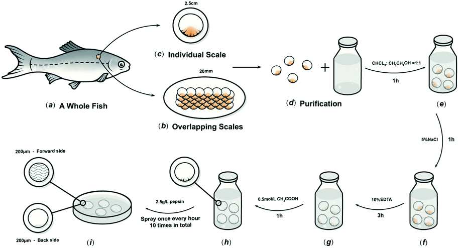 | ||
| Fig. 1 Schematic diagram of the process to prepare FS. | ||
2.2. Characterisation of BG and FS
 , where G1 represents the weight of the wet scaffold, and G0 represents the weight of the dry scaffold.
 .
.2.3. Animal experiments
The study protocol was approved by the Ethics Review Committee of the West China School of Stomatology, Sichuan University (WCHSIRB-D-2017-217). The study group consisted of 36 3 month-old Sprague-Dawley (SD) male rats, each weighing an average of 300 g. Animals were fed standard pellets and water.After the surgical area was scraped aseptically, 10% chloral hydrate (3 mL kg−1) was intraperitoneally injected, and a V-shaped incision was created on the rat cranial bone with a blade. After the cranial bone surface was exposed, a 5 mm-diameter surgical trephine was used to form two bilateral symmetric full-thickness defects that reached the dura mater surface. The BG and FS (5 mm × 5 mm) were then laid above the cranial bone defect. Finally, the skin was sutured for primary closure. Five rats in each group were administered 10% chloral hydrate anaesthesia (3 mL kg−1) at the 4th and 8th week after surgery, separately, for the following tests.18
2.4. Statistical analysis
SPSS 20.0 and Origin 8.5 software were used for data analysis. Three parallel experiments were set up in each experiment, and then the experimental data were analysed by variance (p < 0.05) and represented as X ± S.3. Results
3.1. Morphological observation
FS was transparent and tough, with uniform thickness and smooth surface (Fig. 2A). The longitudinal section of the FS had a multi-layered structure, with a parallel arrangement of fibres (Fig. 2B). The fibres were arranged in parallel (Fig. 2C), while the other part of the FS treated with pepsin was denser (Fig. 2D). BG presented as pale white, translucent, as rectangular patches with a smooth surface (Fig. 2E). The arrangement of fibres in the longitudinal section of BG was regular but scattered in the gap (Fig. 2F). In the cross section, the spongy layer displayed a dynamic distribution of fibres (Fig. 2G). The compact layer consisted of adjacent fibres, imparting a smooth and uniform surface with small collagenous spaces (Fig. 2H).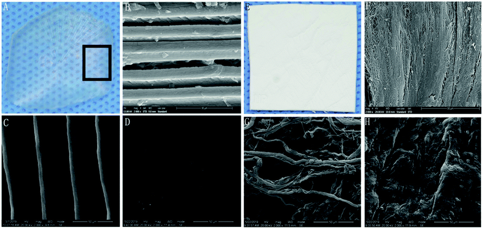 | ||
| Fig. 2 SEM morphological observations. FS: (A) general view; (B) profile view; (C) positive view; (D) negative view. BG: (E) general view; (F) profile view; (G) positive view; (H) negative view; (B–D and F–H). Magnification: 2000×. | ||
3.2. Physicochemical and mechanical tests
As shown in Table 1, BG was 444 ± 2.65 μm, while FS was 344 ± 5.74 μm. Furthermore, FS displayed a higher average tensile strength (16.5 ± 0.43 MPa) compared with that of BG (8.5 ± 1.52 MPa), and slightly lower elongation at break (15.66 ± 2.34%) compared with that of BG (28.25 ± 3.50%) under dry condition; similar results could also be obtained under wet condition. Usually, the decline of tensile strength in wet conditions stems from the absorption of water. The water contact angles of BG and FS were 38.0 ± 4.04° and 60.5 ± 5.62°, respectively, and the water absorption rates were 232.9 ± 15.94% and 143.3 ± 17.34%, respectively (p < 0.05) (Fig. 3).| BG | FS | |
|---|---|---|
| Thickness (μm) | 444 ± 2.65 | 344 ± 5.74 |
| Tensile strength (MPa) dry condition | 8.5 ± 1.52 | 16.5 ± 0.43 |
| Elongation at break (%) dry condition | 28.25 ± 3.50 | 15.66 ± 2.34 |
| Tensile strength (MPa) wet condition | 6.3 ± 0.92 | 15.0 ± 0.32 |
| Elongation at break (%) wet condition | 38.37 ± 2.00 | 20.97 ± 3.20 |
| Contact angle (°) | 38.0 ± 4.04 | 60.5 ± 5.62 |
| Water absorption (%) | 232.9 ± 15.94 | 143.3 ± 17.34 |
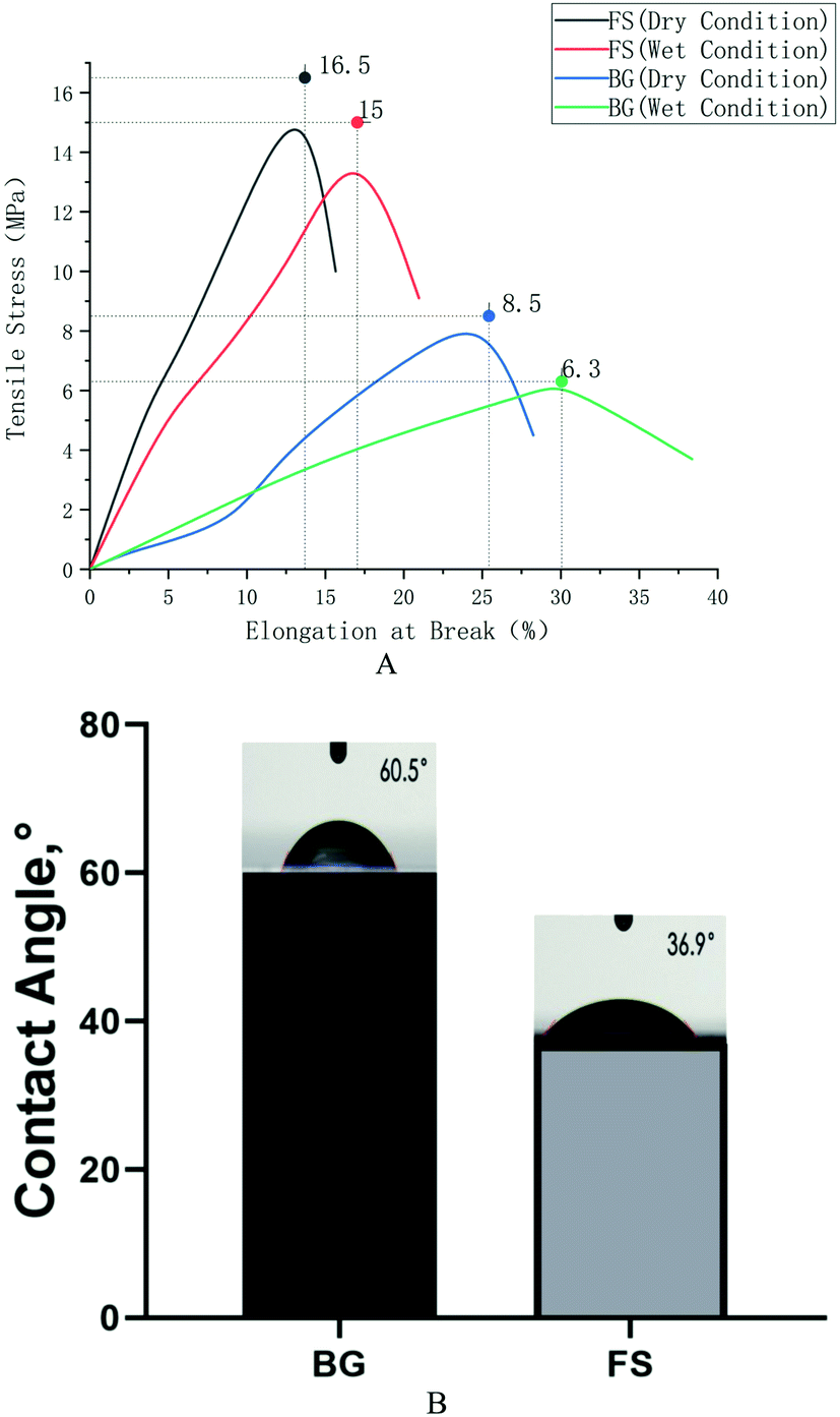 | ||
| Fig. 3 (A) Tensile strength testing; (B) water contents. | ||
As shown in Fig. 4A, the spectra of FS were similar to that of BG. They both had five distinctive absorption bands of type I collagen: amide A, amide B, amide I, amide II, and amide III. The absorption peaks of the amide A band appeared at wavelengths 3308.72 cm−1 and 3322.26 cm−1, separately. The amide B band peaks appeared at wavelengths 2953.59 cm−1 and 2956.40 cm−1, separately, which were related to the stretching vibration of CH2. The C![[double bond, length as m-dash]](https://www.rsc.org/images/entities/char_e001.gif) O stretching vibration of the polypeptide backbone leads to the amide I band with a distinctive absorption peak within the range of 1600–1700 cm−1, while the amide II band was produced by the CN stretching vibration and out-of-phase NH in-plane bending vibration within the range of 1500–1600 cm−1. The amide III band, related to the triple helices of collagen, was a combined absorption peak produced by in-phase NH bending vibration and the CN stretching vibration, appearing within the range of 1200–1360 cm−1.
O stretching vibration of the polypeptide backbone leads to the amide I band with a distinctive absorption peak within the range of 1600–1700 cm−1, while the amide II band was produced by the CN stretching vibration and out-of-phase NH in-plane bending vibration within the range of 1500–1600 cm−1. The amide III band, related to the triple helices of collagen, was a combined absorption peak produced by in-phase NH bending vibration and the CN stretching vibration, appearing within the range of 1200–1360 cm−1.
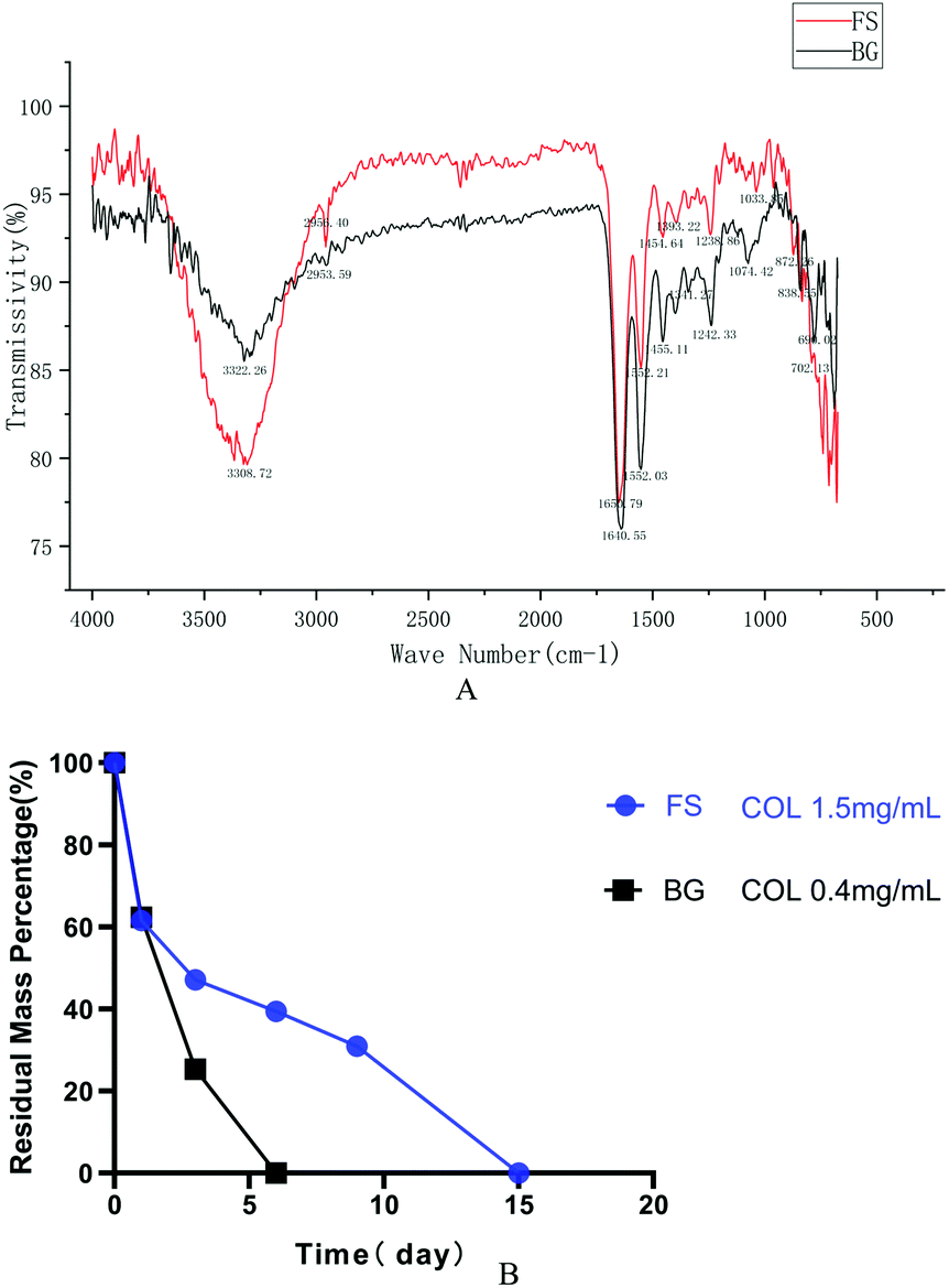 | ||
| Fig. 4 (A) FTIR; (B) degradation rate in vitro. | ||
The degradation rate showed that FS degraded more slowly at each timepoint compared with that of BG. Specifically, the residual weight of FS was 61.61% on the first day, and it degraded completely on the 15th day at a concentration of 1.5 mg mL−1 collagenase, while the residual weight of BG was 62.35% on the first day, and it degraded completely on the 6th day at a concentration of 0.4 mg mL−1 collagenase (Fig. 4B).
3.3. Host immune response in vivo
As shown in Fig. 5, one week after the operation, the rats were in good condition, and no abnormal phenomena, such as swelling, mass, infection, or material excretion were found in the incision. H&E and Masson staining results showed that both BG and FS maintained relatively intact morphology, the fibrous capsule was thin, and a small number of neutrophils were scattered around, whereas no statistical differences were observed in all groups.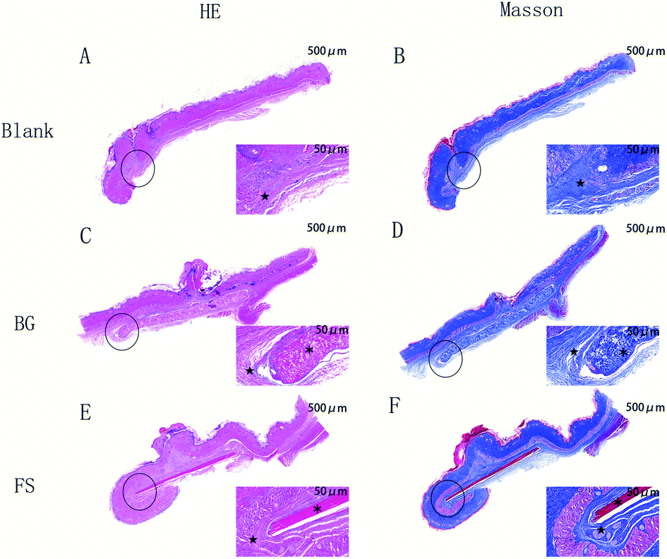 | ||
| Fig. 5 H&E and Masson staining of the rat's subcutaneous tissue. Magnification: left 20×; right: 200×. ★ fibrous capsule; ※ implanted biomaterials. | ||
3.4. Examination of repaired bone defects
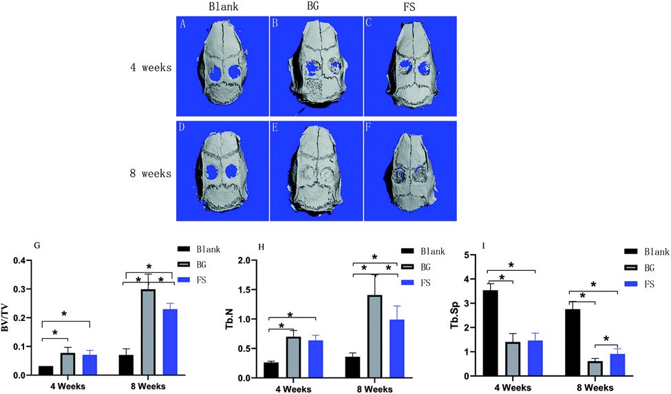 | ||
| Fig. 6 Three-dimensional reconstruction of micro-CT at the (A–C) 4th week and (D–F) 8th week. (G) BV/TV, (H) Tb.N, and (I) Tb.Sp values in the blank group, BG group, and FS group at the 4th week and the 8th week after surgery. *p < 0.05. | ||
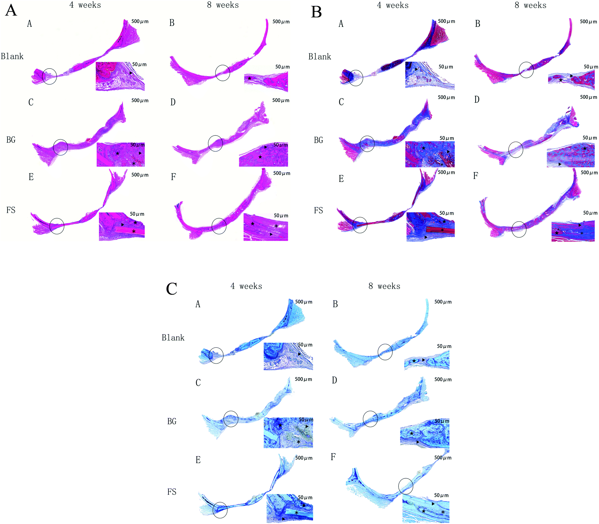 | ||
| Fig. 7 (A) H&E staining of the cranial bone. Magnification: left 20×; right: 200×. ▲ new bone; ★ old bone; ※ implanted biomaterials. (B) Masson staining of the cranial bone. Magnification: left 20×; right: 200×. ▲ new bone; ★ old bone; ※ implanted biomaterials. (C) Toluidine blue staining of the cranial bone. Magnification: left 20×; right: 200×. ▲ new bone; ★ old bone; ※ implanted biomaterials. | ||
4. Discussion
Biomimetic materials are becoming a promising alternative to better mimic the hierarchical structure of natural bone, as the absence of regular structures often leads to poorly functioning tissues.19 Traditional fish-scaled collagen extraction methods, such as acid and enzyme processing, are usually complicated, time-consuming, expensive, and sometimes harmful to the environment.20 The unmodified fish-scaled collagen shows remarkable biological capabilities, but the lack of control of biodegradability and poor mechanical properties have always been a challenge when using it directly as a scaffolding material. In this study, we developed a new strategy to remove mucopolysaccharides, proteins, and fats from fish scales, which was different from the established method for a novel collagen biomaterial preparation.21Mechanical loading plays a critical role in bone remodelling and development due to the promotion of osteogenic major genes and mineral deposition.22 The FS was 0.34 ± 0.05 mm in thickness, without curvature. The results showed that the tensile strength of the FS was significantly higher than that of the BG, while the elongation of the FS was lower than that of the BG, closer to the natural bone.23 The reason may be related to the fact that the lamellar structure of the FS could withstand greater stress tension and was less prone to plastic deformation and stress fatigue than the BG.24 The mechanical properties of collagen biomaterials should match those of natural bone to play a supporting role in the initial stage of bone regeneration. Otherwise, the mismatch of the mechanical properties between biomaterials and natural bone can cause stress shielding, leading to local bone resorption and low bone regeneration, which affects the growth of bone tissue.22,25
According to our previous research, FS is composed of multilayered collagen fibers.17 The FTIR spectra recorded for FS showed typical bands for type I collagen: amide A, amide B, amide I, amide II, and amide III. We observed that the band amide A has been moved to lower frequencies due to the loss of binding water in collagen and the conformational changes of collagen molecules. And the amide II bands appeared at wavelengths of 1552.21 cm−1 and 1552.03 cm−1, respectively, which are consistent with the FTIR absorbance bands for collagen in the amide II wavelength range. These are also higher than those of gelatin in the amide II wavelength range. Therefore, this new method did not destroy the triple helical structure of the collagen molecules. In general, the scaffold degradation rate should match the bone regeneration rate. A very fast degradation rate may result in an inflammatory response due to the accumulation of amino saccharides produced from the degradation.26 The collagenase solutions increased the degradation rate by digesting the fibrous structure of the FS and BG, but the introduction of methyl groups may have caused some spatial hindrance to the interactions between the collagenase and collagen molecules.27 The FS was found to degrade more slowly than the BG because the multi-layered structure of the FS increased connections between collagen molecules, which in turn led to a reduction in hydrolysis reaction and collagenase digestion. However, the degradation of biological scaffolds resembles a double-edged sword: on the one hand, biological scaffolds needed to support bone defect area for a certain time and provide attachment points for osteoblasts to adhere, proliferate, differentiate, or secrete extracellular matrix; on the other hand, biological scaffolds need to be degraded to vacate enough space for the growth of new bone tissue.28,29
The biocompatibility of collagen biomaterials is closely related to their collagen sources, structures, fabrication methods, and physicochemical properties.30–32 The amino acid composition of the fish-scaled protein is similar to that of the porcine protein. To date, there is no strong evidence that fish collagen was associated with any mutagenicity and carcinogenicity in humans. Essen et al.21 demonstrated that a fish scale-derived artificial cornea displayed no cytotoxicity, is not immunogenic, and is well-tolerated in the cornea. Immunogenicity is a critical factor for the biological safety of type I collagen, implantation of the FS subcutaneously in rats allowed us to compare the immune response to the FS with that to the BG that has been safely used in humans as well as with sham surgery. We demonstrated that the FS was biocompatible and had a low immunogenicity, as there was no specific immune response and no signs of sensitisation upon reintroducing the FS subcutaneously in the same animal, accounting from their components, which were type I collagen, regardless of their difference between microstructure, mechanical strength, and contact angle. However, its biocompatibility in vivo requires further observation.
The calvarial bone originates from intramembranous ossification, and it includes two layers of cortical bone on the surface and cancellous bone in the middle of the two layers of cortical bone, similar to the mandibular bone. Moreover, there is a lack of blood supply and self-regeneration. Therefore, the calvarial bone defect is more suitable for bone regeneration testing.33–35 When the FS was implanted in rat critical-sized calvarial defects, there was no obvious inflammation or suppuration in the operation area from the beginning to the end, confirming that the FS had good biocompatibility. Furthermore, the FS showed good osteogenic activity in the early stage of osteogenesis, but the osteogenic activity still needed to be further improved in the late stage. Fish-scaled collagen has good biocompatibility and low immunogenicity, so it could be used as a substitute for mammalian collagen. Tang36 also proved that fish-scaled collagen had good osteogenic activity, similar to that of mammalian collagen. Moreover, the FS had a multi-layered structure, which could better simulate the arrangement of collagen fibres of natural bone, with good mechanical properties, beneficial to maintain osteogenic space, and promote osteoblast differentiation.37 It has been proven that the BG degradation was obviously faster than that of the FS in vivo, so the BG had been completely degraded in the late osteogenic stage, leaving enough osteogenic space for new bone to grow. Meanwhile, the FS still remained in the late stage, the undegraded scaffolds that remain in bone defects may delay the bone regeneration process. Owing to its dense structure, it could quench cells but might block nutrient transportation, and its osteogenic activity still needs to be improved further in the late stage of osteogenesis. Meanwhile, its dense structure may have considerable advantages in the retention of bacteria in comparison to porous membranes.38
Therefore, an ideal membrane does not exist to date; the FS has low porosity, and the attachment of bacteria and cells to the surface of the membrane is hindered, allowing the exposition to the oral environment. In addition, the FS demonstrated good mechanical properties, biocompatibility, osteogenic ability, easy handling, and low cost.
Some limitations existed in our study. First, the degradation rate of the FS was not quantitatively determined in vivo. Another limitation is that the FS needs to be modified to improve its osteogenic activity. Therefore, we are working to adjust the FS using new technologies, such as electrospinning and 3D biological printing, achieving a balance between its ultra-structure, degradation rate, mechanical properties, and osteogenic activity. In the future, the modified FS can be used in alveolar ridge preservation, repair of bone wall defects after jaw cysts, and so on.
5. Conclusions
In this study, through the characterisation of a multi-layered collagen material from fish scales, it was proven that the multi-layered collagen structure material has sufficient mechanical properties, biocompatibility, and osteogenic ability. At the same time, it also shows that there is a gap in clinical needs between the guided tissue regeneration membrane and the one being used. This study provides useful insights into the efforts being made to design and adjust the microstructure to balance its mechanical properties, degradation rate, and osteogenic activity.Conflicts of interest
The authors declare no conflicts of interest.Acknowledgements
This study was supported by the Science Foundation of Sichuan Provincial Department of Science and Technology, China (2019YFG0260 and 2020YJ0242).References
- S. Nyman, J. Lindhe, T. Karring and H. Rylander, J. Clin. Periodontol., 1982, 9, 290–296 CrossRef CAS.
- D. Buser, K. Dula, U. Belser, H. P. Hirt and H. Berthold, International Journal of Periodontics and Restorative Dentistry, 1993, 13, 29–45 CAS.
- D. N. Tatakis, A. Promsudthi and U. M. Wikesjö, Periodontology, 1999, 19, 59–73 CrossRef CAS.
- M. C. Bottino, V. Thomas, G. Schmidt, Y. K. Vohra, T. M. Chu, M. J. Kowolik and G. M. Janowski, Dent. Mater., 2012, 28, 703–721 CrossRef CAS.
- O. Omar, I. Elgali, C. Dahlin and P. Thomsen, J. Clin. Periodontol., 2019, 46, 103–123 CrossRef.
- W. Florjanski, S. Orzeszek, A. Olchowy, N. Grychowska, W. Wieckiewicz, A. Malysa and J. Smardz, Polymers, 2019, 11, 782 CrossRef CAS.
- S. Al-Maawi, C. Herrera-Vizcaíno, A. Orlowska, I. Willershausen, R. Sader, R. J. Miron and J. Choukroun, Materials, 2019, 12, 3993 CrossRef CAS.
- P. Bunyaratavej and H. L. Wang, J. Periodontol., 2001, 72, 215–229 CrossRef CAS.
- A. M. Ferreira, P. Gentile, V. Chiono and G. Ciardelli, Acta Biomater., 2012, 8, 3191–3200 CrossRef CAS.
- A. Kozlovsky, G. Aboodi, O. Moses, H. Tal, Z. Artzi, M. Weinreb and C. E. Nemcovsky, Clin. Oral Implants Res., 2009, 20, 1116–1123 CrossRef.
- F. Pati, P. Datta, B. Adhikari, S. Dhara, K. Ghosh and P. K. Das Mohapatra, J. Biomed. Mater. Res., Part A, 2012, 100, 1068–1079 CrossRef.
- A. M. Deliormanli, X. Liu and M. N. Rahaman, Biomater. Appl., 2014, 28, 643–653 CrossRef.
- D. Arola, S. Murcia, M. Stossel, R. Pahuja, T. Linley, A. Devaraj, M. Ramulu, E. A. Ossa and J. Wang, Acta Biomater., 2018, 67, 319–330 CrossRef CAS.
- Z. Y. Qiu, Y. Cui, C. S. Tao, Z. Q. Zhang, P. F. Tang, K. Y. Mao, X. M. Wang and F. Z. Cui, Materials, 2015, 8, 4733–4750 CrossRef CAS.
- A. Kara, S. Tamburaci, F. Tihminlioglu and H. Havitcioglu, Int. J. Biol. Macromol., 2019, 130, 266–279 CrossRef CAS.
- J. Wang, Q. Cheng and Z. Tang, Chem. Soc. Rev., 2012, 41, 1111–1129 RSC.
- H. H. Feng, X. Li, X. M. Deng, X. L. Li, J. T. Guo, K. Ma and B. Jiang, RSC Adv., 2020, 10, 875 RSC.
- P. P. Spicer, J. D. Kretlow, S. Young, J. A. Jansen, F. K. Kasper and A. G. Mikos, Nat. Protoc., 2012, 7, 1918–1929 CrossRef CAS.
- W. Shao, J. He, Q. Han, F. Sang, Q. Wang, L. Chen, S. Cui and B. Ding, Mater. Sci. Eng., C, 2016, 67, 599–610 CrossRef CAS.
- C. Y. Huang, J. M. Kuo, S. J. Wu and H. T. Tsai, Food Chem., 2016, 190, 997–1006 CrossRef CAS.
- T. H. van Essen, L. van Zijl, T. Possemiers, A. A. Mulder, S. J. Zwart, C. H. Chou, C. C. Lin, H. J. Lai, G. P. M. Luyten, M. J. Tassignon, N. Zakaria, A. El-Ghalbzouri and M. J. Jager, Biomaterials, 2016, 81, 36–45 CrossRef CAS.
- B. P. Hung, D. L. Hutton and W. L. Grayson, Stem Cell Res. Ther., 2013, 4, 10 CrossRef CAS.
- F. Zou, R. Li, J. Jiang, X. Mo, G. Gu, Z. Guo and Z. Chen, J. Biomater. Sci., Polym. Ed., 2017, 28, 2255–2270 CrossRef CAS.
- A. A. Zadpoor, J. Mech. Behav. Biomed. Mater., 2017, 70, 1–6 CrossRef CAS.
- P. Bhattacharjee, B. Kundu, D. Naskar, H. W. Kim, T. K. Maiti, D. Bhattacharya and S. C. Kundu, Acta Biomater., 2017, 63, 1–17 CrossRef CAS.
- X. Hu, S. H. Park, E. S. Gil, X. X. Xia, A. S. Weiss and D. L. Kaplan, Biomaterials, 2011, 32, 8979–8989 CrossRef CAS.
- I. D. Gaudet and D. I. Shreiber, Biointerphases, 2012, 7, 25 CrossRef CAS.
- L. Roseti, V. Parisi, M. Petretta, C. Cavallo, G. Desando, I. Bartolotti and B. Grigolo, Mater. Sci. Eng., C, 2017, 78, 1246–1262 CrossRef CAS.
- R. Shi, Y. Huang, C. Ma, C. Wu and W. Tian, Front. Med., 2019, 13, 160–188 CrossRef.
- M. Meyer, Biomed. Eng. Online, 2019, 18, 24 CrossRef.
- L. C. Mozdzen, A. Vucetic and B. A. C. Harley, J. Mech. Behav. Biomed. Mater., 2017, 66, 28–36 CrossRef CAS.
- B. D. Walters and J. P. Stegemann, Acta Biomater., 2014, 10, 1488–1501 CrossRef CAS.
- K. A. Schlegel, F. J. Lang, K. Donath, J. T. Kulow and J. Wiltfang, Oral Surgery, Oral Medicine, Oral Pathology, Oral Radiology, and Endodontology, 2006, 102, 7–13 CrossRef.
- Y. Su, Q. Su, W. Liu, M. Lim, J. R. Venugopal, X. Mo, S. Ramakrishna, S. S. Al-Deyab and M. El-Newehy, Acta Biomater., 2012, 8, 763–771 CrossRef CAS.
- J. H. Zeng, P. Qiu, L. Xiong, S. W. Liu, L. H. Ding, S. L. Xiong, J. T. Li, Z. B. Xiao and T. Zhang, Int. J. Artif. Organs, 2019, 42, 325–337 CrossRef CAS.
- J. Tang and T. Saito, BioMed Res. Int., 2015, 2015, 139476 Search PubMed.
- D. B. Bhuiyan, J. C. Middleton, R. Tannenbaum and T. M. Wick, J. Biomater. Sci., Polym. Ed., 2016, 27, 1139–1154 CrossRef CAS.
- A. S. Monteiro, L. G. Macedo, N. L. Macedo and I. Balducci, Med. Oral Patol. Oral Cir. Bucal, 2010, 15, e401–e406 CrossRef.
Footnote |
| † Guoping Cheng and Shujuan Guo contributed equally and are co-first authors. |
| This journal is © The Royal Society of Chemistry 2020 |