
Multiple target chemosensor: a fluorescent sensor for Zn(II) and Al(III) and a chromogenic sensor for Fe(II) and Fe(III)†
Yong Sung Kim,
Gyeong Jin Park,
Jae Jun Lee,
Sun Young Lee,
Seong Youl Lee and
Cheal Kim*
Department of Fine Chemistry and Department of Interdisciplinary Bio IT Materials, Seoul National University of Science and Technology, Seoul 139-743, Korea. E-mail: chealkim@seoultech.ac.kr; Fax: +82-2-973-9149; Tel: +82-2-970-6693
First published on 5th January 2015
Abstract
A multifunctional fluorescent and colorimetric chemosensor 1, based on two julolidine moieties as a binding and signaling unit, has been synthesized in a one-step procedure. Receptor 1 showed prompt responses toward Zn2+ and Al3+ ions through selective fluorescence enhancement in dimethylformamide (DMF), while the presence of 5% water made 1 detect only Zn2+. Moreover, with 1 the “naked eye” could sense iron by a clear color change. Upon the addition of Fe2+ and Fe3+ into each solution of 1, the color of the solutions changed from pale yellow to dark green for both Fe2+ and Fe3+. The binding modes of the complexes were determined to be a 1![[thin space (1/6-em)]](https://www.rsc.org/images/entities/char_2009.gif) :1 complexation stoichiometry from a Job plot, 1H NMR titration and ESI-mass spectrometry analysis.
:1 complexation stoichiometry from a Job plot, 1H NMR titration and ESI-mass spectrometry analysis.
Introduction
As the second most abundant transition metal ion in the human body, the zinc ion (Zn2+) plays an important role in gene transcription, regulation of metalloenzymes, neural signal transmission and apoptosis.1–11 However, the imbalance in zinc may cause several health problems, including superficial skin disease, prostate cancer, diabetes, and brain diseases such as Alzheimer's disease, Friedreich's ataxia, and Parkinson's disease.12–19 Therefore, it is very significant to efficiently detect zinc ion. Nevertheless, many of the reported Zn2+ sensors suffer from a limited choice of spectroscopic instruments due to its inherent d10 shell, insufficient selectivity or sensitivity, and interference from other transition metal ions, especially the cadmium ion, which is in the same group of the periodic table and shows similar properties to those of zinc ion.20,21Aluminum is the most abundant (8.3% by weight) metallic element and the third most abundant of all elements (after oxygen and silicon) in the earth's crust.22 Compounds of aluminum are widely dispersed in various ways; textile industry, medicines (antacids), bleached flour, paper industry, food additives, aluminum-based pharmaceuticals, storage/cooking utensils, and production of light alloys.23–27 However, high amounts of aluminum ion are not only harmful to plant growth but also damage the human nervous system to induce Alzheimer's disease, Parkinson's disease, and amyotrophic lateral sclerosis.28–31 Thus, the development of a chemo-sensor for aluminum (Al3+) still progresses with considerable attention. Nevertheless, the detection of Al3+ is difficult because of the lack of spectroscopic characteristics and poor coordination ability compared to those of other transition metals. Therefore, the development of new sensors of high selectivity for Al3+ is much required for environmental and biological fields.32
Iron is the most abundant transition metal for both plants and animals. It plays an important role in cellular metabolism, enzyme catalysis, as an oxygen carrier in hemoglobin and as a cofactor in many enzymatic reactions.33–35 However, less iron in the body has been reportedly linked to diabetes, anemia, liver and kidney damage, and heart diseases.36 Accordingly, the development of methods to detect iron in environmental and biological fields is of considerable significance.37
For these reasons, the development of chemosensors for the detection of these metal ions (Zn2+, Al3+, Fe2+, and Fe3+) has been considered as very worthy research. Moreover, single probes for multiple targets are being actively considered due to the benefits, such as a less expensive and more efficient analysis, while most chemosensors developed to date are based on single-ion responsive systems.38–42
Herein, we report on the development and application of chemosensor 1 for multiple analytes based on the julolidine moiety, well-known as a good fluorophore and chromophore.43–45 1 detected effectively the most abundant and fundamental ions (Zn2+, Al3+, Fe2+/3+) in the ecosystem through two different sensing mechanisms (fluorescent and colorimetric responses).
Experimental section
Materials and instrumentation
All the starting materials (analytical grade and spectroscopic grade) for the syntheses were commercially available and used as received. 1H NMR and 13C NMR measurements were performed on a Varian 400 MHz spectrometer and chemical shifts are recorded in ppm. Electrospray ionization mass spectra (ESI-MS) were collected on a Thermo Finnigan (San Jose, CA, USA) LCQTM Advantage MAX quadrupole ion trap instrument. Elemental analysis for carbon, nitrogen, and hydrogen was carried out by using a Flash EA 1112 elemental analyzer (thermo) in the Organic Chemistry Research Center of Sogang University, Korea. Absorption spectra were recorded at room temperature using a Perkin-Elmer model Lambda 2S UV-vis spectrometer. Fluorescence measurements were performed on a Perkin-Elmer model LS45 fluorescence spectrometer.Synthesis of 1
A solution of 8-hydroxyjulolidine-9-carboxaldehyde (0.69 g, 3.04 mmol) in ethanol was added to a solution containing 2, 2′-thiobis(ethylamine) (199 μL, 1.60 mmol) in ethanol. The reaction mixture was stirred for 12 h at room temperature. After evaporation, the product was recrystallized by ether, filtered, and dried under vacuum. Yield: 0.47 g (59.6%). 1H NMR (400 MHz, CDCl3) δ (ppm): 13.75 (s, 2H), 7.93 (s, 2H), 6.59 (s, 2H), 3.64 (t, J = 6.8 Hz, 4H), 3.22–3.16 (m, 8H), 2.81–2.77 (t, J = 6.8 Hz, 4H), 2.70 (t, J = 6.6 Hz, 4H), 2.65 (t, J = 6.2 Hz 4H), 1.96–1.89 (m, 8H). 13C NMR (CDCl3, 400 MHz): δ 165.40, 160.46, 146.59, 129.54, 112.22, 107.87, 106.34, 57.37, 50.00, 49.62, 33.44, 27.35, 22.32, 21.38, 20.79 ppm. LRMS (ESI) m/z [M + H+]: calcd, 519.279; found, 519.267. Anal. calcd for C30H38N4O2S (518.272): C, 69.46; H, 7.38; N, 10.80. Found: C, 69.39; H, 7.63; N, 10.54%.Fluorescence chemosensor
For the Al3+ ion in DMF, 1 (3.1 mg, 0.003 mmol) was dissolved in DMF (2 mL). 10 μL of 1 (3 mM) were diluted in 2.990 mL DMF to make the final concentration of 10 μM. Al(NO3)3 (22.5 mg, 0.02 mmol) was dissolved in DMF (3 mL). 1.5–16.5 μL of the Al(NO3)3 solution (20 mM) were transferred to the receptor solution (10 μM) prepared above. After mixing them for two minutes, fluorescence spectra were taken at room temperature.
For the Zn2+ ion in aqueous media, 1 (3.1 mg, 0.003 mmol) was dissolved in DMF (2 mL). The receptor solution (10 μL, 3 mM) was diluted in 2.990 mL DMF–buffer solution (95:5, v/v, 10 mM, bis-tris, pH 7.0) to make the final concentration of 10 μM. Zn(NO3)2 (18.2 mg, 0.02 mmol) was dissolved in DMF (3 mL). 1.5–18.0 μL of the Zn(NO3)2 solution (20 mM) were transferred to each receptor solution (10 μM) prepared above. After mixing them for two minutes, fluorescence spectra were taken at room temperature.
For the Al3+ ion in DMF, 1 (3.1 mg, 0.003 mmol) was dissolved in DMF (2 mL). 10 μL of 1 (3 mM) were diluted with 2.990 mL DMF to make the final concentration of 10 μM. Al(NO3)3 (22.5 mg, 0.02 mmol) was dissolved in DMF (3 mL). 0.75–6.0 μL of the Al(NO3)3 solution (20 mM) were transferred to the receptor solution (10 μM) prepared above. After mixing them for two minutes, UV-vis absorption spectra were taken at room temperature.
For the Zn2+ ion in aqueous media, 1 (3.1 mg, 0.003 mmol) was dissolved in DMF (2 mL). 10 μL of 1 (3 mM) were diluted with 2.990 mL DMF–buffer solution (95:5, v/v, 10 mM, bis-tris, pH 7.0) to make the final concentration of 10 μM. Zn(NO3)2 (18.2 mg, 0.02 mmol) was dissolved in DMF (3 mL). 0.75–6.0 μL of the Zn(NO3)2 solution (20 mM) were transferred to the receptor solution (10 μM) prepared above. After mixing them for two minutes, UV-vis absorption spectra were taken at room temperature.
For the Al3+ ion in DMF, 1 (3.1 mg, 0.003 mmol) was dissolved in DMF (2 mL). 10 μL of 1 (3 mM) were diluted with 2.990 mL DMF to make the final concentration of 10 μM. MNO3 (M = Na, K, 0.02 mmol), M(NO3)2 (M = Mg, Ca, Mn, Ni, Cu, Zn, Cd, Hg, 0.02 mmol), M(NO3)3 (M = Al, Cr, Fe, Ga, In, 0.02 mmol) and Fe(ClO4)2 (15.6 mg, 0.02 mmol) were dissolved in DMF (3 mL), respectively. 16.5 μL of each metal solution (20 mM) were taken and added into 3 mL of each receptor solution (10 μM) prepared above to make 11 equiv. Then, 16.5 μL of Al(NO3)3 solution (20 mM) were added into the mixed solution of each metal ion and 1 to make 11 equiv. After mixing them for two minutes, fluorescence spectra were taken at room temperature.
For the Zn2+ ion in aqueous media, 1 (3.1 mg, 0.003 mmol) was dissolved in DMF (2 mL). 10 μL of 1 (3 mM) were diluted with 2.990 mL DMF–buffer solution (95:5, v/v, 10 mM, bis-tris, pH 7.0) to make the final concentration of 10 μM. MNO3 (M = Na, K, 0.02 mmol), M(NO3)2 (M = Mg, Ca, Mn, Ni, Cu, Zn, Cd, Hg, 0.02 mmol), M(NO3)3 (M = Al, Cr, Fe, Ga, In, 0.02 mmol) and Fe(ClO4)2 (15.6 mg, 0.02 mmol) were dissolved in DMF (3 mL), respectively. 18 μL of each metal solution (20 mM) were taken and added into 3 mL of each receptor solution (10 μM) prepared above to make 12 equiv. Then, 18 μL of Zn(NO3)2 solution (20 mM) were added into the mixed solution of each metal ion and 1 to make 12 equiv. After mixing them for two minutes, fluorescence spectra were taken at room temperature.
For the Al3+ ion in DMF, 1 (3.1 mg, 0.003 mmol) was dissolved in DMF (2 mL). 100, 90, 80, 70, 60, 50, 40, 30, 20, 10 and 0 μL of the solution of 1 were taken and transferred to vials. Each vial was diluted with DMF to make a total volume of 2.9 mL. Al(NO3)3 (3.4 mg, 0.003 mmol) was dissolved in DMF (3 mL). 0, 10, 20, 30, 40, 50, 60, 70, 80, 90, and 100 μL of the Al3+ solution were added to each diluted solution of 1. Each vial had a total volume of 3 mL. After shaking them for two minutes, fluorescence spectra were taken at room temperature.
For the Zn2+ ion in aqueous media, 1 (3.1 mg, 0.003 mmol) was dissolved in DMF. 100, 90, 80, 70, 60, 50, 40, 30, 20, 10 and 0 μL of the solution of 1 were taken and transferred to vials. Each vial was diluted with DMF–buffer solution (95:5, v/v, 10 mM, bis-tris, pH 7.0) to make a total volume of 2.9 mL. Zn(NO3)2 (2.7 mg, 0.003 mmol) was dissolved in DMF (3 mL). 0, 10, 20, 30, 40, 50, 60, 70, 80, 90, and 100 μL of the Zn2+ solution were added to each diluted solution of 1. Each vial had a total volume of 3 mL. After shaking them for two minutes, fluorescence spectra were taken at room temperature.
For 1H NMR titrations of 1 with aluminum ions, three NMR tubes of 1 (3.2 mg, 0.01 mmol) dissolved in DMF-d7 (700 μL) were prepared and three different concentrations (0, 0.6 and 1 equiv.) of Al(NO3)3·6H2O dissolved in DMF were added to each solution of 1. After shaking them for two minutes, 1H NMR spectra were taken at room temperature.
:5, v/v, 10 mM, bis-tris, pH 7.0) to make a final concentration of 10 μM. Zn(NO3)2·5H2O (18.2 mg, 0.02 mmol) was dissolved in DMF (3 mL) and 18 μL of the Zn2+ ion solution (20 mM) were added to the solution of 1 (10 μM) prepared above. After mixing it for two minutes, the fluorescence spectrum was taken at room temperature. Ethylenediaminetetraacetic acid disodium salt dehydrate (EDTA, 0.4 mmol) was dissolved in buffer solution (5 mL) and 9 μL of the EDTA solution (40 mM) were added to the solution of the 1–Zn2+ complex (10 μM) prepared above. After mixing it for two minutes, the fluorescence spectrum was taken. For the reversibility study, 18 μL of the Zn2+ ion solution (20 mM) were added to the above solution. After mixing it for two minutes, a fluorescence spectrum was taken at room temperature.Colorimetric chemosensor
:1, v/v, 10 mM, bis-tris, pH 7.0) to make the final concentration of 10 μM. Fe(ClO4)2 (15.6 mg, 0.02 mmol) was dissolved in MeOH (3 mL). 0.3–3.0 μL of the Fe(ClO4)2 solution (20 mM) were transferred to the receptor solution (10 μM) prepared above. After mixing them for two minutes, UV-vis absorption spectra were taken at room temperature.For the Fe3+ ion, 1 (3.1 mg, 0.003 mmol) was dissolved in MeOH. 10 μL of 1 (3 mM) were diluted with 2.990 mL MeOH–buffer solution (9:1, v/v, 10 mM, bis-tris, pH 7.0) to make the final concentration of 10 μM. Fe(NO3)3 (24.7 mg, 0.02 mmol) was dissolved in MeOH (3 mL). 0.3–1.65 μL of the Fe(NO3)3 solution (20 mM) were transferred to the receptor solution (10 μM) prepared above. After mixing them for two minutes, UV-vis absorption spectra were taken at room temperature.
:1, v/v, 10 mM, bis-tris, pH 7.0) to make the final concentration of 10 μM. MNO3 (M = Na, K, 0.02 mmol), M(NO3)2 (M = Mg, Ca, Mn, Ni, Cu, Zn, Cd, Hg, 0.02 mmol), M(NO3)3 (M = Al, Cr, Fe, Ga, In, 0.02 mmol) and Fe(ClO4)2 (15.6 mg, 0.02 mmol) were dissolved in MeOH (3 mL), respectively. 3.0 μL of each metal solution (20 mM) were taken and added into 3 mL of each receptor solution (10 μM) prepared above to make 2.0 equiv. Then, 3.0 μL of Fe(ClO4)2 solution (20 mM) were added into the mixed solution of each metal ion and 1 to make 2.0 equiv. After mixing them for two minutes, UV-vis absorption spectra were taken at room temperature.For the Fe3+ ion, 1 (3.1 mg, 0.003 mmol) was dissolved in MeOH (2 mL). 10 μL of 1 (3 mM) were diluted with 2.990 mL MeOH–buffer solution (9:1, v/v, 10 mM, bis-tris, pH 7.0) to make the final concentration of 10 μM. MNO3 (M = Na, K, 0.02 mmol), M(NO3)2 (M = Mg, Ca, Mn, Ni, Cu, Zn, Cd, Hg, 0.02 mmol), M(NO3)3 (M = Al, Cr, Fe, Ga, In, 0.02 mmol) and Fe(ClO4)2 (15.6 mg, 0.02 mmol) were dissolved in MeOH (3 mL), respectively. 1.8 μL of each metal solution (20 mM) were taken and added into 3 mL of each receptor solution (10 μM) prepared above to make 1.8 equiv. Then, 3.0 μL of Fe(NO3)3 solution (20 mM) were added into the mixed solution of each metal ion and 1 to make 1.8 equiv. After mixing them for two minutes, UV-vis absorption spectra were taken at room temperature.
:1, v/v, 10 mM, bis-tris, pH 7.0) to make a total volume of 2.9 mL. Fe(ClO4)2 (2.3 mg, 0.003 mmol) was dissolved in MeOH (3 mL). 0, 10, 20, 30, 40, 50, 60, 70, 80, 90, and 100 μL of the Fe2+ solution were added to each diluted solution of 1. Each vial had a total volume of 3 mL. After shaking them for two minutes, fluorescence spectra were taken at room temperature.For Fe3+, 1 (3.1 mg, 0.003 mmol) was dissolved in MeOH (2 mL). 100, 90, 80, 70, 60, 50, 40, 30, 20, 10 and 0 μL of the solution of 1 were taken and transferred to vials. Each vial was diluted with MeOH–buffer solution (9:1, v/v, 10 mM, bis-tris, pH 7.0) to make a total volume of 2.9 mL. Fe(NO3)3 (3.7 mg, 0.003 mmol) was dissolved in MeOH (3 mL). 0, 10, 20, 30, 40, 50, 60, 70, 80, 90, and 100 μL of the Fe3+ solution were added to each diluted solution of 1. Each vial had a total volume of 3 mL. After shaking them for two minutes, fluorescence spectra were taken at room temperature.
Results and discussion
Synthesis of 1
A new chemosensor 1 was synthesized by the condensation reaction of 8-hydroxyjulolidine-9-carboxaldehyde with 2,2′-thiobis-(ethylamine) in ethanol at room temperature (Scheme 1), and characterized by 1H and 13C NMR, ESI-mass spectrometry and elemental analysis. | ||
| Scheme 1 Synthesis of 1. | ||
Fluorogenic sensing for Zn2+ and Al3+ in DMF
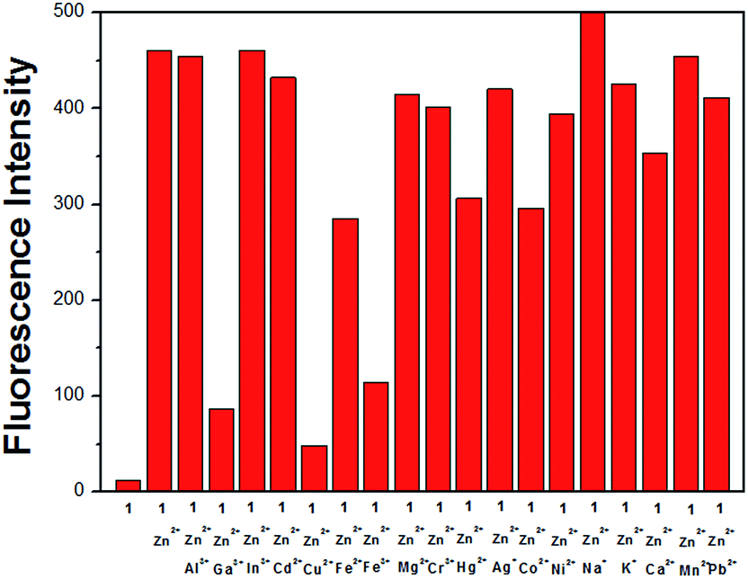
The receptor 1 alone has a very weak fluorescence emission with an excitation of 355 nm in DMF. When 11 equiv. of various metal ions such as Al3+, Ga3+, In3+, Zn2+, Cd2+, Cu2+, Fe2+, Fe3+, Mg2+, Cr3+, Hg2+, Ag+, Co2+, Ni2+, Na+, K+, Ca2+, Mn2+ and Pb2+ were added to 1, the solution of 1 exhibited no or a slight increase of the fluorescence except for Zn2+ and Al3+ (Fig. 1). The addition of Zn2+ and Al3+ resulted in significant enhancements of the emission intensities at 448 nm (32-fold) and 418 nm (35-fold), respectively. These two emissions at different wavelengths indicate that 1 could be used as a dual chemosensor for Zn2+ and Al3+ in the same solvent environment. | ||
| Fig. 1 (a) Fluorescence spectra of 1 (10 μM) upon the addition of metal salts (9 equiv.) of Al3+, Ga3+, In3+, Zn2+, Cd2+, Cu2+, Fe2+, Fe3+, Mg2+, Cr3+, Hg2+, Ag+, Co2+, Ni2+, Na+, K+, Ca2+, Mn2+ and Pb2+ in DMF (λex = 355 nm). (b) Bar graph representing the change of the relative emission intensity of 1 at 460 nm upon treatment with various metal ions (λex = 355 nm). | ||
The changes in the emission spectra of 1 as a function of the concentration of Zn2+ and Al3+ are shown in Fig. 2. Upon the addition of Zn2+, the fluorescence intensity increased gradually and was saturated with 11 equiv. of Zn2+ (Fig. 2(a)). When the fluorescent titration was performed with Al3+, the emission intensity increased up to 11 equiv. and then no further change was observed (Fig. 2(b)).
 | ||
| Fig. 2 (a) Fluorescence spectra of 1 (10 μM; λex = 355 nm) after the addition of increasing amounts of Zn2+ ions (1, 2, 3, 4, 5, 6, 7, 8, 9, 10, 11, 12 and 13 equiv.) at room temperature. Inset: Plot of the fluorescence intensity at 445 nm as a function of Zn2+ concentration. (b) Fluorescence spectra of 1 (10 μM; λex = 355 nm) after the addition of increasing amounts of Al3+ ions (1, 2, 3, 4, 5, 6, 7, 8, 9, 10, 11, 12 and 13 equiv.) at room temperature. Inset: plot of the fluorescence intensity at 418 nm as a function of Al3+ concentration. | ||
The significant increase of fluorescence by the addition of Zn2+ and Al3+ to 1 could be explained by the inhibition of both the C![[double bond, length as m-dash]](https://www.rsc.org/images/entities/char_e001.gif) N isomerization and excited-state proton transfer (ESPT). Imines are generally known to be poorly fluorescent, in part due to isomerization of the CN double bond in the excited state46 and in part due to ESPT involving the phenolic protons of the julolidine moiety.47 Upon stable chelation with a certain metal, CN isomerization and ESPT are inhibited (Scheme 2), thus leading to fluorescence enhancement. Also, we consider the chelation-enhanced fluorescence (CHEF) effect as the responsive mechanism for fluorescence enhancements of the 1–Zn2+ complex and the 1–Al3+ complex. The chelating of 1 with Zn2+ and Al3+ induced rigidity in the complexes, leading to a large CHEF effect with a drastic enhancement of fluorescence.48
N isomerization and excited-state proton transfer (ESPT). Imines are generally known to be poorly fluorescent, in part due to isomerization of the CN double bond in the excited state46 and in part due to ESPT involving the phenolic protons of the julolidine moiety.47 Upon stable chelation with a certain metal, CN isomerization and ESPT are inhibited (Scheme 2), thus leading to fluorescence enhancement. Also, we consider the chelation-enhanced fluorescence (CHEF) effect as the responsive mechanism for fluorescence enhancements of the 1–Zn2+ complex and the 1–Al3+ complex. The chelating of 1 with Zn2+ and Al3+ induced rigidity in the complexes, leading to a large CHEF effect with a drastic enhancement of fluorescence.48
 | ||
| Scheme 2 Proposed structures of 1–Mn+ complexes. | ||
To further explore the interaction between 1 and the two metal ions Zn2+ and Al3+, UV-vis titrations were carried out (Fig. S1†). Upon the addition of Zn2+ ions to a solution of 1, the absorption band at 351 nm decreased and the absorbance intensity at 374 nm increased with an isosbestic point at 358 nm, which indicates a clean conversion of 1 into the 1–Zn2+ complex. Similarly, the addition of Al3+ ions to a solution of 1 resulted in a decrease of the absorption peak at 352 nm and the appearance of a new peak at 380 nm with a clear isosbestic point at 363 nm, which indicates the clean formation of the 1–Al3+ complex.
The binding modes between 1 and the two metal ions, Zn2+ and Al3+, were determined by using Job plot analysis. As shown in Fig. 3, the Job plots for the 1–Zn2+ and 1–Al3+ complexes exhibited 1:1 complexation stoichiometry, respectively.
 | ||
| Fig. 3 Job plots of (a) 1–Zn2+ and (b) 1–Al3+ complexes. The total concentration of 1 and metal ions (Zn2+ and Al3+) was 40 μM, fluorescence intensity at 449 nm, respectively. | ||
From the results of fluorescence titration, the association constants of the 1–Zn2+ and 1–Al3+ complexes were determined as 2.9 × 10⁴ M−1 and 8.5 × 10³ M−1 on the basis of the Benesi–Hildebrand equation (Fig. S2†). These values are comparable to those reported for Zn2+-chemosensors (101 to 107 M−1) and Al3+-chemosensors (103 to 1014 M−1).47,48 For practical applications, the detection limit is also an important parameter. Thus, the detection limits of 1 for the analysis of Zn2+ and Al3+ were calculated to be 1.59 μM and 1.34 μM, respectively, on the basis of 3σ/K (Fig. S3†).49
To further check the practical applicability of 1 as a selective fluorescence sensor for Zn2+, competition experiments were conducted in the presence of Zn2+ mixed with other relevant metal ions, such as Al3+, Ga3+, In3+, Cd2+, Cu2+, Fe2+, Fe3+, Mg2+, Cr3+, Hg2+, Ag+, Co2+, Ni2+, Na+, K+, Ca2+, Mn2+ and Pb2+. When 1 was treated with 11 equiv. of Zn2+ in the presence of the same concentration of the other metal ions (Fig. 4), Ga3+ and Fe3+ quenched about 83 and 77%, respectively, of the fluorescence obtained with Zn2+ alone. Meanwhile, Cu2+ interfered with the emission intensity of 1–Zn2+. However, the Cd2+ ion hardly inhibited the emission intensity of 1–Zn2+. Similarly, we studied the preferential selectivity of 1 as a fluorescence chemosensor for the detection of Al3+ in the presence of various metal ions (Fig. S4†). Unfortunately, Al3+ complexation with 1 was inhibited completely by Ga3+, In3+, Cu2+, Fe2+ and Fe3+, and considerably by Cr3+ and Co2+.
 | ||
| Fig. 4 Bar graph representing the change of the relative emission intensity of 1 (10 μM) at 440 nm upon treatment with various metal ions: Al3+, Ga3+, In3+, Zn2+, Cd2+, Cu2+, Fe2+, Fe3+, Mg2+, Cr3+, Hg2+, Ag+, Co2+, Ni2+, Na+, K+, Ca2+, Mn2+ and Pb2+ (λex = 355 nm). | ||
The interaction between 1 and Zn2+ was further studied through 1H NMR titration experiments in DMF-d7 (Fig. 5). With increasing Zn2+ concentration, the H1 protons of the hydroxyl groups at 13.7 ppm disappeared due to their deprotonation, and the H2 protons of the CN moieties and the H4 and H5 protons of the ethylene moiety were shifted downfield. These results suggest that the bridge S, the imine N, and the phenol O atoms might coordinate to the Zn ion.50
 | ||
| Fig. 5 (a) 1H NMR titration of 1 with Zn2+ in DMF-d7: (III) only 1; (II) 1 + Zn2+ (0.5 equiv.); and (I) 1 + Zn2+ (1 equiv.). | ||
1H NMR titration experiments of 1 with Al(NO3)3 were also carried out in DMF-d7 (Fig. S5†). Upon the addition of the Al3+ to 1, the O–H peaks at 13.6 ppm disappeared completely. In addition, the protons of the imine and ethylene moieties showed a similar pattern to that observed in the 1–Zn2+ complex, demonstrating that both 1–Al3+ and 1–Zn2+ complexes might have a similar coordination environment.
The formation of 1–Zn2+ and 1–Al3+ complexes was further confirmed by ESI-mass spectrometry analysis. The positive-ion mass spectrum of 1 upon the addition of 1 equiv. of Zn2+ showed the formation of the 1 + Zn2+–H+ complex [m/z: 581.267; calcd, 581.193] (Fig. S6a†). For Al3+, the positive-ion mass spectrum of 1 showed the formation of the 1 + Al3+–2H+ complex [m/z: 543.333; calcd, 543.236] (Fig. S6b†). Based on the Job plot, 1H NMR titration, and ESI-mass spectrometry analysis, we propose the structures of 1–Zn2+ and 1–Al3+ complexes shown in Scheme 2.
Fluorogenic sensing for Zn2+ in aqueous media
For the practical application of receptor 1 toward various metal ions, we increased the amount of the bis-tris buffer in DMF. 1 alone displayed a very weak emission band at 440 nm with excitation at 355 nm in the DMF–buffer solution (95:5, v/v, 10 mM, bis-tris, pH 7.0) (Fig. 6). Upon the addition of various metal ions, the metal ions showed either no or a slight change in the emission spectra relative to the free 1 except for Ga3+, In3+, Al3+ and Zn2+. Surprisingly, only Zn2+ induced a noticeable intensity enhancement among the four metal ions, while the other three metal ions showed a small increase in the emission spectra. Unlike the remarkable fluorescence enhancement for Al3+ in DMF, the slight fluorescence enhancement for Al3+ in aqueous solution might be due to the weak coordination ability of Al3+ to 1 by the strong hydrogen bonding between water and a hard acid Al3+. These results suggest that 1 could be a good fluorescent chemosensor for Zn2+ among various metal ions in aqueous solution.49
 | ||
| Fig. 6 (a) Fluorescence spectra of 1 (10 μM) upon the addition of metal salts (10 equiv.) of Al3+, Ga3+, In3+, Zn2+, Cd2+, Cu2+, Fe2+, Fe3+, Mg2+, Cr3+, Hg2+, Ag+, Co2+, Ni2+, Na+, K+, Ca2+, Mn2+ and Pb2+ in DMF–buffer solution (95:5, v/v, 10 mM, bis-tris, pH 7.0) (λex = 355 nm). (b) Bar graph representing the change of the relative emission intensity of 1 at 460 nm upon treatment with various metal ions (λex = 355 nm). | ||
The fluorescence titration for the binding of 1 with Zn2+ is shown in Fig. 7. The emission intensity of 1 gradually increased with the concentration of Zn2+, and was saturated at 12 equiv. of Zn2+.
 | ||
| Fig. 7 Fluorescence spectra of 1 (10 μM; λex = 355 nm) after the addition of increasing amounts of Zn2+ ions (1, 2, 3, 4, 5, 6, 7, 8, 9, 10, 11, 12 and 13 equiv.) in the DMF–buffer solution (95:5, v/v, 10 mM bis-tris, pH 7.0) at room temperature. Inset: Plot of the fluorescence intensity at 446 nm as a function of Zn2+ concentration. | ||
The Job plot showed 1:1 complexation of 1 and Zn2+ (Fig. 8). From the fluorescence titration, the association constant was calculated to be 7.7 × 10³ M−1 by the Benesi–Hildebrand equation (Fig. S7†). This value is lower than that obtained in DMF, suggesting that water might interfere somewhat with the complexation of 1 and Zn2+ through hydrogen bonding. The detection limit of 1 as a fluorescence chemosensor for the analysis of Zn2+ was found to be 3.74 μM on the basis of 3σ/K (Fig. S8†),33 which is far below the World Health Organization guideline (76 μM). This result indicates that 1 could be an influential device for the detection of zinc in drinking water.
 | ||
| Fig. 8 Job plot of 1 and Zn2+ in DMF–buffer solution (95:5, v/v, 10 mM, bis-tris, pH 7.0). The total concentration of 1 and Zn2+ was 40 μM (fluorescence intensity at 430 nm). | ||
To further check the practical applicability of 1 as a Zn2+-selective fluorescent sensor, we carried out competition experiments in the presence of various metal ions (Fig. S9†). When 1 was treated with 12 equiv. of Zn2+ in the presence of the same concentration of other metal ions (Al3+, Ga3+, In3+, Cd2+, Cu2+, Fe2+, Fe3+, Mg2+, Cr3+, Hg2+, Ag+, Co2+, Ni2+, Na+, K+, Mn2+, Ca2+ and Pb2+), Al3+, Fe3+, Cr3+ and Co2+ ions inhibited about 70% of the interaction with 1, and Fe2+ and Cu2+ inhibited it completely.
To examine the reversibility of 1 toward Zn2+ in DMF–buffer solution (95:5, v/v, 10 mM, bis-tris, pH 7.0), EDTA was added to the mixed solution of 1 and Zn2+ (Fig. S10†). The solution of the 1–Zn2+ complex resulted in the disappearance of its emission intensity, which indicates regeneration of the free 1. Upon a further addition of Zn2+ into the mixture solution, the fluorescence intensity was recovered to the original intensity of the 1–Zn2+ complex. These results indicate that 1 could be recyclable through treatment with a proper reagent such as EDTA.
We also constructed the calibration curve for the determination of Zn2+ by 1 (Fig. S11†). Receptor 1 exhibited a good linear relationship between the fluorescence intensity of 1 and Zn2+ concentration (0.00–120.00 μM) with a correlation coefficient of R2 = 0.9982 (n = 3), which means that 1 is suitable for the quantitative detection of Zn2+. In order to examine the applicability of the chemosensor 1 in environmental samples, 1 was applied to the determination of Zn2+ in a tap water sample by using the calibration curve. As shown in Table S1,† one can see that satisfactory recovery and R.S.D. values of the tap water sample were exhibited.
Chromogenic sensing for Fe2+ and Fe3+ in aqueous solution
The chromogenic sensing ability of 1 was examined with the nitrate salt of various metal ions such as Al3+, Ga3+, In3+, Zn2+, Cd2+, Fe2+, Fe3+, Mg2+, Cr3+, Hg2+, Ag+, Co2+, Ni2+, Na+, K+, Ca2+, Mn2+ Pb2+ and Cu2+ in the MeOH–buffer solution (9:1, v/v, 10 mM, bis-tris, pH 7.0) at room temperature. As shown in Fig. 9, both Fe2+ and Fe3+ ion induced distinct spectral and instant color changes from pale yellow to dark green, while other metal ions did not produce any change. This result indicates that 1 could be used as a “naked-eye” sensor for the Fe2+ and Fe3+ ions in aqueous media. These peaks with molar extinction coefficients in the thousands, 8.0 × 10³ M−1 cm−1 (ε455 nm) for Fe2+ and 7.8 × 10³ M−1 cm−1 (ε455 nm) for Fe3+, are too large to be Fe-based d–d transitions. Thus, these new peaks might be attributed to a metal-to-ligand charge-transfer (MLCT),51 which is responsible for the dark green color of the solutions.
 | ||
| Fig. 9 (a) UV-vis absorption spectra of 1 (10 μM) in the presence of 2 equiv. of different metal ions in the MeOH–buffer solution (9:1, v/v, 10 mM, bis-tris, pH 7.0). (b) Color change of 1 (30 μM) in the presence of 2 equiv. of different metal ions. | ||
On the other hand, Zn2+ and Al3+ ions showed an enhanced fluorescence by the complexation of 1 with them in DMF. These results led us to figure out the UV-vis spectral changes of 1 with the two metal ions Zn2+ and Al3+ in the MeOH–buffer solution (9:1, v/v, 10 mM, bis-tris, pH 7.0). The UV-vis titration experiments for the 1–Zn2+ and 1–Al3+ species showed no absorbance in the visible light region (Fig. S12†), indicating no color changes for them. These results suggest that, although 1–Zn2+ and 1–Al3+ complexes form by the reaction of 1 with the two metal ions Zn2+ and Al3+, they do not have color in the MeOH–buffer solution (9:1, v/v, 10 mM, bis-tris, pH 7.0).
In order to understand the binding properties between 1 and Fe2+ and Fe3+ ions, UV-vis titration experiments were carried out (Fig. 10). Upon the addition of Fe2+ ion to a solution of 1, the absorbance at 456 nm increased while the absorption peak at 378 nm decreased with isosbestic points at 363 nm and 429 nm. The two clear isosbestic points indicate the clean formation of the 1–Fe2+ complex. The 1–Fe3+ complex also showed an almost identical UV-vis variation to that of 1–Fe2+.
 | ||
| Fig. 10 (a) UV-vis spectra of 1 (10 μM) upon the addition of increasing amounts of Fe2+. Inset: plot of the UV-vis absorbance at 457 nm as a function of Fe2+ concentration. (b) UV-vis spectra of 1 (10 μM) upon the addition of increasing amounts of Fe3+. Inset: plot of the UV-vis absorbance at 457 nm as a function of Fe3+ concentration. | ||
Job plot analysis exhibited 1:1 complexation stoichiometries for 1–Fe2+ and 1–Fe3+ complex formations (Fig. S13†), which were further confirmed by ESI-mass spectrometry analysis (Fig. S14†). The positive-ion mass spectrum of 1 upon the addition of 1 equiv. of Fe3+ showed the formation of thee 1–2H+ + Fe3+ complex [m/z: 572.267; calcd, 572.191]. In the case of Fe2+, the formation of the 1–Fe3+ complex was observed [1–2H+ + Fe3+; m/z: 572.200; calcd, 572.191], even though Fe2+ was used as the standard metal ion. This phenomenon could be explained by one of two possibilities: one is that the 1–Fe2+ complex might be oxidized to the 1–Fe3+ complex under ESI-mass experimental conditions, and the other is that, after its formation from the reaction of Fe2+ with 1, the 1–Fe2+ complex is oxidized to the 1–Fe3+ complex. Nearly identical UV-vis titration experiments of 1–Fe2+ and 1–Fe3+ complexes (Fig. 10) suggest that the latter would happen. Based on the Job plot and ESI-mass spectrometry analysis, we propose the structures of 1–Fe2+ and 1–Fe3+ complexes shown in Scheme 2.
The binding constants (K) of 1 with Fe2+ and Fe3+ were calculated as 1.1 × 10⁴ and 1.2 × 10⁴, respectively, on the basis of a Benesi–Hildebrand analysis (Fig. S15†). These values are in the range 104 to 105 and 103 to 105, respectively, of those previously reported for Fe2+ and Fe3+ binding sensors. The absorption titration profiles of 1 with Fe2+ and Fe3+ demonstrate that the detection limits of Fe2+ and Fe3+ were 0.21 μM and 0.22 μM on the basis of 3σ/K (Fig. S16†).43 WHO recommends that the acceptable limit for iron in drinking water should be 5.36 μM.44
The UV-vis competitive studies of 1 with Fe2+ and Fe3+ were investigated in the presence of other metal ions (Fig. 11). A background of most competing metal ions did not interfere with the detection of Fe2+ and Fe3+ by 1.
 | ||
| Fig. 11 (a) Effect of competitive metal ions (20 μM) on the interaction between 1 (10 μM) and the Fe2+ ion (20 μM) (UV-vis absorbance at 450 nm). (b) Effect of competitive metal ions (12 μM) on the interaction between 1 (10 μM) and the Fe3+ ion (12 μM) (UV-vis absorbance at 450 nm). | ||
We also constructed the calibration curve for the determination of Fe3+ by 1 (Fig. S17†). Receptor 1 exhibited a good linear relationship between the UV-vis spectra of 1 and Fe3+ concentration (0.00–15.00 μM) with a correlation coefficient of R2 = 0.9925 (n = 3), which means that 1 is suitable for the quantitative detection of Fe3+. In order to examine the applicability of 1 in environmental samples, we carried out the determination of Fe3+ by using the calibration curve in water samples. First, tap water samples were chosen. As shown in Table S2,† satisfactory recovery and R.S.D. values of water sample were exhibited. Next, we prepared an artificial polluted water sample by adding various metal ions known to be involved in industrial processes into deionized water. The result is also summarized in Table S2,† which exhibits satisfactory recovery and R.S.D. values for the water sample.
Conclusions
We have presented a simple, selective and efficient Schiff base chemosensor 1 for Zn2+ and Al3+ by fluorescence emission spectra and for Fe2+ and Fe3+ by UV-vis spectra. The addition of Zn2+ and Al3+ to 1 showed drastic enhancements of the emission intensities in the different wavelengths, which means that 1 could be used as a dual-sensor in DMF. Also, 1 showed a superb selectivity toward only Zn2+ over competing relevant metal ions in aqueous media. Moreover, 1 could function as a colorimetric sensor for both Fe(II) and Fe(III) with color changes from pale yellow to dark green. Importantly, no interference was observed for the detection of both Fe(II) and Fe(III) in the presence of other metal ions. Therefore, we believe that this highly selective fluorescent and chromogenic sensor could be a good guide to the development of chemosensors for multiple targets.Acknowledgements
Financial support from the Basic Science Research Program through the National Research Foundation of Korea (NRF) funded by the Ministry of Education, Science and Technology (2012001725 and 2012008875) is gratefully acknowledged.References
- J. M. Berg and Y. Shi, Science, 1996, 271, 1081–1085 CAS
.
- D. W. Choi and J. Y. Koh, Annu. Rev. Neurosci., 1998, 21, 347–375 CrossRef CAS PubMed
- C. J. Frederickson and A. I. Bush, BioMetals, 2001, 14, 353–366 CrossRef CAS
- P. D. Zalewski, I. J. Forbes and W. H. Betts, Biochem. J., 1993, 296, 403–408 CAS
- K. Falchuk, Mol. Cell. Biochem., 1998, 188, 41–48 CrossRef CAS
- B. L. Vallee and K. H. Falchuk, Phys. Rev., 1993, 73, 79–118 CrossRef CAS
- W. Maret, C. Jacob, B. L. Vallee and E. H. Fischer, Proc. Natl. Acad. Sci. U. S. A., 1999, 96, 1910–1914 CrossRef
- T. O'Halloran, Science, 1993, 261, 715–725 Search PubMed
- J. J. R. Fraústo da Silva and R. J. P. Williams, The Biological Chemistry of the Element, Oxford University PressOxford, 1991 Search PubMed.
- M. S. Nasir, C. J. Fahrni, D. A. Suhy, K. J. Kolodsick, C. P. Singer and T. V. O'Halloran, JBIC, J. Biol. Inorg. Chem., 1999, 4, 775–783 CrossRef CAS
- S. Sinha, T. Mukherjee, J. Mathew, S. K. Mukhopadhyay and S. Ghosh, Anal. Chim. Acta, 2014, 822, 60–68 CrossRef CAS PubMed
- F. Qian, C. Zhang, Y. Zhang, W. He, X. Gao, P. Hu and Z. Guo, J. Am. Chem. Soc., 2009, 131, 1460–1468 CrossRef CAS PubMed
- W. Maret, Biometals, 2001, 14, 187–190 CrossRef CAS
- J. Kim, I. Hwang, S. Jang, J. Kang, S. Kim, I. Noh, Y. Kim, C. Kim and R. G. Harrison, Dalton Trans., 2013, 5500–5507 RSC
- H. Lee, J. Lee, S. Jang, H. Park, S. Kim, Y. Kim, C. Kim and R. G. Harrison, Tetrahedron, 2011, 67, 8073–8078 CrossRef CAS PubMed
- K. B. Kim, H. Kim, E. J. Song, S. Kim, I. Noh and C. Kim, Dalton Trans., 2013, 16569–16577 RSC
- J. Y. Choi, D. Kim and J. Yoon, Dyes Pigm., 2013, 96, 176–179 CrossRef CAS PubMed
- H. Lee, J. Lee, S. Jang, I. Hwang, S. Kim, Y. Kim, C. Kim and R. G. Harrison, Inorg. Chim. Acta, 2013, 394, 542–551 CrossRef CAS PubMed
- A. Truong-Tran, J. Carter, R. Ruffin and P. Zalewski, BioMetals, 2001, 14, 315–330 CrossRef CAS
- L. Xue, C. Liu and H. Jiang, Org. Lett., 2009, 11, 1655–1658 CrossRef CAS PubMed
- L. Xue, C. Liu and H. Jiang, Org. Lett., 2009, 11, 3454–3457 CrossRef CAS PubMed
- J. H. Kim, J. Y. Noh, I. H. Hwang, J. Kang, J. Kim and C. Kim, Tetrahedron Lett., 2013, 54, 2415–2418 CrossRef CAS PubMed
- P. Jiang, L. Chen, J. Lin, Q. Liu, J. Ding, X. Gao and Z. Guo, Chem. Commun., 2002, 1424–1425 RSC
-
(a) S. Goswami, S. Paul and A. Manna, RSC Adv., 2013, 3, 10639–10643 RSC
- D. Karak, S. Lohar, A. Sahana, S. Guha, A. Banerjee and D. Das, Anal. Methods, 2012, 4, 1906–1908 RSC
- G. D. Fasman, Chem. Rev., 1996, 149, 125–165 CAS
- P. Nayak, Environ. Res., 2002, 89, 101–115 CrossRef CAS
- T. P. Flaten, Brain Res. Bull., 2001, 55, 187–196 CrossRef CAS
- C. N. Martyn, C. Osmond, J. A. Edwardson, D. J. P. Barker, E. C. Harris and R. F. Lacey, Lancet, 1989, 333, 61–62 CrossRef
- J. Savory, O. Ghribi, M. S. Forbes and M. M. Herman, J. Inorg. Biochem., 2001, 87, 15–19 CrossRef CAS
- J. R. Walton, Neurotoxicology, 2006, 27, 385–394 CrossRef CAS PubMed
- J. Croom and I. L. Taylor, J. Inorg. Biochem., 2001, 87, 51–56 CrossRef CAS
- J. S. Perlmutter, L. W. Tempel, K. J. Black, D. Parkinson and R. D. Todd, Neurology, 1997, 49, 1432–1438 CrossRef CAS
- K. Soroka, R. S. Vithanage, D. A. Philips, B. Walker and P. K. Dasgupta, Anal. Chem., 1987, 59, 629–636 CrossRef CAS
- P. Aisen, M. Wessling-Resnick and E. A. Leibold, Curr. Opin. Chem. Biol., 1999, 3, 200–206 CrossRef CAS
- E. Beutler, V. Felitti, T. Gelbart and N. Ho, Drug Metab. Dispos., 2001, 29, 495–499 CAS
- D. Touati, Arch. Biochem. Biophys., 2000, 373, 1–6 CrossRef CAS PubMed
- C. Brugnara, Clin. Chem., 2003, 49, 1573–1578 CAS
- C.-Y. Li, C.-X. Zou, Y.-F. Li, J.-L. Tang and C. Weng, Dyes Pigm., 2014, 104, 110–115 CrossRef CAS PubMed
- P. N. Basa and A. G. Sykes, J. Org. Chem., 2012, 77, 8428–8434 CrossRef CAS PubMed
- H. J. Jung, N. Singh, D. Y. Lee and D. O. Jang, Tetrahedron Lett., 2010, 51, 3962–3965 CrossRef CAS PubMed
- A. Liu, L. Yang, Z. Zhang, Z. Zhang and D. Xu, Dyes Pigm., 2013, 99, 472–479 CrossRef CAS PubMed
- L. Wang, H. Li and D. Cao, Sens. Actuators, B, 2013, 181, 749–755 CrossRef CAS PubMed
- M. Wang, J. wang, W. xue and A. Wu, Dyes Pigm., 2013, 97, 475–480 CrossRef CAS PubMed
- D. Maity, A. K. Manna, D. Karthigeyan, T. K. Kundu, S. K. Pati and T. Govindaraju, Chem.–Eur. J., 2011, 17, 11152–11161 CrossRef CAS PubMed
- G. Q. Yang, F. Morlet-Savary, Z. K. Peng, S. K. Wu and J. P. Fouassier, Chem. Phys. Lett., 1996, 256, 536–542 CrossRef CAS
- M. Royzen, A. Durandin, V. G. Young, N. E. Geacintov and J. W. Canary, J. Am. Chem. Soc., 2006, 128, 3854–3855 CrossRef CAS PubMed
- L. Wang, W. Qin, X. Tang, W. Dou and W. Liu, J. Phys. Chem. A, 2011, 115, 1609–1616 CrossRef CAS PubMed
- Z. Xu, J. Yoon and D. R. Spring, Chem. Soc. Rev., 2010, 39, 1996–2006 RSC
- B. N. Ahamed, I. Ravikumar and P. Ghosh, New J. Chem., 2009, 33, 1825–1828 RSC
- A. Vogler and H. Kunkely, Chem. Rev., 2000, 208, 321–329 CAS
Footnote |
| † Electronic supplementary information (ESI) available. See DOI: 10.1039/c4ra13291b |
| This journal is © The Royal Society of Chemistry 2015 |