Nanoconfinement greatly speeds up the nucleation and the annealing in single-DNA collapse†
Liang
Dai
 a,
Jeremy J.
Jones
b,
Alexander R.
Klotz
b,
Stephen
Levy
c and
Patrick S.
Doyle
 *abd
*abd
aBioSystems and Micromechanics IRG, Singapore-MIT Alliance for Research and Technology Centre, Singapore 117543, Singapore. E-mail: pdolye@mit.edu
bDepartment of Chemical Engineering, Massachusetts Institute of Technology (MIT), Cambridge, MA 02139, USA
cDepartment of Physics, Binghamton University, Binghamton, NY 13902, USA
dKoch Institute for Integrative Cancer Research, Massachusetts Institute of Technology (MIT), Cambridge, MA 02139, USA
First published on 29th August 2017
Abstract
Manipulating and measuring single-molecule dynamics and reactions in nanofluidics is a rapidly growing field with broad applications in developing new biotechnologies, understanding nanoconfinement effects in vivo, and exploring new phenomena in confinement. In this work, we investigate the kinetics of DNA collapse in nanoslits using single T4-DNA (165.6 kbp) and λ-DNA (48.5 kbp), with particular focus on the measurement of the nucleation and annealing times. Fixing the ethanol concentration at 35% and varying the slit height from 2000 to 31 nm, the nucleation time dramatically decreases from more than 1 hour to a few minutes or less. The increased collapsed rate results from the larger free energy experienced by coiled DNA in confinement relative to compacted DNA. Our results also shed light on other conformational transitions in confinement, such as protein folding.
1. Introduction
Nanofluidics provides a platform to explore new phenomena in nanoconfinement,1–4 develop biotechnologies to manipulate and measure biomolecules,5–8 as well as understand nanoconfinement effects by membranes and cytoskeleton in vivo.9 The effects of nanoconfinement are usually caused by spatial constraint,10 high surface–volume ratio,1 and altered hydrodynamics as well as transport properties.1 Nanoconfinement may greatly affect conformation,10 dynamics10 and reactions11,12 of molecules, or provide conveniences in observing conformations and reactions13 through molecular stretching.DNA molecules in nanofluidic devices have been extensively investigated to develop genome mapping technology, explore new scaling regimes of confined polymers,14–17 and discover novel ways of manipulating single DNA molecules.7,18–20 Quantitative data for the equilibrium conformation and dynamics of DNA in nanofluidics with well-defined geometries has led to great successes in critical examinations of classic theories about confined polymers and the improvement of those theories.10,14,15,21 In this work, we attempt to extend such quantitative analysis from DNA equilibrium properties to non-equilibrium properties; more specifically, toward describing DNA collapse in nanofluidics. We note that other non-equilibrium DNA behaviors in nano-confinement have also been investigated previously.22–26 The DNA collapse we investigate here is a kinetic process, the effect of nanoconfinement on DNA collapse can provide more general insights about how confinement affects reaction rates. In addition, DNA collapse can be used to reversibly control gene expression.27In vivo, DNA collapse is necessary for dense packaging of the genome.
DNA collapse in confinement has been explored in several experiments. In these experiments, the DNA size rapidly decreases after adding condensing agents (e.g. crowders,3 proteins,28,29 and cationic surfactant13). While these studies used nanofluidic confinement to visualize the collapse process, they did not study the nucleation and annealing processes, which will be described shortly, and the effect of nanoconfinement on these processes.
In this work, we subject DNA to poor solvent conditions, using a buffer based on a 35% ethanol solution, which is marginally sufficient to initiate DNA collapse in bulk and allows us to study the process in detail. Accordingly, the nucleation can take more than 1 hour30 and the change in nucleation time in nanoconfinement can be readily measured.
2. Materials and methods
2.1 Overview of our experimental setup
Fig. 1 shows the overview of our experimental system to study DNA collapse. We drive a DNA molecule into a nanoslit, i.e. a shallow region confined by two parallel walls, by an electric field, and then turn off the electric field immediately after the DNA enters the slit. The DNA molecules are initially stretched by the electric field gradient at the entrance to the slit, which will be discussed in more detail below. A DNA molecule eventually collapses from an extended to a compact conformation due to the concentration of ethanol in the buffer. To obtain quantitative information about the collapse kinetics, we use fluorescent microscopy to record the DNA conformation in the nanoslits. The fluorescence intensity profile is used to calculate the in-plane radius of gyration R‖. In addition, we also calculate the apparent aspect ratio RM/Rm of every conformation, where RM and Rm are the major and minor principal axes of DNA conformations, respectively. Here, we define and
and  , where λ1 and λ2 are the large and small eigenvalues of the radius of gyration tensor,31 respectively. To obtain the relaxation time of a DNA molecule, we calculate the self-correlation time τrelax for the direction of the major principal axis of the gyration tensor.31 We use an exponential function to fit the self-correlation C(t) starting from C(t) ∼ 0.3. We calculate the relaxation time only for coiled conformations, not for globular conformations. See the ESI† for more details about the calculation of R‖ and τrelaxvia image analysis.
2.2 Nanoslit fabrication and DNA sample preparation
The nanoslits are fabricated from fused-silica wafers (Mark Optics) using two layers of contact photolithography and reactive ion etching (CHF3/O2). The detail of nanoslit fabrication was described previously.32 The slit height h is 31, 89, 250, 549, or 2000 nm. We also perform DNA collapse experiments in bulk using a channel with the height of 100 μm.In our experiments, λ-DNA (48.502 kbp, New England Biolabs) or T4 DNA (165.6 kbp, Nippon gene) suspended at a concentration of 0.69 μg mL−1 is stained with YOYO-1 (Invitrogen) intercalating dye at a base pair to dye ratio of 4![[thin space (1/6-em)]](https://www.rsc.org/images/entities/char_2009.gif) :
:![[thin space (1/6-em)]](https://www.rsc.org/images/entities/char_2009.gif) 1 and allowed to sit overnight. YOYO-1 staining significant increases DNA length,33 yielding λ-DNA with a length ∼22 μm and T4-DNA with a length of ∼75 μm. Immediately before experiments, DNA samples are diluted to produce workable concentrations of DNA. The ethanol concentration is always 35% for slit experiments, but varies for the bulk experiments as shown in Fig. 4. The experimental buffer consisted of 1.5 × TBE (134 mM Tris base, 134 mM boric acid, and 3 mM EDTA), 4 vol% β-mercaptoethanol (BME, Cabiochem) and ethanol. The ionic strength is approximately 57 mM.31
1 and allowed to sit overnight. YOYO-1 staining significant increases DNA length,33 yielding λ-DNA with a length ∼22 μm and T4-DNA with a length of ∼75 μm. Immediately before experiments, DNA samples are diluted to produce workable concentrations of DNA. The ethanol concentration is always 35% for slit experiments, but varies for the bulk experiments as shown in Fig. 4. The experimental buffer consisted of 1.5 × TBE (134 mM Tris base, 134 mM boric acid, and 3 mM EDTA), 4 vol% β-mercaptoethanol (BME, Cabiochem) and ethanol. The ionic strength is approximately 57 mM.31
2.3 Data acquisition and analysis
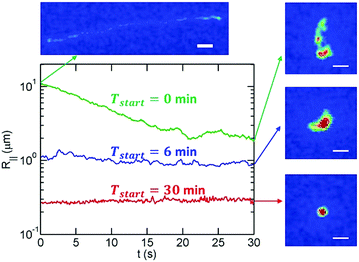
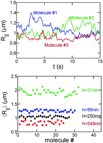
In order to avoid photobleaching over the experiment's duration, we turn on the fluorescent lamp and record 15–30 seconds of video every few minutes until a molecule has fully collapsed. Note that the slit confinement restricts DNA motion in the z-direction, and hence DNA molecules stay in focus during the imaging period. Sample experimental videos are included in the ESI.† For the bulk/free space experiments, DNA molecules move out of focus in a few seconds as indicated by the decrease of the total fluorescence intensity of an individual DNA. Accordingly, for the bulk experiments, we measure the hydrodynamic size of DNA based on the in-plane (X–Y) diffusivity as described below.Fig. 2 shows examples of the time evolution of R‖ for a T4-DNA molecule at different time intervals. We analyze about 20–30 different molecules for each channel height h at various times Tstart. We find that the variance of R‖ among these molecules is small (Fig. 3). We further average R‖ for these molecules with the same h and Tstart. Eventually, we obtain the time evolution of R‖ for each slit height as shown in Fig. 5. Similar data analysis is also applied to obtain the apparent aspect ratio RM/Rm.
Direct calculation of the DNA size from the intensity profile is limited in resolution and accuracy due to the point-spread function of the microscope.31 This problem is more profound when DNA assumes a highly compact conformation. As a result, we also measure the DNA diffusion coefficient D, and then calculate the DNA hydrodynamic radius through Rh = kBT/(6πηD). The viscosity η for different ethanol concentrations are determined by measuring the mean squared displacement of polystyrene beads of known radius. We use this method for our measurement of DNA size in bulk, i.e. free space.
It is worth mentioning that a given DNA molecule is stretched by the electric field when driven into a nanoslit as shown by the top-left image in Fig. 2. The green curve in Fig. 2 indicates that the effect of initial stretching in T4-DNA vanishes after about 20 seconds for the slit height of 89 nm. Our previous experiments34 systematically measured the duration τstr for a stretched DNA molecule to reach its equilibrium conformation in a nanoslit and found the empirical formula τstr ∼ LDNA2.2h−0.5. We estimate that the effect of initial stretching by the electric field will vanish in less than 1 minute for all slit heights used in the current study.
The electric field that is used to drive DNA into a nanoslit may introduce concentration polarization at the microchannel–nanochannel–microchannel interfaces wherein the concentrations of ions in the buffer do not distribute homogenously.35 Prior work has shown that concentration polarization primarily depends on an inverse Dukhin number Fhzc/σ, where F is Faraday's constant, h is the slit height, z is the valence of the ion, c is the concentration of the ion, and σ is the surface charge density of slit walls.35 Substituting z = 1, c ≈ 57 mM, and σ ≈ 60 mC m−2,36 we obtain the inverse Dukhin numbers of approximately 183, 50, 23, 8.2 and 2.8 for h = 2000, 549, 250, 89 and 31 nm, respectively. These inverse Dukhin numbers are greater than unity, which suggests that concentration polarization is not significant in our experiments.
3. Results and discussions
3.1 DNA collapse in free space
We first present our experimental results of DNA collapse in free space. The free-space experiments are used to estimate the critical ethanol concentration for DNA collapse, and later we pick an ethanol concentration in the vicinity of the critical concentration so that the duration of DNA collapse is sufficiently long and we can measure the effect of confinement on DNA collapse more conveniently.Fig. 4a shows the time evolution of the hydrodynamic radius, Rh, of T4-DNA for a range of ethanol concentration cethanol from 0% to 60%. For cethanol ≲ 30%, T4-DNA molecules always assume coiled conformations in 24 hours and show significant conformational fluctuations (Fig. 4b). Accordingly, we consider that T4-DNA or λ-DNA collapse do not take place for cethanol ≲ 30%. Note that the typical relaxation time of DNA conformations is on the order of seconds and our maximal observation time 24 hours is many orders of magnitude longer than the relaxation time. For 30% ≲ cethanol ≲ 45%, T4-DNA molecules collapse towards globular conformations (Fig. 4b). For globular conformations, we can still observe conformational fluctuations, such as a short DNA fragment occasionally stretching out from a globular conformation (see experimental videos in ESI†). For cethanol ≳ 45%, T4-DNA molecules eventually reach more compact conformations with little conformational fluctuations (Fig. 4b). These compact conformations are expected to be toroidal based on previous experimental37,38 and simulation39,40 studies of condensed DNA. The duration until DNA collapse depends on the ethanol concentration. For 30% ≲ cethanol ≲ 50%, DNA collapse takes hours.
Single-molecule DNA collapse under a wide range of ethanol concentration has also been investigated using magnetic tweezers and Atomic Force Microscopy (AFM) by Wang et al.37 They found that λ-DNA collapse occurs within a few hours incubation time for cethanol ≳ 30%, and the condensed λ-DNA structures become more compact with the increasing ethanol concentration. At cethanol = 50%, they found that DNA reaches condensed states between two and three hours. All of these observations are in agreement with our results.
Based on these results for DNA in bulk, we pick the ethanol concentration of 35% for nanoslit experiments. With 35% ethanol, T4-DNA is fully collapsed after about 220 minutes and eventually reaches globular conformations.
3.2 DNA collapse in nanoslits
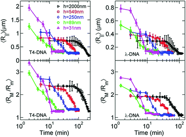
Now, we move to the experimental results of DNA collapse in nanoslits using the fixed ethanol concentration of 35%. To monitor the conformation change during DNA collapse, we calculate both the average in-plane radius of gyration 〈R‖〉 and the apparent aspect ratio 〈RM/Rm〉 of single DNA conformations based on the DNA intensity profiles, as shown in Fig. 5. Recall that RM and Rm are the major and minor principal axes of DNA conformation.41 The observations of 〈R‖〉 (two top plots in Fig. 5) and 〈RM/Rm〉 (two bottom plots in Fig. 5) are consistent with each other. During DNA collapse, the DNA size becomes smaller and the apparent aspect ratio decreases towards unity, which indicates nearly isotropic conformations.With the decrease of the slit height from h = 2000 nm to h = 31 nm, the collapse time decreases from more than one hour to about 10 minutes. To elaborate the process of DNA collapse, we re-plot the time evolution of 〈R‖〉 for T4-DNA in Fig. 6 but with a linear time-scale for h = 2000 nm, h = 250 nm, and h = 31 nm. For h = 2000 nm, the collapse process exhibits three stages: a nucleation state where 〈R‖〉 remains approximately constant, an annealing stage where 〈R‖〉 rapidly decreases, and a final stage where the DNA is collapsed. These stages are similar to those observed in previous experiments of single-DNA collapse.30 The nucleation in our experiments is reasonable considering that the DNA–DNA attraction induced by 35% ethanol is marginally larger than the critical attraction and the condensed state is stable only when its size exceeds a certain value, i.e. the nucleation size. We define the nucleation time τnucl for the first stage and the annealing time τanneal for the second stage as shown in Fig. 6a. We also define the collapse time τcollapse = τnucl + τanneal for the entire process.
 | ||
| Fig. 6 Plot of 〈R‖〉 for T4-DNA in (same as in Fig. 5), but with a linear scale for the incubation time for (a) h = 2000 nm, (b) h = 250 nm, and (c) h = 31 nm. For (a), the collapse process exhibits three stages. The intersection of the two green lines is used to determine the nucleation time. Three inset images in (b) show one T4-DNA molecule at t = 10, 25 and 70 min. The error bars indicate the standard deviations among the 〈R‖〉interval of 20–30 DNA molecules under a certain condition, where 〈R‖〉interval is the average size of a given DNA over an interval of 15–30 seconds. | ||
Now we proceed to more quantitative analysis of the time scales τnucl, τanneal, and τcollapse for DNA collapse. We determine τcollapse as follows. We calculate the average value and standard deviation of R‖ over the time interval that the DNA is clearly in the collapsed stage 3; e.g. t ≥ 230 minutes for T4-DNA with h = 2000 nm, and t ≥ 14 minutes for T4-DNA with h = 31 nm. We refer the average value as 〈R3〉 and the standard deviation as σ3. Then, τcollapse is defined as the earliest time that R‖ is less than 〈R3〉 + σ3. The collapse times are listed in Table 1 and plotted in Fig. 7. We determine τnucl as follows. We approximate that the evolution of R‖ in stage 1 and stage 2 can be fit by two straight lines, respectively, and the intersection of these two straight lines correspond to τnucl. We adjust the lines to minimize the error in the fit (details provided in the ESI†). This method yields τnucl = 70 min for h = 2000 nm, τnucl = 35 min for h = 549 nm, and τnucl = 10 min for h = 250 nm in the case of T4-DNA. We stress that this is an approximate method to estimate the time at which the DNA transitions from stage 1 to stage 2 that we define as τnucl. For T4-DNA with h = 89 nm and h = 31 nm, there is no clear distinction between stages 1 and 2, which can be caused by three possible reasons: (i) no nucleation exists for these two slit heights (see more discussion in Section 3.3); (ii) the nucleation is less than a few minutes, which cannot be resolved by our data sampling rate; (iii) the nucleation is less than a few minutes, and the plateau in the evolution of 〈R‖〉 is convoluted with the initial DNA stretching due to the electric field (see Fig. 2 for an example). The third possible reason is unlikely, because the effect of the initial DNA stretching vanishes in less than one minute as discussed in the methods section. It is worth pointing out that the data in Fig. 6 show a clear trend that the nucleation time is shorter for a smaller slit height from h = 2000 nm to 89 nm, and the transition from stage 2 to 3 is clearly seen. The speeds of DNA collapse for h = 89 nm and h = 31 nm are close and so it is unclear which one is faster. Note that it is also difficult to precisely identify the spatial location of the nucleation core directly from image analysis, because the high-intensity spots seen in nucleation cores are hard to distinguish from similar spots caused by thermal fluctuations in the projected DNA conformation (see an example of the coil in Fig. 4b and more images in the ESI†). Only in a few experiments are we able to observe the nucleation process as shown by the imaging of Fig. 6b. In the previous AFM experiments by Wang et al.,37 DNA collapse sometimes starts from non-local contacts of DNA segments, which are separated by a big loop that assumes a loose conformation. If the nucleation occurs due to non-local contacts, it is even more difficult to identify the nucleation core location from the image analysis.
| τ collapse (min) | τ nucl (min) | τ anneal (min) | |
|---|---|---|---|
| h = 2000 nm | 220 [95] | 70 [35] | 150 [60] |
| h = 549 nm | 80 [45] | 35 [24] | 45 [21] |
| h = 250 nm | 45 [21] | 10 [9] | 35 [12] |
| h = 89 nm | 16 [12] | 0 [0] | 16 [12] |
| h = 31 nm | 12 [9] | 0 [0] | 12 [9] |
 | ||
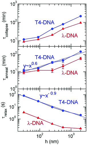
| Fig. 7 The collapse time, annealing time and rotational relaxation time as a function of the slit height for T4 DNA and λ-DNA. | ||
The five slit heights in our experiments vary from h = 2000 nm to h = 31 nm. To evaluate the strength of confinement by these slits, we compare the slit height with the DNA persistence length in 35% ethanol and the DNA size in bulk. The DNA persistence length decreases from Lp ≈ 50 nm to Lp ≈ 22 nm after adding 35% ethanol, which was determined by previous experiments by Wang et al.37 The smallest slit height h = 31 nm is close to Lp ≈ 22 nm. To calculate the size of DNA coils in bulk, we first analyze the effective DNA diameter w in 35% ethanol. The value of w should be close to zero, because 35% ethanol concentration is close to the critical value for DNA collapse, and the solvent quality is close to the θ-condition. As such, we approximate YOYO-1 stained DNA in 35% ethanol as a wormlike chain with Lp ≈ 22 nm and w ≈ 0 nm. We obtain λ-DNA size 〈Rλ-DNA‖,bulk〉 ≈ 0.33 μm and T4-DNA size 〈RT4-DNA‖,bulk〉 ≈ 0.61 μm after using Lλ-DNA ≈ 22 μm, LT4-DNA ≈ 75 μm and applying the theoretical formula  , where
  is used for the in-plane rather than three dimensional radius of gyration. The largest slit height h = 2000 nm is about six times of 〈Rλ-DNA‖,bulk〉 ≈ 0.33 μm, and hence λ-DNA experiences negligible confinement, and the initial λ-DNA size for h = 2000 nm is 〈R‖〉 ≈ 0.30 μm, which is close to 〈Rλ-DNA‖,bulk〉. For T4-DNA, The largest slit height h = 2000 nm is about three times of 〈RT4-DNA‖,bulk〉 ≈ 0.61 μm, and T4-DNA experiences weak confinement. The initial T4-DNA size for h = 2000 nm is 〈R‖〉 ≈ 0.72 μm, which is larger than 〈RT4-DNA‖,bulk〉 due to DNA compression by slit walls. Note that our previous experiment31 obtained the λ-DNA size 〈Rλ-DNA‖,bulk〉 ≈ 0.52 μm in the absence of ethanol, which is larger than 0.33 μm in the current study.
Our experiments employ two types of DNA molecules: T4-DNA with L ≈ 75 μm and λ-DNA with L ≈ 22 μm. Despite of the difference in the contour length by a factor of 3.41, both DNA molecules exhibit the same trend in confinement. Quantitatively, the difference in the contour length leads to the differences in confinement free energy and the relaxation time τrelax, and then the difference in time scales of DNA collapse. Based on the blob-model scaling τrelax ∼ L−5/2 in confinement,31 the relaxation of T4-DNA is expected to be slower than λ-DNA by a factor of 21, which is close to τT4-DNArelax/τλ-DNArelax ≈ 24 observed in our experimental measurement of rotational relaxation time over the range from h = 31 nm to 549 nm. The relaxation time of T4-DNA appears to follow a scaling τrelax ∼ h−0.9 from h = 31 to 2000 nm, while the relaxation time of λ-DNA significantly deviates from this scaling at h = 2000 nm, because λ-DNA experiences negligible confinement at h = 2000 nm due to the insufficiency in the contour length. Although T4-DNA and λ-DNA differ in the relaxation time by a factor of about 24, the difference in the collapse time is only by a factor of about 1.8, and the difference in the annealing times is by a factor of about 2.1 (see ESI†). As previously investigated by Yoshinaga,42 the annealing speed may be determined by the motion of the collapsed domain or the coil domain depending on the stage of collapse process. If it is the former, the annealing time is insensitive to the chain length, while if it is the latter, the annealing time is related to the relaxation time of the entire length. The mixture of both motions in the collapse process may result in a weak dependence of the annealing time or collapse time on the chain length.
3.3 Discussion about how confinement speeds up DNA collapse
Now we proceed to the theoretical explanation for the effect of confinement on DNA collapse. We highlight that as the slit becomes shallower, the relaxation dramatically slows down, but conversely the collapse kinetics greatly speeds up (Fig. 7). These two trends indicate that the faster collapse in confinement is driven by the effect of confinement on the free energy landscape and not by hydrodynamic effects. Fig. 8 illustrates the effect of confinement on the free energy landscape with respect to the compactness of DNA conformations. DNA collapse corresponds to a transition from a coiled conformation to a globular conformation. The confinement free energy experienced by the coiled conformation Fslitcoil is larger than the one by the globular conformation Fslitglobule, because the coiled conformation is larger and more likely to be restricted by slit walls. The difference ΔFslit = Fslitcoil − Fslitglobule reshapes the free energy landscape and thermodynamically promotes the coil-globule transition in confinement. In our previous simulation study,43 we have calculated the free energy landscape for the coil-globule transition in slit confinement using computer simulations and obtained similar curves as shown in Fig. 8. More generally, confinement promotes any reaction or conformation change that compacts the polymer conformation, such as protein folding44,45 and knot formation in confinement.46Due to the existence of plateaus in the time evolution of 〈R‖〉, we expect that there is a free energy barrier along the path of DNA collapse. The free energy barrier is akin to that found in typical crystallization of small molecules, namely that the small nuclei are unstable due to a relatively large surface energy. Here, the surface energy refers to an energy increase because the surface monomers have a smaller number of attractive neighbors than the monomers in the core. The dependence of confinement free energy on the compactness of DNA conformation also reduces the free energy barrier for DNA collapse. The reduction of free energy barrier by confinement can explain our observation that the nucleation process becomes shorter or disappears in shallower slits.
Our experimental data can be understood quantitatively to a certain extent. We focus on the data of T4-DNA rather than λ-DNA, because T4-DNA is longer and it suffers less finite-length effects when applying scaling relationships. We estimate the confinement free energy per unit length fconfine by the scaling relationship15
 | (1) |
τnucl ∼ τrelax![[thin space (1/6-em)]](https://www.rsc.org/images/entities/char_2009.gif) exp(Fslitbarrier/kBT), exp(Fslitbarrier/kBT), | (2) |
| Fslitbarrier = Fbulkbarrier + fconfineLnucl | (3) |
We also adopt a crude approximation to quantitatively analyze the annealing time. We assume the annealing speed is proportional to the slope of free energy landscape as labeled in Fig. 8:
| τanneal ∼ τrelax/εslit, | (4) |
| εslit = εbulk + (π2/3)h−2Lp | (5) |
| τanneal ∼ τrelax/h−2. | (6) |
Using the de Gennes scaling τrelax ∼ h−7/6, we obtain τanneal ∼ h5/6. Considering that many assumptions are made, the predicted exponent of 5/6 agrees fairly well with the exponent 0.6 ± 0.2 obtained by the fit to our experiment results of τanneal. In particular, we ignore εbulk in eqn (5). The mixture of εbulk ∼ h0 and (π2/3)h−2Lp would result in an apparent scaling exponent larger than −2, and then an exponent in τanneal less than 5/6.
Previous simulations by Das and Chakraborty have also quantitatively investigated the effect of slit confinement on polymer collapse, however, only for short flexible chains.47 After including hydrodynamic interactions, the slit confinement can speed up polymer collapse by a factor from 2.5 to 4 for the chain length from 80 to 200. If we extrapolate the confinement effect to longer chains, it may explain our experimental observation that the slit confinement speeds up DNA collapse by 18 times. Recall that T4-DNA used in our experiments has a contour length ∼75 μm, which is about 3400 times of DNA the persistence length ∼22 nm in 35% ethanol.
3.4 Discussion about the surface charge of the slit walls
In addition to the steric repulsion, the slit walls also affect DNA behavior through the surface charge.36 The silica nanoslits used here have negative surface charges. The electrostatic repulsion between slit walls and DNA makes the accessible height by DNA less than the actual slit height, i.e. hactual = h − 2δ, where δ is the effective thickness of the surface layer inaccessible by DNA. The calculation of δ is rather complicated, but δ depends on the Debye length and is on the order of nanometres in our studies.2,48 Accordingly, we expect the difference between hactual and h is small in our studies and does not affect our overall conclusions.The counterions (more specifically, H+ in the current study) of surface charge also change the pH value of the buffer in nanoslits. Previous experiments by Bottenus et al.49 measured the change in the pH value in nanochannels over a range of buffer conditions. In a channel with a cross-section of 100 nm × 400 nm, the pH value shift is about 0.2 at an ionic strength of 57 mM.49 This prior work suggests that the changes in pH value for h = 2000, 549, 250 and 89 nm are smaller than or around 0.2. Such a small change in pH value should not change DNA properties, such as charge density.
4. Conclusions
In summary, single molecule experiments show that nanoconfinement greatly speeds up the nucleation and the annealing of single-DNA collapse, despite the fact that DNA dynamics is slowed down by confinement. If we view DNA collapse as a reaction from a metastable coiled state to a globule state, then nanoconfinement can be considered as a catalyst to accelerate this reaction by reshaping the free energy landscape and promoting smaller conformations. What is different from a traditional catalyst is that the relative free energy between the coil (reactant) and the globule (product) is also altered by confinement. However, if DNA is driven into confinement before reaction and driven out of confinement after reaction, then confinement essentially acts as a catalyst by changing the free energy barrier. Such catalysis by confinement may be used by cells to speed up protein folding. Experiments44,50 and simulations45 suggest that the nanocage-confinement by chaperonin may be a key factor, among others,51 facilitating protein folding. Confinement is also suggested to affect other conformation transitions, e.g. DNA denaturation,52 polymer looping53 and protein dimerization.54 The direct visualization of long DNA in our experiment allows for quantitative measurements and sheds light on conformation transitions in confinement.Conflicts of interest
There are no conflicts to declare.Acknowledgements
This work is supported by the Singapore-MIT Alliance for Research and Technology (SMART) and the National Science Foundation (NSF) grant CBET-1602406.References
- R. B. Schoch, J. Han and P. Renaud, Rev. Mod. Phys., 2008, 80, 839 CrossRef CAS.
- W. Reisner, J. N. Pedersen and R. H. Austin, Rep. Prog. Phys., 2012, 75, 106601 CrossRef PubMed.
- C. Zhang, P. G. Shao, J. A. van Kan and J. R. van der Maarel, Proc. Natl. Acad. Sci. U. S. A., 2009, 106, 16651 CrossRef CAS PubMed.
- D. J. Mai, C. Brockman and C. M. Schroeder, Soft Matter, 2012, 8, 10560 RSC.
- E. T. Lam, A. Hastie, C. Lin, D. Ehrlich, S. K. Das, M. D. Austin, P. Deshpande, H. Cao, N. Nagarajan, M. Xiao and P. Y. Kwok, Nat. Biotechnol., 2012, 30, 771 CrossRef CAS PubMed.
- R. Marie, J. N. Pedersen, D. L. Bauer, K. H. Rasmussen, M. Yusuf, E. Volpi, H. Flyvbjerg, A. Kristensen and K. U. Mir, Proc. Natl. Acad. Sci. U. S. A., 2013, 110, 4893–4898 CrossRef CAS PubMed.
- W. Reisner, N. B. Larsen, H. Flyvbjerg, J. O. Tegenfeldt and A. Kristensen, Proc. Natl. Acad. Sci. U. S. A., 2009, 106, 79–84 CrossRef CAS PubMed.
- W. Reisner, N. B. Larsen, A. Silahtaroglu, A. Kristensen, N. Tommerup, J. O. Tegenfeldt and H. Flyvbjerg, Proc. Natl. Acad. Sci. U. S. A., 2010, 107, 13294–13299 CrossRef CAS PubMed.
- H.-X. Zhou, G. Rivas and A. P. Minton, Annu. Rev. Biophys., 2008, 37, 375 CrossRef CAS PubMed.
- W. Reisner, K. J. Morton, R. Riehn, Y. M. Wang, Z. Yu, M. Rosen, J. C. Sturm, S. Y. Chou, E. Frey and R. H. Austin, Phys. Rev. Lett., 2005, 94, 196101 CrossRef PubMed.
- A. Küchler, M. Yoshimoto, S. Luginbühl, F. Mavelli and P. Walde, Nat. Nanotechnol., 2016, 11, 409 CrossRef PubMed.
- T. E. Sintra, S. P. Ventura and J. A. Coutinho, J. Mol. Catal. B: Enzym., 2014, 107, 140 CrossRef CAS.
- G. Henkin, D. Berard, F. Stabile, M. Shayegan, J. S. Leith and S. R. Leslie, Anal. Chem., 2016, 88, 11100 CrossRef CAS PubMed.
- Y. Wang, D. R. Tree and K. D. Dorfman, Macromolecules, 2011, 44, 6594 CrossRef CAS PubMed.
- L. Dai, J. R. C. Van der Maarel and P. S. Doyle, Macromolecules, 2014, 47, 2445 CrossRef CAS.
- L. Dai, C. B. Renner and P. S. Doyle, Adv. Colloid Interface Sci., 2016, 232, 80 CrossRef CAS PubMed.
- L. Dai, D. R. Tree, J. R. C. van der Maarel, K. D. Dorfman and P. S. Doyle, Phys. Rev. Lett., 2013, 110, 168105 CrossRef PubMed.
- D. J. Berard, F. Michaud, S. Mahshid, M. J. Ahamed, C. M. McFaul, J. S. Leith, P. Bérubé, R. Sladek, W. Reisner and S. R. Leslie, Proc. Natl. Acad. Sci. U. S. A., 2014, 111, 13295 CrossRef CAS PubMed.
- A. R. Klotz, M. Mamaev, L. Duong, H. W. de Haan and W. W. Reisner, Macromolecules, 2015, 48, 4742 CrossRef CAS.
- J.-W. Yeh, A. Taloni, Y.-L. Chen and C.-F. Chou, Nano Lett., 2012, 12, 1597 CrossRef CAS PubMed.
- C. Zhang, F. Zhang, J. A. van Kan and J. R. C. van der Maarel, J. Chem. Phys., 2008, 128, 225109 CrossRef PubMed.
- J. Tang, D. W. Trahan and P. S. Doyle, Macromolecules, 2010, 43, 3081–3089 CrossRef CAS PubMed.
- A. C. Klepinger, M. K. Greenier and S. L. Levy, Macromolecules, 2015, 48, 9007–9014 CrossRef CAS.
- A. Khorshid, S. Amin, Z. Zhang, T. Sakaue and W. W. Reisner, Macromolecules, 2016, 49, 1933–1940 CrossRef CAS.
- M. J. Ahamed, S. Mahshid, D. J. Berard, F. O. Michaud, R. Sladek, W. W. Reisner and S. R. Leslie, Macromolecules, 2016, 49, 2853–2859 CrossRef CAS.
- D. Kim, C. Bowman, J. T. Del Bonis-O'Donnell, A. Matzavinos and D. Stein, Phys. Rev. Lett., 2017, 118, 048002 CrossRef PubMed.
- A. Estevez-Torres and D. Baigl, Soft Matter, 2011, 7, 6746 RSC.
- C. Zhang, D. Guttula, F. Liu, P. P. Malar, S. Y. Ng, L. Dai, P. S. Doyle, J. A. van Kan and J. R. van der Maarel, Soft Matter, 2013, 9, 9593 RSC.
- C. Zhang, K. Jiang, F. Liu, P. S. Doyle, J. A. van Kan and J. R. van der Maarel, Lab Chip, 2013, 13, 2821 RSC.
- K. Yoshikawa and Y. Matsuzawa, J. Am. Chem. Soc., 1996, 118, 929 CrossRef CAS.
- J. Tang, S. L. Levy, D. W. Trahan, J. J. Jones, H. G. Craighead and P. S. Doyle, Macromolecules, 2010, 43, 7368 CrossRef CAS.
- E. A. Strychalski, S. L. Levy and H. G. Craighead, Macromolecules, 2008, 41, 7716 CrossRef CAS.
- K. Günther, M. Mertig and R. Seidel, Nucleic Acids Res., 2010, 38, 6526 CrossRef PubMed.
- A. Balducci, C.-C. Hsieh and P. Doyle, Phys. Rev. Lett., 2007, 99, 238102 CrossRef CAS PubMed.
- T. A. Zangle, A. Mani and J. G. Santiago, Chem. Soc. Rev., 2010, 39, 1014 RSC.
- D. Stein, M. Kruithof and C. Dekker, Phys. Rev. Lett., 2004, 93, 035901 CrossRef PubMed.
- Y. Wang, S. Ran, B. Man and G. Yang, Soft Matter, 2011, 7, 4425 RSC.
- V. A. Bloomfield, Curr. Opin. Struct. Biol., 1996, 6, 334 CrossRef CAS PubMed.
- A. Montesi, M. Pasquali and F. MacKintosh, Phys. Rev. E: Stat., Nonlinear, Soft Matter Phys., 2004, 69, 021916 CrossRef PubMed.
- H. Noguchi, S. Saito, S. Kidoaki and K. Yoshikawa, Chem. Phys. Lett., 1996, 261, 527 CrossRef CAS.
- C. C. Hsieh, A. Balducci and P. S. Doyle, Nano Lett., 2008, 8, 1683 CrossRef CAS PubMed.
- N. Yoshinaga, Phys. Rev. E: Stat., Nonlinear, Soft Matter Phys., 2008, 77, 061805 CrossRef PubMed.
- L. Dai, C. B. Renner, J. Yan and P. S. Doyle, Sci. Rep., 2015, 5, 18438 CrossRef CAS PubMed.
- A. Brinker, G. Pfeifer, M. J. Kerner, D. J. Naylor, F. U. Hartl and M. Hayer-Hartl, Cell, 2001, 107, 223 CrossRef CAS PubMed.
- J. Mittal and R. B. Best, Proc. Natl. Acad. Sci. U. S. A., 2008, 105, 20233 CrossRef CAS PubMed.
- L. Dai, C. B. Renner and P. S. Doyle, Macromolecules, 2015, 48, 2812–2818 CrossRef CAS.
- S. Das and S. Chakraborty, J. Chem. Phys., 2010, 133, 174904 CrossRef PubMed.
- G. K. Cheong, X. Li and K. D. Dorfman, Phys. Rev. E, 2017, 95, 022501 CrossRef PubMed.
- D. Bottenus, Y.-J. Oh, S. M. Han and C. F. Ivory, Lab Chip, 2009, 9, 219–231 RSC.
- Y.-C. Tang, H.-C. Chang, A. Roeben, D. Wischnewski, N. Wischnewski, M. J. Kerner, F. U. Hartl and M. Hayer-Hartl, Cell, 2006, 125, 903 CrossRef CAS PubMed.
- A. I. Jewett and J.-E. Shea, Cell. Mol. Life Sci., 2010, 67, 255 CrossRef CAS PubMed.
- H. Li, Z. Wang, N. Li, X. He and H. Liang, J. Chem. Phys., 2014, 141, 044911 CrossRef PubMed.
- J. Shin, A. G. Cherstvy and R. Metzler, ACS Macro Lett., 2015, 4, 202 CrossRef CAS.
- W. Wang, W.-X. Xu, Y. Levy, E. Trizac and P. Wolynes, Proc. Natl. Acad. Sci. U. S. A., 2009, 106, 5517 CrossRef CAS PubMed.
Footnote |
| † Electronic supplementary information (ESI) available: More information about experiments, data analysis and images and videos of DNA collapse. See DOI: 10.1039/c7sm01249g |
| This journal is © The Royal Society of Chemistry 2017 |


