Locating microcalcifications in breast histopathology sections using micro CT and XRF mapping
Abstract
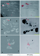
Spectroscopic measurement of microcalcification chemistry holds great promise as a rapid, quantitative, and non-invasive aid to diagnosis of early stage breast cancer. Previous work has shown that carbonate substitution in hydroxyapatite is highly correlated to breast cancer grade. A deeper understanding of the chemistry–pathology relationships is important in the development of spectroscopic aids to diagnosis. However, investigation of calcification chemistry is hampered by the difficulty of quickly and systematically locating microcalcifications within tissue specimens. We have demonstrated two simple methods based on micro-CT and XRF mapping which can achieve this in sections cut from wax embedded breast tissue from diagnostic archives.

- This article is part of the themed collection: Clinical Diagnostics

 Please wait while we load your content...