Investigating the use of Raman and immersion Raman spectroscopy for spectral histopathology of metastatic brain cancer and primary sites of origin
Abstract
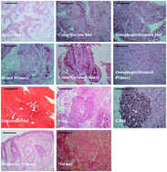
It is estimated that approximately 13 000 people in the UK are diagnosed with brain cancer every year; of which 60% are metastatic. Current methods of diagnosis can be subjective, invasive and have long diagnostic windows. Raman spectroscopy provides a non-destructive, non-invasive, rapid and economical method for diagnosing diseases. The aim of this study was to investigate the use of Raman and immersion Raman spectroscopy for diagnosing metastatic brain cancer and identifying primary sites of origin using brain tissue. Through spectral examination, the Raman peaks at 721 cm−1 and 782 cm−1 were identified as being the most distinct for discriminating between the glioblastoma multiforme (GBM), metastatic and normal brain tissue spectra. A ratio score plot of these peaks calculated the classification sensitivities and specificities as 100% and 94.44% for GBM, 96.55% and 100% for metastatic brain, and 85.71% and 100% for normal brain tissue. Principal Component-Linear Discriminant Analysis (PC-LDA) also showed discrimination between normal, GBM and metastatic brain tissue spectra. We also present, for the first time, the use of Raman spectroscopy to investigate primary site of origin for metastatic brain cancer and any biochemical differences between different primary and metastatic cancer using linked samples. This study revealed interesting spectral differences in the amide regions showing changes in the biochemistry of the metastatic brain cancer from the primary cancer.

- This article is part of the themed collection: Clinical Diagnostics

 Please wait while we load your content...