
 Open Access Article
 This Open Access Article is licensed under a Creative Commons Attribution-Non Commercial 3.0 Unported Licence
Enhancing the luminescence intensity of Eu3+-activated NaYb(MoO4)2 phosphors through bismuth doping: Judd–Ofelt analysis, lighting, and temperature-sensing applications†
Yosra
Bahrouni
a,
Ikhlas
Kachou
a,
Kamel
Saidi
 a,
Tarak
Kallel
a,
Mohamed
Dammak
*a,
Irene
Mediavilla
b and
Juan
Jiménez
b
a,
Tarak
Kallel
a,
Mohamed
Dammak
*a,
Irene
Mediavilla
b and
Juan
Jiménez
b
aLaboratoire de Physique Appliquée, Faculté des Sciences de Sfax, Département de Physique, Université de Sfax, BP 1171 Sfax, Tunisia. E-mail: madidammak@yahoo.fr; mohamed.dammak@fss.usf.tn
bGdS Optronlab, Department of Condensed Matter Physics, LUCIA Building, University of Valladolid, Paseo de Belén 19, 47011, Valladolid, Spain
First published on 17th January 2025
Abstract
In this work, we investigate the impact of Bi3+ doping on the luminescence properties of Eu3+-activated NaYb(MoO4)2 phosphors synthesized via the conventional solid-state reaction method. Rietveld refinement of X-ray diffraction data confirmed the tetragonal crystal structure (space group I41/a) for all samples. UV-visible absorption spectroscopy revealed an indirect bandgap of approximately 3.25 eV for the 5% Bi3+-doped sample. Under UV excitation, intense red emissions originating from the 5D0 → 7F transitions of Eu3+ ions were observed at 589 nm, 613 nm, 652 nm, and 700 nm, along with near-infrared emission from Yb3+ at 997 nm, sensitized by the MoO42− group. Photoluminescence (PL) analysis demonstrated an enhancement in the Eu3+ emission intensity with increasing Bi3+ concentration, reaching an optimum at 5% Bi3+ doping. Chromaticity coordinates confirmed a significant enhancement in the red emission intensity upon Bi3+ incorporation. Judd–Ofelt parameters and crystal field parameters were determined, revealing that Bi3+ doping influences the local environment of Eu3+ ions, impacting the luminescence properties. Furthermore, we explored the potential of Bi3+/Eu3+ codoped NaYb(MoO4)2 for optical thermometry based on the fluorescence intensity ratio (FIR) technique, achieving a high relative sensitivity (Sr = 1.14% K−1). This work demonstrates the influence of Bi3+ doping on the luminescence properties of Eu3+ in NaYb(MoO4)2 and explores its potential for applications in temperature sensing and other optoelectronic devices.
Introduction
The field of luminescence thermometry is experiencing intense growth, with significant advances in detection, imaging, diagnostics, and therapy, among others. This interest has been stimulated mainly by the fact that many of today's technological requirements in fields such as micro- and nanoelectronics, photonics, nanomedicine, micro- and nanofluidics1 have reached a point where the use of traditional contact thermal probes is unable to provide reliable measurements when the spatial resolution enters the submicron range. This limitation of conventional thermometers for small systems has stimulated the development of luminescent micro- and nano-thermometers.2 Moreover, from an industrial point of view, this expected miniaturization should make it possible to bring new nanoscale thermal probes to the market. Although the recently developed luminescent nanothermometers are radically more sophisticated and involve complex synthesis procedures, the fundamental problems and applications addressed are similar to those reported at the beginning of the field: understanding heat and energy transfer mechanisms, optimizing temperature readout, and developing efficient cost-effective sensors for advanced medical and engineering tools. Among the various emitting centers used in luminescence thermometry, one can include proteins, nucleic acids and other biomolecules, thermosensitive polymers,3,4 organic dyes and QDs.5 Systems include molecular complexes, MOFs, organic–inorganic polymers and hybrids, multifunctional thermometer nanoplatforms, and UC, DC, and DS materials. This article describes the use of Ln3+ lanthanide and transition-metal ion co-doped phosphors as luminescent ratiometric thermometers aiming for the use of luminescence thermometry for thermal imaging.6 Lanthanide ions (Ln3+) (such as Eu3+, Tb3+, Dy3+, and Sm3+) are used as the reference signals, while transition-metal ions (such as Mn4+ and Bi3+) are chosen for detection signals due to their different thermal quenching mechanisms7–10 for early tumor detection, but also as a tool to unveil the properties of the thermometers themselves or their local environment.11 Numerous applications in lighting devices have been found when doped with rare-earths.12 The Eu3+ ion, which generally exhibits intense red fluorescence corresponding to the 5D0 → 7F2 emission transition at around 620 nm, is often used as the active center in red phosphors in many industrial applications. Spectroscopy of the Eu3+ ion is also of particular interest because of its extreme sensitivity to the crystalline environment, enabling it to be used as a point probe in many materials, especially those containing rare-earth cations whose ionic radii are close to that of Eu3+.13–15 Bismuth is a non-rare-earth metal ion. It is considered the least toxic of the heavy metals, which has been the subject of intensive research in recent years due to its special optical characteristics.16As a typical non-rare earth activator, Bi3+ emits light via the 3P1 → 1S0 transition.17 This emission is highly dependent on the crystal field environment and structural symmetries. Therefore, phosphors doped with Bi3+ can emit luminescence at different wavelengths, such as ultraviolet (UV), visible, and infrared light.18 It can also be used as a sensitizer in order to efficiently transfer the absorbed NUV or UV energy to Eu3+ or Sm3+ ions. Besides, the selection of appropriate host materials is important as the host has a significant influence on the emission of the Ln3+ ions. Some of the hosts that have been used as a part of the Ln3+ doped red emitting phosphors are fluorides, germanates, vanadates, tungstates and molybdates.19,20 According to the existing literature, many reports have been devoted to synthesizing and characterizing the luminescence properties of lanthanide doped molybdate microcrystals or phosphors. Moreover, molybdates have been extensively studied for their remarkable properties such as high chemical stability, brilliance, high melting point, low chemical toxicity and long persistence without radioactive irradiation.21–23
However, molybdate phosphors present several problems. For example, SrZnMoO6 and NaY(MoO4)2 phosphors have low efficiencies of self-activating emission, which is not suitable for solid-state lighting.24 In addition, some molybdate phosphors such as NaY(MoO4)2:Eu3+ have low energy transfer (ET) efficiency from MoO42− groups to rare earth ions,25 therefore, tunable luminescence is difficult to achieve. In contrast to these MoO42− weaknesses, SrZnMoO6 vanadate presents high luminescence efficiency and a high ET efficiency. Many reports show that codoping with Bi3+ permits to enhance the luminescence performance of phosphors, such as CaMoO4:Bi3+/Eu3+, CaY4(SiO4)3O:Bi3+/Eu3+ and BaLa2Si3O10:Eu3+/Bi3+ phosphors,26,27 because of the sensitizer role of Bi3+ ions. While this progress is promising, a need remains for materials with improved thermal stability, improved luminescence, and precise temperature sensitivity for a broader range of applications.
Many research groups are constantly searching for new ratiometric thermometers with improved thermometric performance. Conventional FIR analysis requires expensive equipment, time-consuming measurements followed by complicated data processing, and considering a variety of rare earth ions and potential hosts. However, it is difficult to control all these materials from a thermometric point of view using a traditional approach. In order to solve this problem, Ciric et al. developed an extension of the Judd–Ofelt theory to the field of lanthanide thermometry, which allows calculation of the thermometric performances from the single emission spectrum measured at room temperature.28 The theoretically calculated parameters may differ slightly from those obtained experimentally due to imperfections in the Judd–Ofelt theory and in the acquisition and treatment of spectroscopic data. However, Ciric et al. demonstrated the applicability of the model for the difficult case of Eu3+ doped Y2O3 samples, which have the largest discrepancy between theoretical and experimental parameters due to the largest energy gap among rare earth ions.15
We aim here to investigate the effects of Bi3+ on the structural, optical and thermometric properties of Bi3+-codoped Eu3+-activated NaYb(MoO4)2: (NYMO) phosphors, in order to optimize its performance for optoelectronic and temperature sensing applications.
Experimental section
Materials and preparation
A series of NYMO compounds doped with Eu3+ and codoped with Bi3+ (NYMO:0.05Eu3+/yBi3+, where y = 0, 0.05, 0.15, 0.2) were successfully synthesized via a solid-state reaction. The precursors Na2CO3, MoO3, Bi2O3, Y2O3, and Eu2O3 were weighed in stoichiometric ratios and thoroughly mixed in an agate mortar for 15 minutes. The resulting mixture was dried in a furnace at 350 °C. After regrinding for 1 hour, the precursor powder was placed in alumina crucibles and sintered at 600 °C for 12 hours in an air atmosphere. Then, they undergo post-calcination under an oxygen flow to ensure appropriate oxygen stoichiometry. The annealing duration was sufficient for grain formation and growth, ensuring the structural integrity of the final product.Characterization
X-ray diffraction radiation (XRD) was used to check the crystalline structure and phase purity of the samples. The morphology and microstructures of the phosphors were analyzed by scanning electron microscopy (SEM) images which were photographed on a Hitachi SU-4800 field-emission SEM. UV-Vis-NIR absorption measurements are carried out with a UV-Vis-NIR spectrometer (PerkinElmer Lambda 950). Added to this, the photoluminescence excitation (PLE) and photoluminescence (PL) spectra as a function of temperature were recorded by using a Fluoromax-4P fluorometer (HORIBA) spectrophotometer.Results and discussion
Structural study and morphology
The synthesized particles were subjected to extensive analysis to confirm their composition and morphology. The structure and phase purity of the NYMO:0.05Eu3+/yBi3+ (y = 0, 0.05, 0.15 and 0.2) were determined by XRD analysis (Fig. 1). The particles exhibited high crystallinity and an ordered tetragonal phase, according to the NaYb(MoO4)2 reference model in JCPDS No. 04005-9926.29 Furthermore, the crystal structure of the resulting samples using Rietveld structure refinements of NYMO:0.05Eu3+ with different Bi3+ fractions (y = 0, 0.05, 0.15 and 0.2), was obtained, Fig. S1 (ESI†). The Yb3+ and Mo6+ ions are coordinated with six and four oxygen atoms respectively. The Yb3+ radius is 0.0985 nm (0.985 Å, CN = 8), the Bi3+ radius is 0.117 nm (1.17 Å, CN = 8), and the Eu3+ radius is 0.1066 nm (1.066 Å, CN = 8).30 Additionally, the crystallographic structure of the NYMO unit cell along with the coordination geometry of oxygen ions surrounding the Yb3+, Eu3+ and Bi3+ ions is drawn and presented in Fig. 1. The refined lattice parameters, cell volume as well as structural parameters are given in Table S1 (ESI†). However, as indicated in Table S2 (ESI†), the length of the Mo → O2− bond changes slightly with the increase of Bi3+.31 Meanwhile, the percentage differences between the dopants and the substituted ions are obtained using the following equation:32 | (1) |
 | (2) |
 | ||
| Fig. 1 (a) XRD powder patterns of NYMO:0.05Eu3+/yBi3+ samples. The standard data for NaYb(MoO4)2 (JCPDS card 04-005-9926) are also presented for comparison and (b) the view of the structure of NYMO:0.05Eu3+/0.05Bi3. | ||
The results are illustrated in Fig. S2 (ESI†). The distortion of the samples decreases with the Bi3+ doping concentration, which can lead to the crystal field splitting. Furthermore, the lattice distortion is inversely proportional to the variation of the volume.
The morphology of the samples doped with Eu3+ and codoped Eu3+/Bi3+ in NYMO phosphors was observed by SEM. Fig. 2 illustrates the SEM images of NYMO:0.05Eu3+ (a), and NYMO:0.05Eu3+/0.05Bi3+ (b). Moreover, it can be concluded from the SEM images that the grain conglomeration is not obvious. The particle sizes in sample NYMO:0.05Eu3+/0.05Bi3+ are smaller than those shown in Fig. 2(a), with both being well below 10 μm.
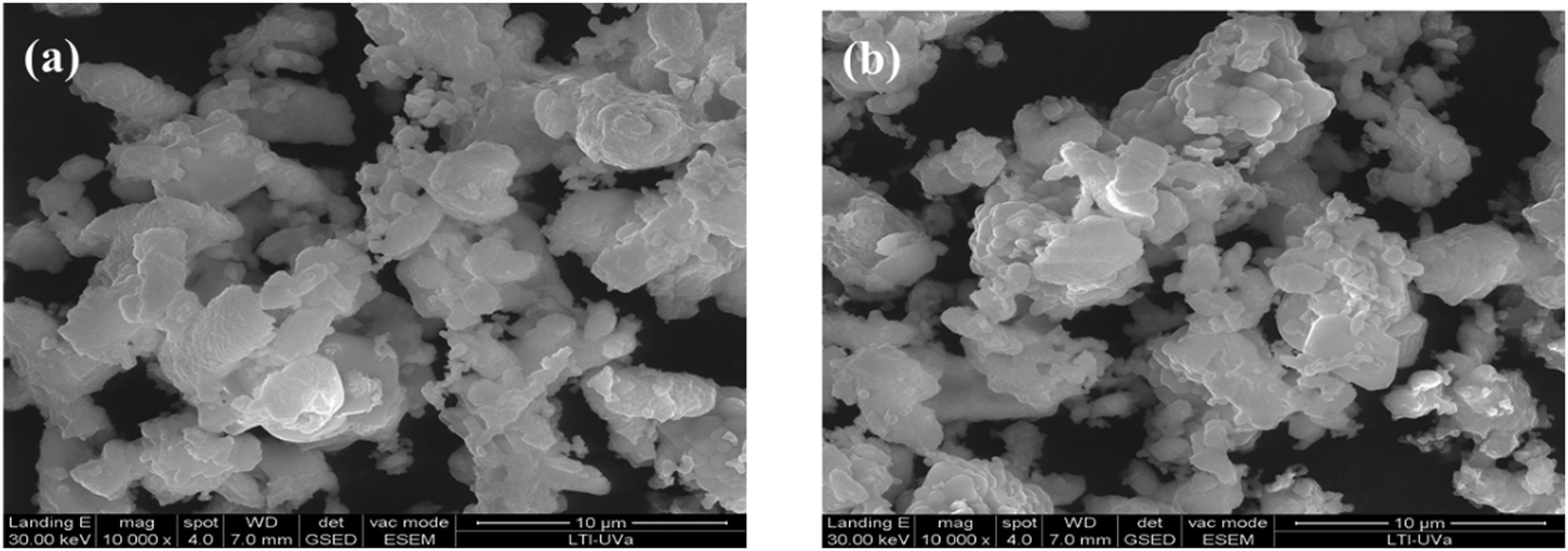 | ||
| Fig. 2 SEM images of (a) NYMO:0.05Eu3+, and (b) NYMO:0.05Eu3+/0.05Bi3+. | ||
Optical characterization
It is worth pointing out that another factor can influence the luminescence emission of lanthanide ions, particularly in systems doped with Eu3+. Due to its 4f6 electronic configuration, the Eu3+ ion must absorb an extra electron to reach the more stable 4f7 configuration. When Eu3+ is bound to O2− ligands, electron transfer can occur from the 2p oxygen orbital to the 4f7 orbitals. Two intense bands peaking at 212 and 253 nm are assigned to the charge transfer band (CTB), which can then be transferred non-radiatively to energy levels associated with Eu3+. The corresponding band in the excitation spectrum is known as the Eu–O charge transfer band (CTB).34 Furthermore, the band detected at 360 nm is attributed to the 1S0 → 1P1 Bi3+ transition.17 Doping with Bi3+ ions leads to the emergence of additional energy levels in the form of a continuous band. In addition, this doping process generates extra valence electrons from the 6s2 state.18 One observes that the optical band gap increases with respect to the host, and it is estimated to vary from 3.02 eV to 3.25 eV when the concentration of Bi3+ ions increase from 0% up to 20%.
Photoluminescence properties
To examine the sensitizing effect of Bi3+ ions on NYMO:0.05Eu3+ samples, a series of NYMO samples co-doped with Bi3+ and Eu3+ ions were successfully synthesized. Fig. 3a illustrates the PL excitation spectra of the host NYMO monitored at 615 nm emission wavelength. The spectrum displays a broad band in the range of 285 to 350 nm and a sequence of sharp bands in the range of 350 to 500 nm. The broadband with a maximum at 330 nm is related to the charge transfer from O2− → Eu3+, Mo6+ ions. There is a marked improvement in the charge transfer band when different concentrations of bismuth are added.35,36 Moreover the sharp peaks located at 363 nm (7F0 → 5L7), 382 nm (7F0 → 5D4), 394 nm (7F0 → 5L6), 416 nm (7F0 → 5D3), 464 nm (7F0 → 5D2) and 535 nm (7F0 → 5D1) are attributed to the 4f–4f transitions of Eu3+ ions.37,38 Based on the excitation spectra, the two peaks centered at 464 and 535 nm exhibit stronger intensities, suggesting that NYMO:Eu3+ phosphors can be efficiently pumped by UV light. On the other hand, Fig. 3b shows the photoluminescence emission spectra of NYMO:0.05Eu3+/yBi3+ microcrystals under excitation with UV light at 325 nm from a He–Cd laser. Emission bands at 702 nm (5D0 →7F4), 654 nm (5D0 →7F3), 613 nm (5D0 →7F2), 589 nm (5D0 → 7F1) and 533 nm (5D1 → 7F2) are associated with Eu3+ transitions.39,40 To understand these luminescence transitions, it is important to consider the crystal field splitting of the energy levels. The most intense transitions occur from the 5D0 level onwards, which remains non-degenerate (J = 0) by the crystal field. According to Judd–Ofelt theory, transitions to even-numbered J levels are considerably more intense than those to their odd-numbered counterparts. Thus, the dominant red peak at around 614 nm is attributed to the hypersensitive 5D0 →7F2 transition with ΔJ = 2.41,42 Additionally, one emission peak at 993 nm is associated with transitions related to Yb3+.43 No discernible peaks corresponding to the presence of Bi3+ ions were observed.25 Moreover, the emission intensity is gradually enhanced with the doping Bi3+ ions, which gives support to the successful incorporation of Bi3+ ions into the NYMO lattice. The Bi3+ ion concentration was y = 0, 5, 15, 20%, and it appears that the optimal emission corresponds to 5% Bi3+. | ||
| Fig. 3 Excitation and emission spectra of the NYMO:0.05Eu3+/yBi3+ phosphors monitored at λem = 615 nm, and λex = 325 nm respectively. | ||
Photometric characterization (CIE)
Based on the detected PL emission spectra, the phosphor's emission color can be mathematically expressed using the Commission International de I′Eclairage (CIE) chromaticity coordinates,44 and the CIE coordinates for Eu3+/yBi3+ codoped NaYb(MoO4)2 phosphors were evaluated from the respective emission spectra monitored at 325 nm excitation. The CIE chromaticity diagram is illustrated in Fig. 4. The color coordinates of Eu3+/yBi3+ codoped NaYb(MoO4)2 phosphors are summarized in Table S3 (ESI†). It is seen that the CIE coordinates are close to each other. They all fall within the red region. The phosphor exhibits excellent light-color conversion efficiency.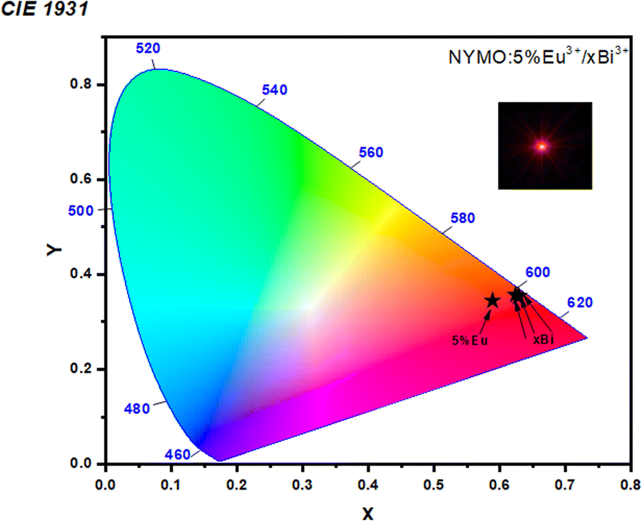 | ||
| Fig. 4 CIE 1931 chromaticity diagram of NYMO phosphors codoped with 0.05Eu3+/yBi3+ excited at λex = 325 nm. | ||
The correlated color temperature (CCT), which characterizes the color appearance of the light emitted by a light source, is another essential measure for evaluating the overall emission of phosphors. The CCT value of the phosphor can be determined from the CIE color coordinates (x, y) using the McCamy relationship.45
It should be noted that phosphors with CCT values above 4000 K are generally considered to be cold light sources, while those with CCT values below 3200 K are regarded as warm light sources in appearance.46 NYMO phosphors doped with 0.05Eu3+/yBi3+ have CCT ranging from 1977 to 2262 K. The CCT values obtained are all below 3200 K, indicating that the NYMO:Eu3+/yBi3+ phosphors fall within the warm light category.
The color purity of the phosphors is crucial for enhancing light sources in LED applications. The concept is particularly beneficial for light sources that emit light within a narrow range of wavelengths. For light sources that emit in a wide range of wavelengths (such as white light), the CCT is more advantageous. Therefore, it is essential to determine the color purity of the emitted red light.47
The calculated CCT values for all as-synthesized samples range from 1977 K to 2262 K as illustrated in Table S3 (ESI†). Furthermore, the addition of different concentrations of Bi3+ leads to an improvement in both color purity and CCT values. A pronounced red shift is thus observed. The purity of the color peaks is 87.21% for a Bi3+ concentration of 5%. The results of the chromaticity study confirm that the NYMO:Eu3+/yBi3+ compound has significant potential application in white LEDs as a red-emitting phosphor.
Judd–Ofelt analysis
To further investigate the local environment around the Eu3+ ions in the NYMO host, the Judd–Ofelt (JO) analysis is a practical tool for understanding the detailed site symmetry and luminescence of rare earth ions in a specific coordination environment. Judd–Ofelt parameters are generally derived from absorption spectra, but can be determined from emission spectra for the Eu3+ ion, as it has a simple energy diagram.47In this study, it was possible to calculate the parameters J–O, Ω2 and Ω4 from the emission spectra because the Eu3+ ion has a special energy structure under 325 nm excitation. However, the intensity parameter Ω6 was not determined because the 5D0 → 7F6 transition was not detected in the PL spectra.48 Indeed, Ω2 is strongly influenced by the environment in which Eu3+ ions are found. Thus, the maximum value of Ω2 may be associated with variations in the environment structure of the Eu3+ ion due to its high sensitivity to the 5D0 → 7F2 transition.49 A higher value of Ω2 often indicates that the symmetry of sites containing Eu3+ ions is altered. As for Ω4 (5D0 → 7F4), it is linked to the strength and stability of the matrix surrounding the rare earth ions.47 In this case, the JO parameters for Eu3+-doped NYMO were calculated using JOES software.50 The value of the refractive index used was reported in the literature (n = 1.99).51 The values obtained are listed in Table S4 (ESI†). For europium doping with different Bi3+ concentrations, the trend observed in the J–O parameters (Ω2 > Ω4) confirms that the bond is covalent.52 These theoretical calculations are in good agreement with the experimental results obtained from the XRD profile and photoluminescence spectra.
The analysis of J–O intensity parameters in NYMO phosphors co-doped with varying concentrations of Bi3+ provides insightful conclusions regarding the luminescent properties of the material. Table S4 (ESI†) summarizes the average J–O intensity parameters, specifically Ω2 and Ω4, for different Bi3+ concentrations. As the Bi3+ concentration increases from 0 to 0.05, the Ω2 parameter rises to 15.182 × 10⁻²⁰ cm2, indicating enhanced electric-dipole transition probability associated with the Eu3+ ions. However, at higher concentrations (0.15 and 0.2Bi3+), Ω2 stabilizes around 14.729 and 14.858 × 10⁻²⁰ cm2, respectively, suggesting that the positive influence of Bi3+ on the local symmetry and transition strength reaches saturation. Conversely, the Ω4 parameter shows a decline from 4.186 × 10⁻²⁰ cm2 at 0Bi3+ to 3.351 × 10⁻²⁰ cm2 at 0.05Bi3+, followed by a slight increase at 0.15Bi3+ (3.515 × 10⁻²⁰ cm2) and a subsequent decrease again at 0.2Bi3+ (3.204 × 10⁻²⁰ cm2). This trend indicates that the higher Bi3+ concentration may negatively impact the parity of the transitions, reducing the associated transition probability for the transitions governed by Ω4.
These results suggest that a low concentration of Bi3+ (y = 0.05) optimizes the J–O parameters, enhancing the luminescence of the Eu3+ ions due to improved electric-dipole transitions. However, for further increase in Bi3+ concentration, the effectiveness diminishes, indicating the need for a careful optimization of Bi3+ doping in order to maximize the optical response of the phosphors for applications in optoelectronic devices.
Crystal-field calculation
The second-order crystal field (CF) parameters, B20 and B22, characterize the interaction between the magnetic moment of the Eu3+ ion and the electric crystal field generated by its local environment. These parameters are key to understanding the spectroscopic and luminescent properties of lanthanide-doped materials.In the present case, the CF parameters B20 and B22 have been calculated from the Stark levels of the Eu3+ 7F1 ground level in the S4 symmetry crystal structure.53 This analysis provides precise information on the nature of the crystal field and its influence on the 4f orbitals of the Eu3+ ion.54
In addition to parameters B20 and B22, it is also important to calculate the parameter S2, which represents the strength of the spin–orbit interaction. This parameter is important for understanding the splitting of Eu3+ f–f energy levels due to the interaction between its spin and orbital moment, and we calculate the values of B20, B22 and S2.55 The evolution of the crystal field (CF) parameters B20 and B22 as a function of Bi3+ concentration highlights the influence of this dopant on the crystal structure and luminescent properties as shown in Fig. S4 and Table S5 (ESI†). Since our study focuses on the effect of bismuth, we carried out a comparison with other matrices, Table S6 (ESI†). These results imply that moderate Bi3+ doping (around 5–15%) enhances the crystal field strength, potentially optimizing its luminescence performance. However, high concentrations (20% Bi3+) lead to a more substantial alteration in the crystal field, which seems to affect the luminescence mechanisms and thermometric sensitivity. The variation of these parameters highlights the critical role played by Bi3+ doping in modulating the crystal environment and optimizing the optical properties of the material.
From the above analysis, the value of B20 was estimated at 55 cm−1 and gradually decreases down to an optimal Bi concentration. This decrease suggests that the introduction of Bi3+ into the crystal structure modifies the local symmetry around the Eu3+ ion, resulting in a reduction in the CF interaction between the crystal field and the Eu3+ 4f orbitals. On the other hand, the value of B22 increases up to a concentration of 5% Bi3+ decreasing thereafter. This observation indicates that the effect of Bi3+ on the crystal field is more complex and depends on the dopant concentration. It is possible that the initial increase in B22 is linked to a Bi3+-induced distortion of the crystal structure, while the subsequent decrease reflects a stabilization of the structure.
The overall analysis of the evolution of the CF parameters B20 and B22 suggests that the effect of Bi3+ on the luminescent properties of the material is linked to modification of CF around the Eu3+ ion. This modification may influence the relaxation processes of excited electrons and, consequently, the intensity and efficiency of the luminescence emission. In accordance with the structural results, which confirm that the addition of bismuth reduces the distortion modifying the structure. Therefore, the results obtained can be explained in terms of the distortion of the tetragonal structure, which lifts the local symmetry of the ion. This symmetry reduction splits the triply orbital degeneracy of the 7F1 state into three non-degenerate A + E states. Such a change in point symmetry must increase the probability of radiative transitions of the Eu3+ excited state. This is consistent with the results shown in Table S7 (ESI†).
It is important to note that the interpretation of CF parameter variations requires a detailed analysis of the crystal structure and electronic properties of the material. Theoretical simulations and additional experiments may be required to unveil the underlying mechanisms and establish a precise correlation between Bi3+ concentration, CF parameters, and the luminescent properties of the material.
Temperature sensing
Since the PL peaks of the 5D0,1 levels for Eu3+ and the 2F5/2 transition for Yb3+ are the most intense in NYMO:0.05Eu3+/yBi3+, it was the sample of choice for thermometric measurements. Its temperature dependent PL spectra are presented in Fig. 5. The temperature was varied over a wide range from 298 to 525 K. It can be seen that temperature affects the intensity of the different emission bands in a different way. Specifically, the intensities of the photoluminescence (PL) bands peaking at 534, 589, 614, 702 and 993 nm all decrease significantly with increasing temperature over the entire temperature range studied. This decrease in PL intensity at relatively high temperatures can be attributed to a thermal extinction phenomenon.56 This phenomenon is linked to an increase in the probability of non-radiative transitions between the excited energy levels of the Eu3+ ion and the crystal lattice as temperature increases. | ||
| Fig. 5 Temperature-dependent PL of the NYMO phosphor (a) doped 0.05Eu3+ and (b) codoped 0.05Eu3+/0.05Bi3+ excited at 325 nm. | ||
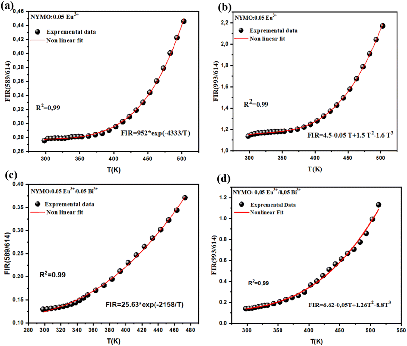
Further analysis of the emission spectra reveals an interesting relationship between the FIR (integrated fluorescence ratio) value of the areas under the emission bands at 589 and 614 nm and temperature. These two emission bands correspond to the 5D0 → 7F1 (MD) and 5D0 →7F2 (ED) transitions of the Eu3+ ion, respectively. The ground state 7F0 is excited to the 5L6 level and subsequently relaxes non-radiatively to the lower energy state 5D0. Radiative transitions then occur to the 7F1 and 7F2 levels, resulting in red emission. The FIR can be expressed as the ratio of the emission intensity of 5D0 →7F1 to that of 5D0→ 7F2. According to the Boltzmann distribution, the populations of these energy levels follow an inverse exponential distribution with temperature57,58:
 | (3) |
Furthermore, as the temperature increases from 298 K to 523 K, an increase in the FIR ratio is observed, and this increase could be attributed to lattice expansion during heating. The FIR ratio is sensitive to the covalent nature of the Eu3+ → O2− bond and the asymmetric nature of the Eu3+ site. Lattice expansion upon heating increases the bond distance of the Eu3+ → O2− bond, leading to an increase in covalency and consequently an increase in FIR.59 Also, this confirms that the populations of the 7F1 and 7F2 energy levels follow a Boltzmann distribution, and that thermal extinction is the primary mechanism responsible for the decrease in PL intensity at high temperatures. The FIR is correctly fitted and plotted as a function of temperature in Fig. 6.
 | ||
| Fig. 6 FIR vs. temperature variations for NYMO:0.05Eu3+ and NYMO:0.05Eu3+/0.05Bi3+. | ||
The emission intensity ratio of Yb3+ (993 nm) and Eu3+ (614 nm) ions, defined as FIR (993/614), is adopted to study the temperature-dependent photoluminescence property. The FIR (IYb/IEu) can be fitted as:60,61
| FIR = A + B × T + C × T2 + D × T3 | (4) |
Absolute sensitivity (Sa) and relative sensitivity (Sr) are two key factors in assessing the performance of optical thermometry. Sa and Sr are defined respectively as the rate of change and the relative rate of change of the FIR as a function of temperature, which can be calculated according to the following equations:62
 | (5) |
 | (6) |
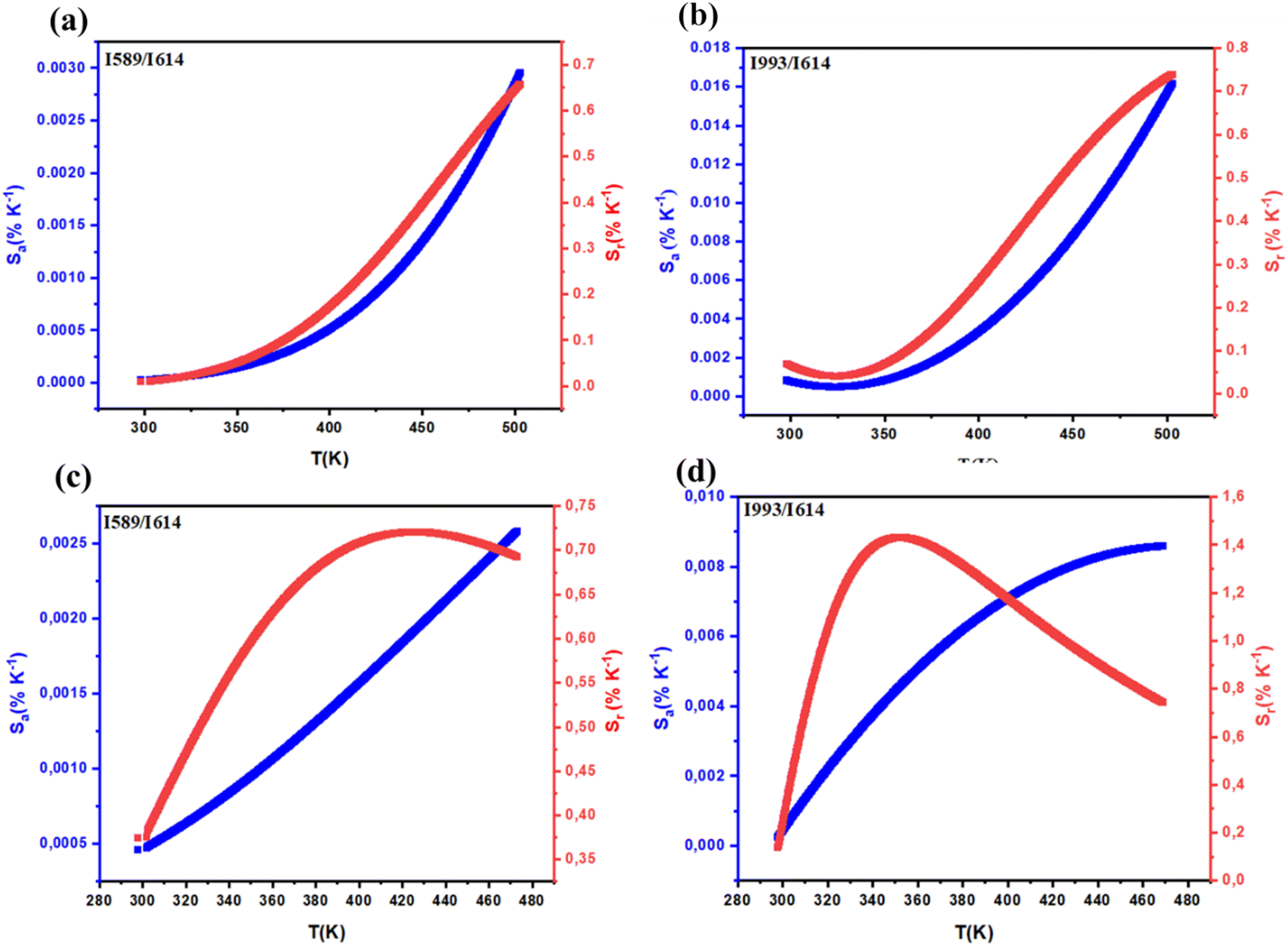 | ||
| Fig. 7 S a absolute and Sr relative sensitivity variations with temperature of NYMO:0.05Eu3+ (a) and (b) and NYMO:0.05Eu3+/0.05Bi3+ (c) and (d) under 325 nm excitation. | ||
However, for the FIR ratio (993/614), the sensitivities increase up to 350 K (Srmax = 1.14% K−1). After this temperature the Sr decreases. When examining the absolute and relative sensitivity trends between NYMO doped with 0.05Eu3+ and NYMO doped with 0.05Eu3+/0.05Bi3+, we observe a clear Sr and Sa value increase with the addition of bismuth. As a result, NYMO codoped with Eu3+ and Bi3+ seems highly suitable for temperature sensing applications.
Thermal resolution is determined by the actual temperature measured by the sensor and the precision of the experimental detection device, as shown below:64,65
 | (7) |
FIR uncertainty (δFIR) was determined as the standard deviation of the statistical distribution for each method. Using eqn (7), it is clear that these values are less than 0.5 K for FIR (589/613) and 0.25 K for FIR (993/613), indicating high accuracy for both FIR techniques over the entire temperature range studied. Fig. S5 (ESI†) shows the calculated values of temperature resolution. Note that the δT value shows a decreasing trend with increasing Bi3+ concentrations and all δT values are below 0.35 K for FIR (589/613) and 0.12 K for FIR (993/613), indicating that the designed phosphors have excellent temperature resolution.
Table 1 evidences a sensitivity of our samples higher than the previously reported values in other host materials. From the comparative analysis presented in Table 1, it is evident that the NaYb(MoO4)2:Eu3+/Bi3+ phosphor synthesized in this work shows unique thermometric properties within the temperature range of 298–503 K. Specifically, although its maximum absolute sensitivity (Samax) of 0.002 K−1 is lower than that of other materials, such as Ca2Y8(SiO4)6O2:Eu3+/Bi3+ (0.0717 K−1) and KBaGd(WO4)33+/Eu3+ (0.033 K−1), it exhibits an exceptionally high relative sensitivity (Srmax) of 1.14 K−1. This Srmax value surpasses that of all other materials in the comparison, making it particularly promising for applications requiring high temperature resolution and accuracy. The combination of moderate temperature range, improved relative sensitivity, and stable luminescence intensity positions NYMO:0.05Eu3+/0.05Bi3+ as a competitive candidate for advanced optical thermometry applications, where high precision is critical.
| Materials | Temperature range (K) | S amax (K−1) | S rmax (K−1) | Ref. |
|---|---|---|---|---|
| SrLu2O2:Eu3+/Bi3+ | 313–543 | 0.011 | 0.87 | 66 |
| Ca2Y8(SiO4)6O2:Eu3+/Bi3+ | 298–523 | 0.0717 | 0.96 | 67 |
| NaCaPO4:Tb3+/Eu3+ | 293–573 | — | 0.66 | 68 |
| Ba2TiGe2O8:Eu3+ | 358–548 | — | 0.93 | 69 |
| La2LiNbO6:Eu3+/Bi3+ | 298–498 | 0.0247 | 0.9 | 70 |
| Gd2Zr2O7:Eu3+ | 300–700 | — | 0.92 | 71 |
| NaEuF4 | 395 | — | 0.24 | 72 |
| Gd2Ti2O7:Eu3+ | 303–423 | 0.015 | 0.95 | 73 |
| Ca8ZnLa(PO4)7:Tb3+/Eu3+ | 298–448 | 0.0025 | 0.53 | 74 |
| KBaGd(WO4)3:Dy3+/Eu3+ | 298–478 | 0.033 | 0.64 | 75 |
| NaYb(MoO4)2:Eu3+/Bi3+ | 298–503 | 0.002 | 1.14 | This work |
Conclusion
A series of Eu3+-activated NYMO phosphors co-doped with Bi3+ were synthesized via solid-state reaction, confirming a tetragonal crystal structure. Under UV excitation, strong emissions from Eu3+ (589, 613, 652, 700 nm) and Yb3+ (997 nm), sensitized by MoO42−, were observed. The introduction of Bi3+ not only increased the energy gap (∼3.25 eV for 5% Bi3+) but also significantly enhanced the luminescence emission, particularly, the red emissions from Eu3+. Chromaticity analysis revealed a shift in CIE coordinates with increasing Bi3+ content, confirming intensified red emission and improved color purity, with the highest purity (87.21%) observed at 5% Bi3+. Additionally, Judd–Ofelt parameters showed that the introduction of Bi3+ affects the electric-dipole transitions of Eu3+ ions, with the Ω2 parameter increasing for low Bi3+ concentrations (up to 0.05) before stabilizing, while Ω4 decreased with increasing Bi3+, suggesting that moderate doping optimizes luminescence. The calculated crystal field parameters demonstrated that moderate Bi3+ doping, especially at 5%, optimizes the crystal field strength, enhancing the luminescence performance. However, higher concentrations (20%) lead to significant alterations of the crystal field, potentially affecting luminescence negatively. Furthermore, the incorporation of Bi3+ also improved the material's optical thermometry performance by increasing Eu3+ emission and temperature sensitivity, with a maximum absolute sensitivity (Samax) of 0.002 K−1 and a notable relative sensitivity (Srmax) of 1.14 K−1, enabling superior temperature resolution. In summary, Bi3+ doping significantly enhances the optical, structural, chromatic, and thermometric properties of the phosphors, making them highly suitable for advanced optoelectronic devices and temperature sensing applications.Data availability
All data underlying the results are available as part of the article and no additional source data are required.Conflicts of interest
There are no conflicts to declare.Acknowledgements
Mediavilla, and J. Jimenez were partly funded by the Spanish Ministry of Science and Innovation (Grants PID2021–126046OB-C22, PID2020-113533RB-C33, TED2021-130786B-I00), and the Regional Government of Castilla y León (Junta de Castilla y León) and by the Ministry of Science and Innovation and the European Union NextGenerationEU/PRTR under the project ‘Programa Complementario de Materiales Avanzados’. I. Kchaou was funded by the Erasmus+ KA171 Project.References
- M. G. Brik and A. M. Srivastava, Luminescent Materials: Fundamentals and Applications, De Gruyter, Berlin, Germany; Boston, MA, USA, 2023 Search PubMed.
- L. D. Carlos and F. Palacio, Thermometry at the Nanoscale: Techniques and Selected Applications, Royal Society of Chemistry, Cambridge, CA, USA, 2016 Search PubMed.
- M. Quintanilla and L. M. Liz-Marzán, Guiding Rules for Selecting a Nanothermometer, Nano Today, 2018, 19, 126–145 CrossRef CAS.
- C. Bradac, S. F. Lim, H.-C. Chang and I. Aharonovich, Optical Nanoscale Thermometry: From Fundamental Mechanisms to Emerging Practical Applications, Adv. Opt. Mater., 2020, 8, 2000183 CrossRef CAS.
- A. Nexha, J. J. Carvajal, M. C. Pujol, F. Díaz and M. Aguiló, Lanthanide doped luminescence nanothermometers in the biological windows: Strategies and applications, Nanoscale, 2021, 13, 7913 RSC.
- F. Liao, B. Shen, W. Wu, Y. Zhang and J. Hu, A Study on the Anti-Thermal Dy3+/Eu3+ Co-Doped BaLa4Si3O13Red Phosphors for White-Light-Emitting Diodes and Optical Thermometry Applications, Ind. Eng. Chem. Res., 2021, 60(7), 2931–2943 CrossRef CAS.
- M. Back, J. Ueda, H. Nambu, M. Fujita, A. Yamamoto, H. Yoshida, H. Tanaka, M. Brik and S. Tanabe, Boltzmann thermometry in Cr3+-doped Ga2O3 polymorphs: the structure matters!, Adv. Opt. Mater., 2021, 9, 2100033 CrossRef CAS.
- J. Stefanska, A. Bednarkiewicz and L. Marciniak, Advancements in excited state absorption-based luminescence thermometry, J. Mater. Chem. C, 2022, 10, 5744–5782 RSC.
- K. Wang, Y. Liu, D. Liu, G. Tan, S. Bai and H. Ren, et al., Enhancing Sm3+ emission of LiLa(MoO4)2:Sm3+, Bi3+ phosphors by non-sensitiza tion of Bi3+, J. Lumin., 2019, 214, 116590 CrossRef CAS.
- P. Du, X. Huang and J. Yu, Yb3+-Concentration dependent upconversion luminescence and temperature sensing behavior in Yb3+/Er3+ codoped Gd2MoO6 nanocrystals prepared by a facile citric-assisted sol–gel method, Inorg. Chem. Front., 2017, 4, 1987–1995 RSC.
- L. Đačanin Far and M. D. Dramićanin, Luminescence Thermometry with Nanoparticles:Luminescence Thermometry with Nanoparticles: A Review, Nanomaterials, 2023, 13, 2904 CrossRef PubMed.
- A. Zhang, Z. Sun, G. Liu, Z. Fu, Z. Hao, J. Zhang and Y. Wei, Ln3+ (Er3+, Tm3+ and Ho3+)-Doped NaYb(MoO4)2 Upconversion Phosphors as Wide Range Temperature Sensors with High Sensitivity, J. Alloys Compd., 2017, 728, 476–483 CrossRef CAS.
- P. Du, X. Huang and J. S. Yu, Facile Synthesis of Bifunctional Eu3+ -Activated NaBiF4 Red-Emitting Nanoparticles for Simultaneous White Light-Emitting Diodes and Field Emission Displays, Chem. Eng. J., 2018, 337, 91–100 CrossRef CAS.
- T. Sakthivel, G. Annadurai, R. Vijayakumar and X. Huang, Synthesis, Luminescence Properties and Thermal Stability of Eu3+-Activated Na2Y2B2O7 Red Phosphors Excited by near-UV Light for Pc-WLEDs, J. Lumin., 2019, 205, 129–135 CrossRef CAS.
- B. Z. Yang, Z. P. Yang and Y. F. Liu, et al. , Ceram. Int., 2012, 38, 4895–4900 CrossRef CAS.
- A. Yousif and H. C. Swart, Colour tuneable emission from (Y1.995−xGax)2O3:Bi3+ phosphor prepared by a sol-gel combustion method, Mater. Lett., 2017, 186, 345–348 CrossRef CAS.
- Bismuth- and erbium-doped optical fiber with ultrabroadband luminescence and high optical gain.
- K. Saidi, I. Kachou, K. S. Carracedo, M. Dammak and I. R. Martín, Ba2YV3O11:Er3+/Yb3+ Nanostructures for Temperature Sensing in the Presence of Bismuth Ions, 2023 DOI:10.1021/acsanm.3c02911.
- K. Saidi, C. Hernández-Álvarez, M. Runowski, M. Dammak and I. R. Martín, Dalton Trans., 2023, 52, 14904–14916 RSC.
- I. Kachou, K. Saidi, R. Salhi and M. Dammak, Synthèse et Optique Spectroscopie de N/A3Y(VO4)2:UE3+ Phosphores pour Applications de thermométrie et d’affichage, RSC Adv., 2022, 12(12), 7529–7539 RSC.
- G. Li, L. Li, M. Li, W. Bao, Y. Song, S. Gan, H. Zou and X. Xu, J. Alloys Compd., 2013, 550, 1–8 CrossRef CAS.
- Y. Wang, C. Lin, H. Zheng, D. Sun, L. Li and B. Chen, J. Alloys Compd., 2013, 559, 123–128 CrossRef CAS.
- S.-F. Wang, K. Koteswara Rao, Y.-R. Wang, Y.-F. Hsu, S.-H. Chen and Y.-C. Lu, J. Am. Ceram. Soc., 2009, 92, 1732–1738 CrossRef CAS.
- L. Xu, X. Yang, H. Lu, C. Hu and W. Hou, NaY(MoO4)2 microcrystals with controlled faceting and their tunable photoluminescence properties after doping with Eu3+, RSC Adv., 2014, 4(26), 13502–13508 RSC.
- B. Yan and J.-H. Wu, NaY(MoO4)2:Eu3+ and NaY0.9Bi0.1(MoO4)2:Eu3+ submicrometer phosphors: Hydrothermal synthesis assisted by room temperature-solid state reaction, microstructure and photoluminescence, Mater. Chem. Phys., 2009, 116(1), 67–71, DOI:10.1016/j.matchemphys.2009.02.042.
- S. Yan, J. Zhang, X. Zhang, S. Lu, X. Ren, Z. Nie and X. Wang, Enhanced Red Emission in CaMoO4:Bi3+,Eu3+, J. Phys. Chem. C, 2007, 111, 13256–13260 CrossRef CAS.
- Correction to: White-light/tunable emissions in single-phased BaLa2Si3O10:Eu3+,Bi3+ phosphor for the simultaneous applications in white light-emitting diodes and luminous cement – ProQuest.
- A. Ćirić, S. Stojadinović and M. D. Dramićanin, An extension of the Judd-Ofelt theory to the field of lanthanide thermometry, J. Lumin., 2019, 216, 116749 CrossRef.
- A. Zhang, M. Jia, Z. Sun, G. Liu, Z. Fu, T. Sheng, P. Li and F. Lin, High concentration Eu3+-doped NaYb(MoO4)2 multifunctional material: Thermometer and plant growth lamp matching phytochrome PR, J. Alloys Compd., 2019, 782, 203–208 CrossRef CAS.
- R. D. Shannon, Revised effective ionic radii and systematic studies of interatomic distances in halides and chalcogenides, Acta Crystallogr., Sect. A, 1976, 751–767 CrossRef CAS.
- J. Liao, P. Xie, W. Li, Z. Chen, Z. Liu and L. Chen, et al., White-light/tunable emissions in single-phased BaLa2Si3O10:Eu3+, Bi3+ phosphor for the simultaneous applications in white light-emitting diodes and luminous cement, J. Mater. Sci.: Mater. Electron., 2019, 31, 1–10 Search PubMed.
- J. Zhang, Y. Liu, L. Li, N. Zhang, L. Zou and S. Gan, Hydrothermal synthesis, characterization, and color-tunable luminescence properties of Bi2MoO6:Eu3+ phosphors, RSC Adv., 2015, 5(37), 29346–29352 RSC.
- H. Ji, L. Wang, M. S. Molokeev, N. Hirosaki, R. Xie and Z. Huang, et al., Structure evolution and photoluminescence of Lu3(Al, Mg)2(Al, Si)3O12:Ce3+ phosphors: new yellow-color converters for blue LEDdriven solid state lighting, J. Mater. Chem. C, 2016, 4(28), 6855–6863 RSC.
- W. T. Zhang, J. F. Li, Y. L. Wang, J. P. Long and K. H. Qiu, Synthesis and luminescence properties of NaLa(MoO4)2−xAGx:Eu3+ (AG = SO42−, BO32−) red phosphors for white light emitting diodes, J. Alloys Compd., 2015, 635, 16–20 CrossRef CAS.
- B. Devakumar, P. Halappa and C. Shivakumara, Dy3+/Eu3+ co-doped CsGd(MoO4)2 phosphor with tunable photoluminescence properties for near-UV WLEDs applications, Dyes Pigm., 2017, 137, 244–255 CrossRef CAS.
- S. Tomar and C. Shivakumara, Enhanced Emission Intensity in (Li+/Ca2+/Bi3+) Ions Co-doped NaLa(MoO4)2:Dy3+ phosphors and Their Judd-Ofelt Analysis for WLEDs Applications, Methods Appl. Fluoresc., 2023, 11, 024001 CrossRef PubMed.
- G. Annadurai, B. Li, B. Devakumar, H. Guo, L. Sun and X. Huang, J. Lumin., 2019, 208, 75–81 CrossRef CAS.
- T. Liu, Q. Y. Meng and W. J. Sun, Electron-phonon coupling properties and energy transfer in NaY(WO4)2:Eu3+ phosphor, J. Alloys Compd., 2015, 647, 830 CrossRef CAS.
- F. W. Kang, Y. Zhang and M. Peng, Controlling the Energy Transfer via Multi Luminescent Centers to Achieve White Light/Tunable Emissions in a Single-Phased X2-Type Y2SiO5:Eu3+,Bi3+ Phosphor For Ultraviolet Converted LEDs, Inorg. Chem., 2015, 54, 1462–1473 CrossRef CAS PubMed.
- J. Huang, Z. W. Yang, C. Y. Yu, Z. Z. Chai, J. B. Qiu and Z. G. Song, Tunable and White Light Emission of a Single-Phased Ba2Y(BO3)2Cl:Bi3+,Eu3+ Phosphor by Energy Transfer for Ultraviolet Converted White LEDs, J. Phys. Chem. C, 2017, 121, 5267–5276 CrossRef.
- R. Lu, X. Zhang, Y. Fang, X. Wu, M. Jia, K. Wang, J. Wu, Q. Li and Z. Sun, Europium Ions Self-Reduction Benefiting from AlO4/Si(Al)O4 Network Structure for Multimode Optical Thermometry Manometry, Laser Photonics Rev., 2024, 2400409 CrossRef CAS.
- S. Tomar and C. Shivakumara, Enhanced Emission Intensity in (Li+/Ca2+/Bi3+) Ions Co-Doped NaLa(MoO4)2:Dy3+ phosphors and Their Judd-Ofelt Analysis for WLEDs Applications, Methods Appl. Fluoresc., 2023, 11, 024001 CrossRef PubMed.
- S. Cui, W. Xu, Y. Zhu, X. Chen, D. Zhou, Z. Yin, H. Song and W. Han, Highly modified spontaneous emission in NaY(MoO4)2:Yb3+/Er3+ inverse opal photonic crystals, RSC Adv., 2015, 5, 104862–104869 RSC.
- P. Singh, S. Modanwal, H. Mishra and S. B. Rai, Intense Photoluminescence in CaTiO3:Sm3+ Phosphors, Effect of Co-Doping Singly, Doubly and Triply Ionized Elements and Their Applications in LEDs, RSC Adv., 2023, 13, 22663–22674 RSC.
- C. S. McCamy, Correlated Color Temperature as an Explicit Function of Chromaticity Coordinates, Color Res. Appl., 1992, 17, 142–144 CrossRef.
- P. Singh, S. Modanwal, H. Mishra and S. B. Rai, Intense Photoluminescence in CaTiO3:Sm3+ Phosphors, Effect of Co-Doping Singly, Doubly and Triply Ionized Elements and Their Applications in LEDs, RSC Adv., 2023, 13, 22663–22674 RSC.
- N. Dhananjaya, C. Shivakumara, R. Saraf and H. Nagabhushana, Red-emitting LaOF:Eu3+ phosphors: Synthesis, structure and their Judd–Ofelt analysis for LED applications, Mater. Res. Bull., 2016, 75, 100–109 CrossRef CAS.
- F. Lei and B. Yan, Hydrothermal synthesis and luminescence of CaMO4:RE3+ (M = W, Mo; RE = Eu, Tb) submicro-phosphors, J. Solid State Chem., 2008, 181(4), 855–862 CrossRef CAS.
- H. George, N. Deopa, S. Kaur, A. Prasad, M. Sreenivasulu and M. Jayasimhadri, et al., Judd-Ofelt parametrization and radiative analysis of Dy3+ ions doped Sodium Bismuth Strontium Phosphate glasses, J. Lumin., 2019, 215, 116693 CrossRef CAS.
- A. Ćirić, S. Stojadinović, M. Sekulić and M. D. Dramićanin, JOES: an application software for Judd–Ofelt analysis from Eu3+ emission spectra, J. Lumin., 2018, S0022-2313(18)31329-2 Search PubMed.
- Thermal, optical and spectroscopic assessment of Yb3+:NaY(MoO4)2 single crystal as a potential diode pumped laser near 1.04 μm, Solid State Commun., 2008 Search PubMed.
- S. G. Prasanna Kumar, R. Hari Krishna, N. Kottam, P. Krishna Murthy, C. Manjunatha and R. Preetham, et al., Understanding the photolumi nescence behaviour in nano CaZrO3:Eu3+ pigments by Judd-Ofelt intensity parameters, Dyes Pigm., 2018, 2018(150), 306–314 CrossRef.
- M. Dolores, C. Cascales, X. Han, C. Zaldo, A. Jezowski, P. Stachowiak, N. Ter-Gabrielyan, V. Fromzel and M. Dubinskii, Thermal Characterization, Crystal Field Analysis and In Band Pumped Laser Performance of Er Doped NaY(WO4)2 Disordered Laser Crystals, PLoS One, 2013, 29255-C02-01 Search PubMed.
- M. Karbowiak, E. Zych and J. H. Is, J. Phys.: Condens. Matter, 2003, 15, 2169 CrossRef CAS.
- Energy levels, fluorescence lifetime and Judd–Ofelt parameters of Eu3+ in Gd2O3 nanocrystals, IOP Science Search PubMed.
- W. Yu, W. Xu, H. Song and S. Zhang, Temperature-dependent upconversion luminescence and dynamics of NaYF4:Yb3+/Er3+ nanocrystals: influence of particle size and crystalline phase, Dalton Trans., 2014, 43(16), 6139–6147 RSC.
- Z. E. A. A. Taleb, K. Saïdi, M. Dammak, D. Przybylska and T. Grzyb, Thermométrie optique ultrasensible utilisant Tb3+ NaSrGd-dopé(Meuglement4)3 Basé sur la luminescence ratiométrique à bande unique, Dalton Trans., 2023, 52, 4954–4963 RSC.
- M. Runowski, S. Goderski, D. Przybylska, T. Grzyb, S. Lis and I. R. Martin, Sr2LuF7:Yb3+−Ho3+–Euh3+ Conversion ascendanteLes nanoparticules comme thermomètres luminescents dans les premier, deuxième et troisième cycleset la troisième fenêtre biologique, ACS Appl. Nano Mater., 2020, 3(7), 6406–6415 CrossRef CAS.
- J. Cheng, J. Zhang, X. Bian, Z. Zhai and J. Shi, Photoluminescence properties, JuddOfelt analysis, and optical temperature sensing of Eu3+-doped Ca3La7(SiO4)5(PO4) O2 luminescent materials, Spectrochim. Acta, Part A, 2020, 230, 118057 CrossRef CAS PubMed.
- N. Kumar Mishra, M. M. Upadhyay, S. Kumar and K. Kumar, Efficient Dual Mode Emission in Ce3+/Yb3+/Er3+ Doped Yttrium Aluminium Gallium Garnet for Led Device and Optical Thermometry, Spectrochim. Acta, Part A, 2022, 282, 12166 CrossRef PubMed.
- W. Xu, X. Y. Gao, L. J. Zheng, Z. G. Zhang and W. W. Cao, An optical temperature sensor based on the upconversion luminescence from Tm3+/Yb3+ co-doped oxyfluoride glass ceramic, Sens. Actuators, B, 2012, 173, 250–253 CrossRef CAS.
- Y. Luo, L. Zhang, Y. Liu, E. Heydari, L. Chen and G. Bai, Designing Dual-Mode Luminescence in Er3+ Doped Y2WO6 Micro? particles for Anticounterfeiting and Temperature Measurement, J. Am. Ceram. Soc., 2022, 105, 1375–1385 CrossRef CAS.
- A. V. Egorysheva, V. D. Volodin, A. A. Chistyakov, Y. A. Kuzishchin, V. M. Skorikov and T. D. Dudkina, Luminescence of europium-doped BaO-Bi2O3-B2O3 glasses, Inorg. Mater., 2010, 46, 1384–1390 CrossRef CAS.
- F. Ayachi, K. Saidi, W. Chaabani and M. Dammak, Synthesis and Luminescence Properties of Er3+ Doped and Er3+ −Yb3+ Codoped Phosphovanadate YP0.5V0.5O4 Phosphors, J. Lumin, 2021, 240, 118451 CrossRef CAS.
- K. Saidi, M. Dammak, K. Soler-Carracedo and I. R. Martín, A Novel Optical Thermometry Strategy Based on Emission of Tm3+/Yb3+ Codoped Na3GdV2O8 Phosphors, Dalton Trans., 2022, 51(13), 5108–5117 RSC.
- X. Chen, Z. Zheng, L. Teng, R. Wei, F. Hu and H. Guo, Self-calibrated optical ther? mometer based on luminescence from SrLu2O4:Bi3+,Eu3+ phosphors, RSC Adv., 2018, 8, 35422–35428 RSC.
- K. Li and R. Van Deun, Site-Bi3+ and Eu3+ dual emissions in color-tunable Ca2Y8(SiO4)6O2:Bi3+,Eu3+ phosphors prepared via sol-gel synthesis for poten? tially ratiometric temperature sensing, J. Alloys Compd., 2019, 787, 86–95 CrossRef CAS.
- L.-Q. Yao, G.-H. Chen, T. Yang, S.-C. Cui, Z.-C. Li and Y. Yang, Energy Transfer, Tunable Emission and Optical Thermometry in Tb3+/Eu3+ Co-Doped Transparent NaCaPO4 Glass Ceramics, Ceram. Int., 2016, 42(11), 13086–13090 CrossRef CAS.
- B. Hou, M. Jia, P. Li, G. Liu, Z. Sun and Z. Fu, Multifunctional Optical Thermometry Based on the Rare-Earth-Ions Doped Up-/Down-Conversion Ba2TiGe2O8:Ln (Ln = Eu3+/Er3+/Ho3+/Yb3+) Phosphors, Inorg. Chem., 2019, 58, 7939–7946 CrossRef CAS PubMed.
- M. Song, W. Ran, Y. Ren, L. Wang and W. Zhao, Characterizations and photoluminescence properties of a dual-functional La2LiNbO6:Bi3+,Eu3+ phosphor for WLEDs and ratiometric temperature sensing, J. Alloys Compd., 2021, 865, 158825 CrossRef CAS.
- A. Kumar and J. Manam, Color tunable emission and temperature dependent photoluminescence properties of Eu3+ co-doped Gd2Zr2O7:Dy3+ phosphors, Opt. Mater., 2019, 96, 109373 CrossRef CAS.
- Y. Tian, B. Tian, C. Cui, P. Huang, L. Wang and B. Chen, Excellent optical thermometry based on single-color fluorescence in spherical NaEuF4 phosphor, Opt. Lett., 2014, 39(14), 4164–4167 CrossRef CAS PubMed.
- V. Lojpur, S. Ćulubrk and M. D. Dramićanin, Ratiometric luminescence thermometry with different combinations of emissions from Eu3+ doped Gd2Ti2O7 nanoparticles, J. Lumin., 2016, 169, 534–538, DOI:10.1016/j.jlumin.2015.01.027.
- L. Li, X. Tang, Z. Wu, Y. Zheng, S. Jiang, X. Tang, G. Xiang and X. Zhou, Simultaneously tuning emission color and realizing optical thermometry via efficient Tb3+ → Eu3+ energy transfer in whitlockite-type phosphate multifunctional phosphors, J. Alloys Compd., 2019, 780, 266–275 CrossRef CAS.
- D. Huang, P. Dang, H. Lian, Q. Zeng and J. Lin, Luminescence and energy-transfer properties in Bi3+/Mn4+-codoped Ba2GdNbO6 double-perovskite phosphors for white-light-emitting diodes, Inorg. Chem., 2019, 58, 15507–15519 CrossRef CAS PubMed.
Footnote |
| † Electronic supplementary information (ESI) available. See DOI: https://doi.org/10.1039/d4ma01167h |
| This journal is © The Royal Society of Chemistry 2025 |