
 Open Access Article
 This Open Access Article is licensed under a
 Creative Commons Attribution 3.0 Unported Licence
Enhanced magnetic susceptibility in Ti3C2Tx MXene with Co and Ni incorporation†
Yizhou
Yang‡
 a,
Mark
Anayee‡
ab,
Ajith
Pattammattel
c,
Mikhail
Shekhirev
ab,
Ruocun (John)
Wang
ab,
Xiaojing
Huang
c,
Yong S.
Chu
c,
Yury
Gogotsi
ab and
Steven J.
May
*a
a,
Mark
Anayee‡
ab,
Ajith
Pattammattel
c,
Mikhail
Shekhirev
ab,
Ruocun (John)
Wang
ab,
Xiaojing
Huang
c,
Yong S.
Chu
c,
Yury
Gogotsi
ab and
Steven J.
May
*a
aDepartment of Materials Science and Engineering, Drexel University, Philadelphia, Pennsylvania 19014, USA. E-mail: smay@drexel.edu
bA.J. Drexel Nanomaterials Institute, Drexel University, Philadelphia, Pennsylvania 19104, USA
cBrookhaven National Laboratory, National Synchrotron Light Source II, Upton, New York, 11973, USA
First published on 16th February 2024
Abstract
Magnetic nanomaterials are sought to provide new functionalities for applications ranging from information processing and storage to energy generation and biomedical imaging. MXenes are a rapidly growing family of two-dimensional transition metal carbides and nitrides with versatile chemical and structural diversity, resulting in a variety of interesting electronic and optical properties. However, strategies for producing MXenes with tailored magnetic responses remain underdeveloped and challenging. Herein, we incorporate elemental Ni and Co into Ti3C2Tx MXene by mixing with dilute metal chloride solutions. We achieve a uniform distribution of Ni and Co, confirmed by X-ray fluorescence (XRF) mapping with nanometer resolution, with Ni and Co concentrations of approximately 2 and 7 at% relative to the Ti concentration. The magnetic susceptibility of these Ni- and Co-incorporated Ti3C2Tx MXenes is one to two orders of magnitude larger than pristine Ti3C2Tx, illustrating the potential for dilute metal incorporation to enhance linear magnetic responses at room temperature.
Introduction
MXenes encompass a rapidly growing and diverse family of two-dimensional (2D) materials, represented by the Mn+1XnTx formula, where M is one or more early transition metals, X is carbon and/or nitrogen, T are surface terminations (including –O, –OH, halogens, and chalcogens), and n ranges from 1–4.1–4 MXenes present a unique combination of properties that render them promising materials for applications such as electromagnetic interference shielding, implantable electrodes, and pseudocapacitive energy storage enabled by their large electronic conductivity and surface redox activity. Moreover, the hydrophilic surfaces of O- and OH-terminated MXenes allow for easy processing and device fabrication.4 However, the ability to tailor the magnetic properties of MXenes is less-understood and lags behind activities focused on electrochemical, electronic, or optical behavior. The development of MXenes with robust magnetic responses, through either enhancing their linear susceptibility in paramagnetic phases or realizing long-range magnetically ordered states, could enable the use of MXenes in magnetic technologies.5The search for MXenes with magnetic ordering has been the subject of extensive computational studies, yielding predictions of both ferromagnetic and antiferromagnetic phases. Density functional theory (DFT) calculations predict that magnetic behavior can be tuned intrinsically by changing the M-site metal,6–11 the surface termination species,7,10,12–17 and extrinsically by applying strains.18–20 The ability to test these computational predictions rests on the synthetic feasibility of the targeted MXene. MXenes are typically derived by selective etching of the A element (e.g., Al) from layered MAX phase precursors,21,22 represented by Mn+1AXn. For example, Cr2CTx MXenes have been predicted to exhibit ordered magnetic states,23–26 and in principle, these materials could be derived from parent MAX phases, such as Cr2AlC and Cr2GeC.27 However, synthesis of the corresponding Cr2CTx MXene is challenging, has only recently been reported, and needs to be further confirmed.28 Another Cr-based MXene that has been predicted to host magnetic ordering is Cr2TiC2Tx.10,11,14,29 This material was successfully synthesized and found to exhibit a spin-freezing transition temperature near 30 K, indicative of local magnetic interactions but lacking an ordered magnetic state.30
An alternative approach for controlling magnetic behavior in MXenes is incorporating secondary phase particles into paramagnetic MXenes, such as the prototypical Ti3C2Tx. Along these lines, the synthesis of MXene-ferromagnetic nanoparticle composites has been widely studied as a route to engineer properties related to electromagnetic shielding.31–35 One commonly used synthetic strategy is to employ metal chlorides mixed in a solution with MXenes as the precursors for the ferromagnetic second phase. For example, a wide range of morphologies have been realized in ferromagnetic Ni phases adhered to MXenes using NiCl2·6H2O-containing solutions, including Ni chains or rods,36,37 spherical particles,38–42 and shells.43 Similarly, CoCl2·6H2O has been utilized to create Co-based nanoparticles that bolster microwave absorption in MXene-based composites.40,44 In these composites, clear ferromagnetic behavior is observed due to the non-MXene ferromagnetic secondary phase. A less-studied strategy for engineering magnetic responses is incorporating magnetically active ions in dilute concentrations into MXenes flakes without forming secondary phases, as dilute atomic concentrations of Ni and Co can impact electromagnetic wave absorption.45,46 For example, dilute concentrations of Mn were incorporated into Ti3C2Tx utilizing a solution of Mn(CH3COO)2·4H2O dissolved in ethanol.47 The resultant MXenes exhibited an increase in magnetic susceptibility compared to Ti3C2Tx without inducing magnetic order. Ni atoms have also been incorporated into Ti3C2Tx by immersing Ti3C2Tx into a KOH solution containing NiCl2·6H2O.48 Multilayer powders of these Ni-Ti3C2Tx were found to exhibit enhanced performance for microwave absorption applications, but the magnetic properties of the materials were not reported, nor was the nanoscale distribution of Ni atoms. Ni-intercalated Mo2TiC2Tx has also been synthesized via mixing MXene ink with a NiCl2·6H2O solution, but the magnetic properties of these materials were not reported.49 The limited reports of single-phase MXenes hosting magnetic elements such as Ni, Co, or Fe point to the need to better understand the magnetic responses of such materials.
In this work, we incorporated Ni and Co into delaminated Ti3C2Tx MXene sheets by mixing with metal chloride solutions under conditions that do not result in the formation of ferromagnetic secondary phase particles. By using a low concentration of metal chloride solutions (<0.03 M), it was possible to prevent aggregation of the negatively charged MXenes sheets by self-assembly with the positively charged metal cations. We show that the incorporation of Ni and Co results in significant increases in room temperature magnetic susceptibility, presenting a promising strategy for modifying linear magnetic responses through dilute ion incorporation in single-phase MXenes.
Results and discussion
Delaminated d-Ti3C2Tx MXene was prepared following standard procedures for MXene synthesis, as described in the Methods section. The incorporation of Ni and Co was realized by dissolving 0.2 g of NiCl2·6H2O and 0.156 g of anhydrous CoCl2 in 35 mL of DI water, respectively. The salt solutions were added to 5 mL (∼2 mg mL−1) of d-Ti3C2Tx dispersions and mixed for 15 min at room temperature. The resulting dispersions were washed with DI water through a series of centrifugation, mechanical shaking, and decantation cycles until the color became transparent. Free-standing Ti3C2Tx, Co-Ti3C2Tx and Ni-Ti3C2Tx films were successfully prepared from the MXene dispersions by vacuum-assisted filtration (Fig. 1). The cross-sectional scanning electron microscopy (SEM) micrographs of the free-standing films show the lamellar stacking of the MXene sheets before and after Ni and Co incorporation (Fig. 1d–f and Fig. S1†). The surface of the pristine MXene film is mostly smooth, with a few defects and titania crystals on the edges of MXene flakes indicative of oxidation and a few wrinkles and bumps indicative of the roughness of the filtration membrane (Fig. 1a and Fig. S1a†). After the incorporation of Co, the surface of the resulting film is similar to that of the pristine MXene (Fig. 1b and Fig. S1b†), while the incorporation of Ni resulted in some wrinkling of the film surface (Fig. 1c and Fig. S1c†). The corresponding energy-dispersive X-ray spectroscopy (EDS) maps of the film surfaces indicate that both Co and Ni are uniformly dispersed throughout the films, although local areas with high Ni concentration exist correlated with areas of high Ti concentration, which suggests some local agglomeration of MXene sheets (Fig. S1†). | ||
| Fig. 1 Scanning electron microscopy (SEM) images of (a and d) pristine and (b and e) Co and (c and f) Ni incorporated Ti3C2Tx MXene free-standing films showing the (top) surface and (bottom) cross-section. Scale bars are 5 μm for (a–c) and 2 μm for (d–f). | ||
The ordered out-of-plane stacking of the MXene sheets in the free-standing films is further indicated by the 00l peaks in the X-ray diffraction (XRD) patterns at a standard Cu Kα X-ray energy (Fig. 2), where the 002 peak at 5.74° indicates that the interlayer spacing is 15.39 Å compared to the d-spacing of completely dried multilayer MXene powder of 15.3 Å. After the Co and Ni metal incorporation, the 002 peak does not shift significantly, appearing at 5.74° and 5.98°, respectively, which further suggests that there is no significant growth of salt crystals in between the MXene sheets. However, the 002 peak broadens where the FWHM increases from 0.48° to 1.06° and 1.70°, respectively. This broadening is likely due to local structural inhomogeneities from the presence of Co and Ni in between the MXene sheets or from stacking disorder induced by Ni/Co incorporation within the M-site of the MXenes. Moreover, no new peaks appear that would suggest the presence of salt crystals.
 | ||
| Fig. 2 X-ray diffraction (XRD) patterns of (a) pristine Ti3C2Tx MXene, and after (b) Co and (c) Ni metal incorporation. The 00l peaks marked with asterisks (*) indicate the out-of-plane alignment of the MXene sheets in the free-standing films. | ||
To better understand the uniformity and the amount of Co and Ni within the samples, X-ray fluorescence mapping with nanometer resolution (nano-XRF) was performed at the National Synchrotron Light Source II (NSLS-II) at the 3-ID Hard X-ray Nanoprobe (HXN) beamline.50 MXene sheets were deposited onto a Si substrate from a colloidal aqueous dispersion, which aggregated once dried as opposed to forming a smooth layered morphology as in the vacuum-assisted filtration process (Fig. S2†). The corresponding nano-XRF maps obtained with 50 nm step sizes show intensity distributions corresponding to the amount of each element present (Fig. 3a and b). In both Co- and Ni-Ti3C2Tx samples, the distributions of Co and Ni follow that of Ti, suggesting that they are distributed throughout the MXene flakes instead of clustered as isolated particles. The variation of elemental intensity is due to the non-uniform deposition of the MXene sheets on the substrate. The uniform intensity across the maps correlating the Ti and Co/Ni distributions, after normalization by the X-ray fluorescence cross-sections (to obtain the number density of atoms per unit area),51 further suggests the uniform distribution of Co and Ni across the flakes (Fig. 3c). The atomic ratios of Co to Ti and Ni to Ti, obtained from the overall integrated fluorescence intensity ratios after compensating for the corresponding XRF cross-sections, were found to be 0.075 and 0.026, respectively, based on the image histograms of the atomic ratios obtained from each pixel of the XRF maps (Fig. 3d). Analysis of six other clusters further supports the uniform distribution of Co and Ni, and lead to similar concentration values (Fig. S3 and S4†).
 | ||
| Fig. 3 X-ray fluorescence (XRF) mapping of MXene flakes drop-cast onto Si substrates. (a) Ti XRF map, (b) XRF map of the incorporated metal, and (c) intensity ratio of the incorporated metal and Ti. Scale bars are 2 μm. (d) Histograms showing the distribution of Co vs. Ti and Ni vs. Ti from the concentration values within each pixel in the XRF maps. | ||
To investigate the coordination of the incorporated metals and their species, we probed the surface of free-standing MXene films using X-ray photoelectron spectroscopy (XPS) (Fig. 4). The Ti 2p core-level spectra indicate that the surface of all films is free of significant oxidation in the form of titania, assigned to the Ti 2p3/2 component at ∼459 eV (Fig. 4a–c). The Co 2p and Ni 2p core-level spectra indicate that both species appear predominantly as zero-valent metals, rather than metal chlorides (Fig. 4d and e), based on the binding energy of the major Co 2p3/2 component at ∼778 eV and the Ni 2p3/2 component at ∼853 eV.52 Further, survey XPS spectra indicate no Cl present (Fig. S5†). The reduction of the metal chlorides suggests that the Ti3C2Tx MXene was partially oxidized and acted as a reducing agent, evidenced by the slight increase in the oxide-related component in the Ti 2p spectra at ∼459 eV. The negative values in the difference spectra of the Ti 2p region further support the shift in the spectra to higher binding energy and, therefore, slight oxidation (Fig. 4f). Moreover, as the concentration of the oxidizing salt (e.g., CoCl2) increased, the MXene was more oxidized, evidenced by the shift in the Ti 2p spectra to higher binding energy (Fig. S6a†), and the yield of reduction decreased, leaving behind some residual material (e.g., CoO, Co3O4, etc.) (Fig. S6b†). A similar reduction of metals upon reaction with MXenes has been reported for U, Cd, Ru and Cu.53–58
 | ||
| Fig. 4 X-ray photoelectron spectroscopy (XPS) of the (a-c) Ti 2p core-level from (a) pristine Ti3C2Tx MXene, and after (b) Co and (c) Ni metal incorporation. Corresponding (d) Ni 2p and (e) Co 2p spectra. (f) Difference spectra, obtained by subtracting the metal-incorporated spectra from the pristine MXene spectra, for the Ti 2p core-level, indicating slight oxidation of the MXene during metal incorporation. | ||
To probe the influence of Ni and Co incorporation on the MXene structure and bonding, we performed Raman spectroscopy analysis (Fig. S7†). The resonant Raman peak, which is related to the coupling between the exciting Raman laser and the surface plasmon resonance in the MXene, shifts from ∼127 cm−1 in pristine and Ni-incorporated MXene to ∼125 cm−1 in Co-incorporated MXene, which suggests there are slight changes to the electronic structure of the material.59 The out-of-plane A1g (Ti, C, Tx) shifts from ∼209 cm−1 in pristine MXene to ∼210 and ∼213 cm−1 in Ni and Co incorporated MXene, which indicates bond stiffening, possibly due to greater disorder in layer stacking and the presence of metal atoms near the MXene surface.59 However, the A1g (C) at ∼734 cm−1, Eg (C) at ∼600 cm−1, and the Eg (Tx) modes at ∼290 and ∼370 cm−1 appear unaffected by the metal incorporation. Moreover, no other species were detected in the Raman spectra, despite the high sensitivity of Raman spectroscopy to various impurities in MXene samples, such as nano-crystalline titania in Ti3C2Tx.
Aside from changes to the structure and chemistry, the slight increase in the Ti oxidation state has a relatively minimal impact on electronic conductivity. For example, the temperature-dependent resistivity measurements exhibit similar shapes with similar values for Δρ/ΔT of 0.76, 1.2, and 0.85 μΩ cm K−1, and room temperature resistivity values of ∼0.96, 0.73, and 1.53 mΩ cm, for pristine, Co and Ni incorporated samples, respectively (Fig. S8†). Thus, the resistivity in Ni-Ti3C2Tx is increased by a factor of approximately 1.5 compared to Ti3C2Tx.
The magnetic properties of the MXenes were investigated using DC magnetometry (Fig. 5). The field-dependent magnetization (M vs. H) curves at different temperatures indicate that pristine Ti3C2Tx MXene is paramagnetic as it exhibits a linear relationship, with very low magnetization of ∼4 × 10⁻⁴ emu g−1 at 300 K and 1.5 × 10⁻³ emu g−1 at 10 K under an applied field of 0.2 T (Fig. 5a). The M vs. H curves for Co and Ni incorporated Ti3C2Tx MXene exhibit non-linear behavior below 50 K and 20 K (Fig. 5b and c), respectively, that indicate deviations from simple paramagnetism at low temperature, which can be fit with the Langevin function (Fig. S9 and Table S1†).60–62 Importantly, the incorporation of these magnetically active transition metals into Ti3C2Tx MXene samples leads to a significant increase in the room temperature magnetization with the susceptibility increasing from 2.0 × 10⁻⁷ emu g−1 Oe−1 for Ti3C2Tx, to 1.1 × 10⁻⁵ emu g−1 Oe−1 and 2.3 × 10⁻⁶ emu g−1 Oe−1 for Co- and Ni-incorporated Ti3C2Tx (Fig. 5d).
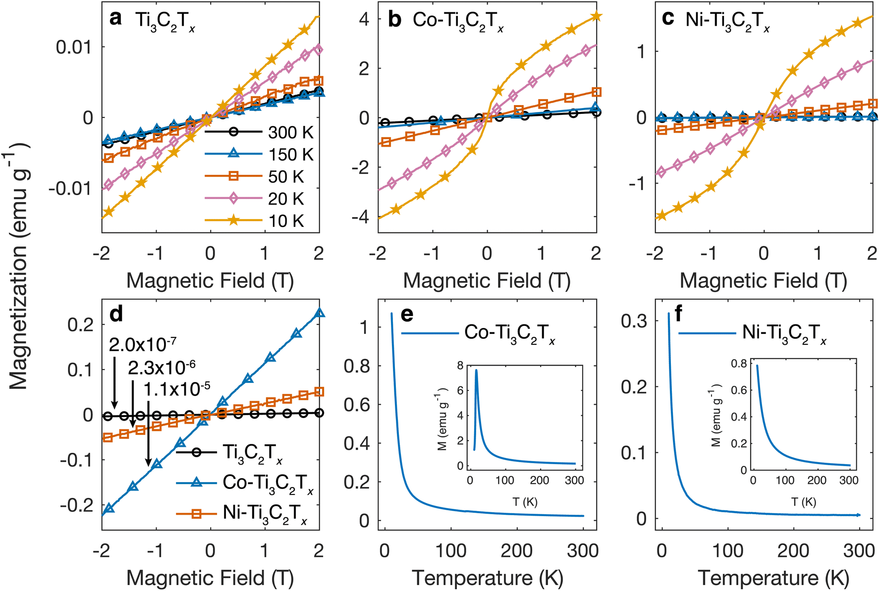 | ||
| Fig. 5 (a–c) Field-dependent magnetization curves at different temperatures for (a) pristine Ti3C2Tx and (b) Co- and (c) Ni-incorporated Ti3C2Tx. (d) Field-dependent magnetization curves at room temperature for Ti3C2Tx MXene before and after metal incorporation. The magnetic susceptibility is indicated in terms of emu g−1 Oe−1. (e and f) Temperature-dependent magnetization curves for (e) Co-Ti3C2Tx and (f) Ni-Ti3C2Tx at an applied field of 0.2 T. The insets are the temperature-dependent magnetization curves for CoCl2 and NiCl2 salt powders. | ||
For both, Co- and Ni-incorporated samples, the temperature-dependent magnetization (M vs. T) curves show that while there is a low-temperature upturn in magnetization, there is no evidence of a ferromagnetic or antiferromagnetic transition (Fig. 5e and f). The M vs. T curves for the chloride salts indicate that CoCl2 exhibits a maximum in magnetization at 15.5 K, associated with an antiferromagnetic transition. The magnetization data from Co-Ti3C2Tx does not exhibit a local maximum or anomaly near 15.5 K, providing further evidence that the MXene samples do not contain residual metal chloride salts. By comparing the magnetization values between the Co and Ni-incorporated MXene samples and the respective salts, we estimate it would require 10 wt% of salt incorporated in the sample to induce the change in the magnetization herein measured in the MXenes (Fig. 5e and f). Such a significant mass fraction of a secondary phase would be detectable by one of the characterization techniques presented earlier. The difference in the magnetic behavior observed for the salt and MXene sample, along with the large increase in magnetic susceptibility despite the low concentration of incorporated transition metal atoms, further support that the magnetization enhancement originates from a relatively uniform distribution of dilute Ni and Co atoms, rather than the presence of residual salt impurities. While this work establishes the general feasibility and utility of incorporating dilute concentrations of magnetic metal atoms in MXenes, future efforts are needed to understand the location of these atoms within or on the surface of MXenes and the upper limits to the Ni and Co concentrations that can be incorporated without the formation of ferromagnetic non-MXene secondary phases. Atomically resolved scanning transmission electron microscopy coupled with electron energy loss spectroscopy would be especially helpful in understanding whether the magnetic elements are incorporated within Ti vacancies or other defect sites in the MXenes.
Conclusions
In summary, we demonstrated a method to incorporate a secondary metal element into delaminated Ti3C2Tx MXene. While only Co and Ni are reported in this work, we anticipate that other transition metal elements with magnetic activity can also be incorporated into Ti3C2Tx and other MXenes by mixing MXene dispersions with salt solutions at low concentrations to prevent significant oxidation of the MXene and neutralizing the negative surface charge of the MXene and cause agglomeration. Further, this work provides evidence that Ti3C2Tx MXene can reduce CoCl2 and NiCl2 to Co and Ni metal, while maintaining its structure, electronic conductivity, and resulting film morphology. Using nano-XRF, we find that the Co and Ni are uniformly distributed across the MXene flakes, but future studies are required to identify the specific location of the Ni and Co atoms with respect to the MXene lattice. The Co and Ni incorporation results in a 57× and 11× enhancement, respectively, of the magnetic susceptibility at room temperature, which can be utilized for applications such as electromagnetic energy absorption.Experimental section
Material synthesis
Pristine Ti3C2Tx MXene was prepared by selective etching of Al from Ti3AlC2 MAX (<40 μm, Carbon-Ukraine). Briefly, 1 g of Ti3AlC2 was added to a solution of 30% hydrofluoric acid (48–50%, Acros Organics) and stirred for 3 hours at 35 °C. The resulting dispersion was washed with DI water through a series of centrifugation (2500 RCF for 3 min) and decantation cycles until the supernatant reached a pH > 6. Delaminated d-Ti3C2Tx MXene was prepared by exfoliation of multilayered ml-Ti3C2Tx using tetramethylammonium hydroxide (TMAOH). Briefly, 3 mL of 25 wt% TMAOH solution was added to a dispersion of 1 g ml-Ti3C2Tx in 20 mL of DI water, followed by stirring for 20 hours at 35 °C. The resulting dispersion was washed with DI water through a series of centrifugation (12,000 RCF for 5 min) and decantation cycles until the supernatant reached a pH < 8. Stock solutions of the metal salts were prepared by dissolving 0.2 g of NiCl2·6H2O (Acros Organics, CAS#7791-20-0) and 0.156 g of anhydrous CoCl2 (Alfa Aesar, CAS#7646-79-9) in 35 mL of DI water, respectively. The salt solutions were respectively added to 5 mL (∼2 mg mL−1) of d-Ti3C2Tx dispersions and mixed for 15 min at room temperature. The resulting dispersions were washed with DI water through a series of centrifugation (12![[thin space (1/6-em)]](https://www.rsc.org/images/entities/char_2009.gif) 000 RCF for 5 min), mechanical shaking (5 min), and decantation cycles until the color became transparent from the initial purple (CoCl2) or green (NiCl2) colors. The washing process continued until the MXene flakes were well dispersed. Free-standing Co-Ti3C2Tx and Ni-Ti3C2Tx films were prepared by vacuum-assisted filtration through a porous polypropylene membrane (Celgard, 3501). The films were dried in a vacuum desiccator at room temperature for over 48 hours before characterization.
000 RCF for 5 min), mechanical shaking (5 min), and decantation cycles until the color became transparent from the initial purple (CoCl2) or green (NiCl2) colors. The washing process continued until the MXene flakes were well dispersed. Free-standing Co-Ti3C2Tx and Ni-Ti3C2Tx films were prepared by vacuum-assisted filtration through a porous polypropylene membrane (Celgard, 3501). The films were dried in a vacuum desiccator at room temperature for over 48 hours before characterization.
Materials characterization
For XPS analysis, free-standing MXene films were mounted on conductive carbon tape and measured using a PHI VersaProbe 5000 spectrometer (Physical Electronics) with a 200 μm, 50 W monochromatic Al Kα X-ray source. Samples were sputtered using Ar+ ions for 1 min at 2 kV and 2 μA across a 2 × 2 mm area. The pass energy and step size were set at 23.5 eV and 0.05 eV, respectively. Curve fitting and quantification were conducted using the CasaXPS V2.3.19 software. SEM analysis was performed using a Zeiss Supra 50VP microscope operating at 3 kV accelerating voltage in secondary electron mode. XRD patterns were collected using a Rigaku SmartLab diffractometer (Rigaku Co. Ltd) with a Cu Kα source operating at 40 kV and 15 mA, within the 2θ range of 3° to 60°, at 1° min−1, in 0.02° steps. Raman spectra were collected using a Renishaw InVia confocal Raman microscope using a 785 nm laser at 5% power (∼0.15 mW). Magnetometry measurements were carried out using vibrating sample magnetometry in a Physical Property Measurement System (Quantum Design). For XRF mapping measurements, we used a nanofocused X-ray beam of size ∼40 nm at 8.4 keV, produced by a Fresnel zone plate with an outermost zone width of 30 nm.50,63 The XRF spectra were fitted using PyXRF software to generate elemental intensity maps.51Author contributions
Material synthesis was carried out by YY and MA. YY carried out XRD, XPS, resistivity, and magnetometry measurements. MS performed SEM and EDS. RW carried out Raman spectroscopy measurements and analysis. AP, XH, and YSC contributed to the nano-XRF measurements and data analysis. YG and SJM helped to conceptualize and supervised the project. All authors have contributed to the preparation of the manuscript and given approval to the final version of the manuscript.Conflicts of interest
The authors have no conflicts to disclose.Acknowledgements
This work was supported by the U.S. Department of Energy (DOE), Office of Science, Office of Basic Energy Sciences, grant No. DE-SC0018618. Nano-XRF measurements were conducted at the Hard X-ray Nanoprobe (HXN) beamline at the National Synchrotron Light Source II (NSLS-II), a U.S. Department of Energy (DOE) Office of Science User Facility operated for the DOE Office of Science by Brookhaven National Laboratory (BNL) under contract No. DE-SC0012704. M. A. was supported by the National Science Foundation Graduate Research Fellowship under grant No. DGE-1646737 and the U.S. Department of Education Graduate Assistance in Areas of National Need (GAANN) fellowship. Any opinions, findings, and conclusions or recommendations expressed in this material are those of the author(s) and do not necessarily reflect the views of the National Science Foundation. XRD, XPS, and SEM analysis were conducted using instruments in the Drexel University Materials Characterization Core (MCC) facilities.References
- M. Naguib, M. Kurtoglu, V. Presser, J. Lu, J. Niu, M. Heon, L. Hultman, Y. Gogotsi and M. W. Barsoum, Adv. Mater., 2011, 23, 4248–4253 CrossRef CAS PubMed.
- V. Kamysbayev, A. S. Filatov, H. Hu, X. Rui, F. Lagunas, D. Wang, R. F. Klie and D. V. Talapin, Science, 2020, 369, 979–983 CrossRef CAS PubMed.
- Y. Li, H. Shao, Z. Lin, J. Lu, L. Liu, B. Duployer, P. O. Å. Persson, P. Eklund, L. Hultman, M. Li, K. Chen, X.-H. Zha, S. Du, P. Rozier, Z. Chai, E. Raymundo-Piñero, P.-L. Taberna, P. Simon and Q. Huang, Nat. Mater., 2020, 19, 894–899 CrossRef CAS PubMed.
- A. VahidMohammadi, J. Rosen and Y. Gogotsi, Science, 2021, 372, eabf1581 CrossRef CAS PubMed.
- V. Shukla, Mater. Adv., 2020, 1, 3104–3121 RSC.
- Y. Yue, J. Magn. Magn. Mater., 2017, 434, 164–168 CrossRef CAS.
- L. Dong, H. Kumar, B. Anasori, Y. Gogotsi and V. B. Shenoy, J. Phys. Chem. Lett., 2017, 8, 422–428 CrossRef CAS PubMed.
- B. Anasori, Y. Xie, M. Beidaghi, J. Lu, B. C. Hosler, L. Hultman, P. R. C. Kent, Y. Gogotsi and M. W. Barsoum, ACS Nano, 2015, 9, 9507 CrossRef CAS.
- B. Anasori, C. Shi, E. J. Moon, Y. Xie, C. A. Voigt, P. R. Kent, S. J. May, S. J. Billinge, M. W. Barsoum and Y. Gogotsi, Nanoscale Horiz., 2016, 1, 227–234 RSC.
- J. Yang, X. Zhou, X. Luo, S. Zhang and L. Chen, Appl. Phys. Lett., 2016, 109, 203109 CrossRef.
- W. Sun, Y. Xie and P. R. C. Kent, Nanoscale, 2018, 10, 11962–11968 RSC.
- S. Li, J. He, L. Grajciar and P. Nachtigall, J. Mater. Chem. C, 2021, 9, 11132–11141 RSC.
- N. C. Frey, A. Bandyopadhyay, H. Kumar, B. Anasori, Y. Gogotsi and V. B. Shenoy, ACS Nano, 2019, 13, 2831–2839 CrossRef CAS.
- J. Yang, S. Zhang, A. Wang, R. Wang, C.-K. Wang, G.-P. Zhang and L. Chen, Nanoscale, 2018, 10, 19492–19497 RSC.
- Q. Tang, Z. Zhou and P. Shen, J. Am. Chem. Soc., 2012, 134, 16909–16916 CrossRef CAS PubMed.
- M. Zhao, J. Chen, S.-S. Wang, M. An and S. Dong, Phys. Rev. Mater., 2021, 5, 094408 CrossRef CAS.
- R. Ponce-Pérez, J. Guerrero-Sanchez, S. J. Gutierrez-Ojeda and M. G. Moreno-Armenta, ACS Appl. Electron. Mater., 2021, 3, 4967–4976 CrossRef.
- Y. Yue, B. Wang, N. Miao, C. Jiang, H. Lu, B. Zhang, Y. Wu, J. Ren and M. Wang, Ceram. Int., 2021, 47, 2367–2373 CrossRef CAS.
- J. Yang, X. Luo, X. Zhou, S. Zhang, J. Liu, Y. Xie, L. Lv and L. Chen, Comput. Mater. Sci., 2017, 139, 313–319 CrossRef CAS.
- J. He, G. Ding, C. Zhong, S. Li, D. Li and G. Zhang, Nanoscale, 2019, 11, 356–364 RSC.
- M. W. Barsoum, Prog. Solid State Chem., 2000, 28, 201–281 CrossRef CAS.
- M. Sokol, V. Natu, S. Kota and M. W. Barsoum, Trends Chem., 2019, 1, 210–223 CrossRef CAS.
- C. Si, J. Zhou and Z. Sun, ACS Appl. Mater. Interfaces, 2015, 7, 17510–17515 CrossRef CAS PubMed.
- M. Je, Y. Lee and Y.-C. Chung, Thin Solid Films, 2016, 619, 131–136 CrossRef CAS.
- Q. Sun, Z. Fu and Z. Yang, J. Magn. Magn. Mater., 2020, 514, 167141 CrossRef CAS.
- X. Feng, Z. He, R. Peng, Y. Dai, B. Huang and Y. Ma, Phys. Rev. Mater., 2022, 6, 044001 CrossRef CAS.
- M. Jaouen, M. Bugnet, N. Jaouen, P. Ohresser, V. Mauchamp, T. Cabioc'h and A. Rogalev, J. Phys.: Condens. Matter, 2014, 26, 176002 CrossRef CAS PubMed.
- O. Akinola, I. Chakraborty, H. Celio, D. Akinwande and J. A. C. Incorvia, J. Mater. Res., 2021, 36, 1980–1989 CrossRef CAS.
- Y. Zhang, Z. Cui, B. Sa, N. Miao, J. Zhou and Z. Sun, Nanoscale Horiz., 2022, 7, 276–287 RSC.
- K. Hantanasirisakul, B. Anasori, S. Nemsak, J. L. Hart, J. Wu, Y. Yang, R. V. Chopdekar, P. Shafer, A. F. May, E. J. Moon, J. Zhou, Q. Zhang, M. L. Taheri, S. J. May and Y. Gogotsi, Nanoscale Horiz., 2020, 5, 1557–1565 RSC.
- P. Liu, V. M. H. Ng, Z. Yao, J. Zhou and L. B. Kong, Mater. Lett., 2018, 229, 286–289 CrossRef CAS.
- X. Li, W. You, L. Wang, J. Liu, Z. Wu, K. Pei, Y. Li and R. Che, ACS Appl. Mater. Interfaces, 2019, 11, 44536–44544 CrossRef CAS PubMed.
- J. He, D. Shan, S. Yan, H. Luo, C. Cao and Y. Peng, J. Magn. Magn. Mater., 2019, 492, 165639 CrossRef CAS.
- Q. Zhang, D. Xu, Y. Si, R. Xu, B. Luo, S. He and D. Liu, ACS Appl. Nano Mater., 2022, 5, 7175–7186 CrossRef CAS.
- X. Li, M. Zhang, W. You, K. Pei, Q. Zeng, Q. Han, Y. Li, H. Cao, X. Liu and R. Che, ACS Appl. Mater. Interfaces, 2020, 12, 18138–18147 CrossRef CAS PubMed.
- Y. Cui, Z. Liu, Y. Zhang, P. Liu, M. Ahmad, Q. Zhang and B. Zhang, Carbon, 2021, 181, 58–68 CrossRef CAS.
- L. Liang, G. Han, Y. Li, B. Zhao, B. Zhou, Y. Feng, J. Ma, Y. Wang, R. Zhang and C. Liu, ACS Appl. Mater. Interfaces, 2019, 11, 25399–25409 CrossRef CAS.
- N. Li, X. Xie, H. Lu, B. Fan, X. Wang, B. Zhao, R. Zhang and R. Yang, Ceram. Int., 2019, 45, 22880–22888 CrossRef CAS.
- L. Liang, R. Yang, G. Han, Y. Feng, B. Zhao, R. Zhang, Y. Wang and C. Liu, ACS Appl. Mater. Interfaces, 2020, 12, 2644–2654 CrossRef CAS PubMed.
- Y. Peng, L. Deng, H. Luo and S. Huang, Results Phys., 2020, 19, 103516 CrossRef.
- J. He, X. Liu, Y. Deng, Y. Peng, L. Deng, H. Luo, C. Cheng and S. Yan, J. Alloys Compd., 2021, 862, 158684 CrossRef CAS.
- X. Li, Z. Wu, W. You, L. Yang and R. Che, Nano-Micro Lett., 2022, 14, 73 CrossRef CAS.
- C. Wen, X. Li, R. Zhang, C. Xu, W. You, Z. Liu, B. Zhao and R. Che, ACS Nano, 2022, 16, 1150–1159 CrossRef CAS PubMed.
- C. Zhou, X. Wang, H. Luo, L. Deng, S. Wang, S. Wei, Y. Zheng, Q. Jia and J. Liu, Appl. Surf. Sci., 2019, 494, 540–550 CrossRef CAS.
- H. Liang, G. Chen, D. Liu, Z. Li, S. Hui, J. Yun, L. Zhang and H. Wu, Adv. Funct. Mater., 2023, 33, 2212604 CrossRef CAS.
- H. Liu, X. Li, X. Zhao, M. Zhang, X. Liu, S. Yang, H. Wu and Z. Ma, Adv. Funct. Mater., 2023, 33, 2304442 CrossRef CAS.
- H. Jin, K. Wang, Z. Mao, L. Tang, J. Zhang and X. Chen, J. Phys.: Condens. Matter, 2022, 34, 015701 CrossRef CAS.
- W. Feng, H. Luo, S. Zeng, C. Chen, L. Deng, Y. Tan, X. Zhou, S. Peng and H. Zhang, Mater. Chem. Front., 2018, 2, 2320–2326 RSC.
- M. W. Hakim, S. Fatima, R. Tahir, M. Z. Iqbal, H. Li and S. Rizwan, J. Energy Storage, 2023, 61, 106662 CrossRef.
- E. Nazaretski, H. Yan, K. Lauer, N. Bouet, X. Huang, W. Xu, J. Zhou, D. Shu, Y. Hwu and Y. S. Chu, J. Synchrotron Radiat., 2017, 24, 1113–1119 CrossRef CAS PubMed.
- L. Li, Y. Hanfei, X. Wei, Y. Dantong, H. Annie, L. Wah-Keat, I. C. Stuart and S. C. Yong, In PyXRF: Python-based X-ray fluorescence analysis package, Proc. SPIE 10389, X-Ray Nanoimaging: Instruments and Methods III, 2017, p. 103890U.
- M. C. Biesinger, B. P. Payne, A. P. Grosvenor, L. W. M. Lau, A. R. Gerson and R. S. C. Smart, Appl. Surf. Sci., 2011, 257, 2717–2730 CrossRef CAS.
- Y. Long, Y. Tao, T. Shang, H. Yang, Z. Sun, W. Chen and Q.-H. Yang, Adv. Sci., 2022, 9, 2200296 CrossRef CAS PubMed.
- L. Wang, H. Song, L. Yuan, Z. Li, Y. Zhang, J. K. Gibson, L. Zheng, Z. Chai and W. Shi, Environ. Sci. Technol., 2018, 52, 10748–10756 CrossRef CAS PubMed.
- Y. Yi, Y. Ma, F. Ai, Y. Xia, H. Lin and G. Zhu, Chem. Commun., 2021, 57, 7790–7793 RSC.
- W. Peng, M. Luo, X. Xu, K. Jiang, M. Peng, D. Chen, T.-S. Chan and Y. Tan, Adv. Energy Mater., 2020, 10, 2001364 CrossRef CAS.
- H. Gu, X. Li, J. Zhang and W. Chen, Small, 2022, 18, 2105883 CrossRef CAS.
- K. Eid, Q. Lu, S. Abdel-Azeim, A. Soliman, A. M. Abdullah, A. M. Abdelgwad, R. P. Forbes, K. I. Ozoemena, R. S. Varma and M. F. Shibl, J. Mater. Chem. A, 2022, 10, 1965 RSC.
- A. Sarycheva and Y. Gogotsi, Chem. Mater., 2020, 32, 3480–3488 CrossRef CAS.
- “Magnetic Data Storage”, in Magnetic Materials: Fundamentals and Applications, ed. N. A. Spaldin, Cambridge University Press, Cambridge, 2nd edn, 2010, pp. 177–188 Search PubMed.
- C. Binns, in Frontiers of Nanoscience, ed. C. Binns, Elsevier, 2014, vol. 6, pp. 1–32 Search PubMed.
- D. X. Chen, A. Sanchez, E. Taboada, A. Roig, N. Sun and H. C. Gu, J. Appl. Phys., 2009, 105, 083924 CrossRef.
- H. Yan, N. Bouet, J. Zhou, X. Huang, E. Nazaretski, W. Xu, A. P. Cocco, W. K. S. Chiu, K. S. Brinkman and Y. S. Chu, Nano Futures, 2018, 2, 011001 CrossRef.
Footnotes |
| † Electronic supplementary information (ESI) available. See DOI: https://doi.org/10.1039/d3nr05685f |
| ‡ These authors contributed equally to the work. |
| This journal is © The Royal Society of Chemistry 2024 |