An intelligent peptide recognizes and traps Mycobacterium tuberculosis to inhibit macrophage phagocytosis†
Gui-Yuan
Wang‡
ab,
Bin
Lu‡
c,
Xu
Cui
*c,
Guang
Li
c,
Kuo
Zhang
 b,
Qing-Shi
Zhang
b,
Xin
Cui
ab,
Gao-Feng
Qi
ab,
Qi-Lin
Liang
b,
Xiao-Bo
Luo
c,
Huan-Ge
Xu
b,
Li
Xiao
d,
Lei
Wang
b,
Qing-Shi
Zhang
b,
Xin
Cui
ab,
Gao-Feng
Qi
ab,
Qi-Lin
Liang
b,
Xiao-Bo
Luo
c,
Huan-Ge
Xu
b,
Li
Xiao
d,
Lei
Wang
 *b and
Litao
Li
*c
aDepartment of Graduate, Hebei North University, Zhangjiakou, 075000, Hebei Province, China
bCAS Center for Excellence in Nanoscience, CAS Key Laboratory for Biomedical Effects of Nanomaterials and Nanosafety, National Center for Nanoscience and Technology (NCNST), Beijing, 100190, China. E-mail: wanglei@nanoctr.cn
cDepartment of Orthopedics, The 4th Medical Center of Chinese PLA General Hospital, No. 51 Fucheng road, Beijing, 100091, China. E-mail: cuixuprossor@163.com; lltop_1@sina.com
dInstitute of Respiratory and Critical Medicine, the Eighth Medical Center of PLA General Hospital, Beijing 100091, China
First published on 28th November 2022
Abstract
Tuberculosis is a major public health concern worldwide, and it is a serious threat to human health for a long period. Macrophage phagocytosis of Mycobacterium tuberculosis (M. tuberculosis) is a crucial process for granuloma formation, which shelters the bacteria and gives them an opportunity for re-activation and spread. Herein, we report an intelligent anti-microbial peptide that can recognize and trap the M. tuberculosis, inhibiting the macrophage phagocytosis process. The peptide (Bis-Pyrene-KLVFF-WHSGTPH, in abbreviation as BFH) first self-assembles into nanoparticles, and then forms nanofibers upon recognizing and binding M. tuberculosis. Subsequently, BFH traps M. tuberculosis by the in situ formed nanofibrous networks and the trapped M. tuberculosis are unable to invade host cells (macrophages). The intelligent anti-microbial peptide can significantly inhibit the phagocytosis of M. tuberculosis by macrophages, thereby providing a favorable theoretical basis for inhibiting the formation of tuberculosis granulomas.
Introduction
Tuberculosis remains one of the world's largest infectious “killers”.1 The WHO Global Tuberculosis Report 2021 states that the number of deaths from tuberculosis has increased for the first time in nearly a decade due to the COVID-19 pandemic, and it has aggravated an already sub-optimal global tuberculosis response.2 Almost all Mycobacterium tuberculosis (M. tuberculosis) infections are spread by inhalation of aerosols, making lungs the most common site of infection.3,4 At the infected site, alveolar macrophages phagocytose M. tuberculosis.4 More importantly, following the phagocytosis of M. tuberculosis, macrophages secrete a variety of chemoattractant cytokines, which help recruit uninfected macrophages, monocytes and lymphocytes from the circulation to the site of infection, initiating the formation of tuberculous granulomas, which is the histological hallmark of tuberculosis.5–7Tuberculous granulomas have long been endowed with the role of host protective structures that can “isolate” infecting mycobacteria.7–11 Tuberculosis is caused by bacterium-host interactions involving a complex series of processes.12M. tuberculosis can replicate after being phagocytosed by macrophages in the lung because of their ability to subvert phagocytic endocytic trafficking and resist innate defense mechanisms.13–15 Infected macrophages migrate into tissues and aggregate into tuberculous granulomas.15 The formation of granulomas in turn allows mycobacteria to proliferate and spread within the host.7,16–18 When the human body is affected by factors such as malnutrition, HIV co-infection and immunodeficiency, the host's immunity weakens. This ultimately leads to granuloma destruction through necrolysis of mycobacterial-infected macrophages, thereby causing tuberculosis infection and recurrence.19–21 Therefore, the inhibition of phagocytosis of M. tuberculosis by macrophages is the first and crucial step to prevent the formation of granulomas against tuberculosis infection and recurrence.
Nanomedicine is the medical application of nanotechnology and nanomaterial.22–27 The development of nanotechnology and nanomaterial will bring a profound revolution to the medical field. Supramolecular chemistry, also known as ‘chemistry beyond the molecule’, relies on non-covalent interactions between molecules to self-assemble into molecular assemblies.28 Based on the concept of supramolecular chemistry, peptide may self-assemble into nanomaterials that function as nanomedicine.29 In recent years, the development of biomimetic binding-induced fibrillogenesis (BIF) peptides showed great significance in the fight against cancer and bacteria.30–37 For example, we previously designed a human defensin-6 mimic peptide (HDMP), which recognizes S. aureus through BIF process from nanoparticles (NPs) to nanofibers (NFs) to capture S. aureus, inhibiting the invasion into host cells.38
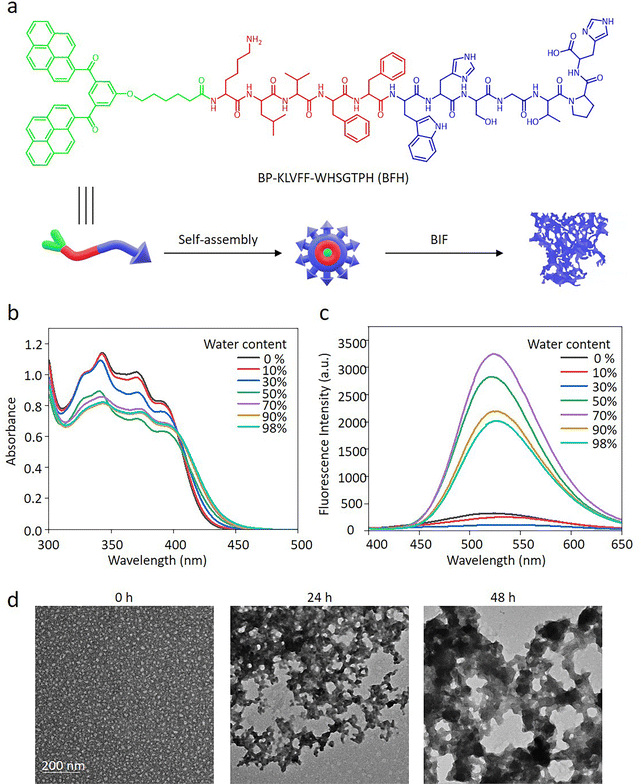
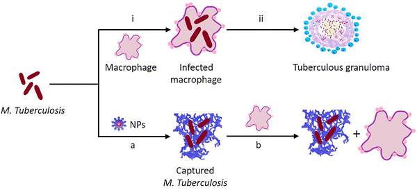
Herein, we design a biomimetic BIF peptide, Bis-Pyrene-KLVFF-WHSGTPH (BFH), which consists of three modules. (i) A ligand peptide sequence WHSGTPH can target and bind M. tuberculosis.39 (ii) The KLVFF sequence is a peptide skeleton for β-sheet fibrous structures.40 (iii) Bis-pyrene (BP) enables the formation of BFH NPs (Fig. 1a). Moreover, the fluorescence arising from the aggregation-induced emission (AIE) of BP can be used to indicate BFH.41 Mechanistically, BFH first self-assembles into NPs, which subsequently bind to M. tuberculosis, Bacillus Calmette–Guérin (BCG) and transform into NFs to form fibrous networks. The BCG are trapped by fibrous networks of BFH, achieving effective resistance to macrophage phagocytosis (Scheme 1).
Results and discussion
Preparation and characterization of BFH and BAH NPs
The BFH peptide and the control peptide BP-KAAGG-WHSGTPH (BAH) with the replacement of β-sheeting forming KLVFF by KAAGG (Fig. S1 and S2, ESI†) were dissolved in DMSO, respectively, and subsequently injected into water. As a result, the BFH and BAH self-assembled into NPs (Fig. 1a), which was confirmed by the UV-vis absorption and fluorescence spectra. The absorption peaks of BFH and BAH monomers around 320 to 400 nm decreased with the emerging bathochromic absorption peak, indicating the aggregation of BP (Fig. 1b and Fig. S3a, ESI†). Meanwhile, as the water ratio in the mixed solution increased, the fluorescence intensity at 525 nm increased gradually, indicating the aggregation of BP (Fig. 1c and Fig. S3b, ESI†). Through the particle size analysis from dynamic light scattering (DLS), BFH and BAH were well-dispersed NPs with diameters of 24.03 ± 1.00 nm and 28.21 ± 4.77 nm, respectively (Fig. S4, ESI†). The experimental results showed that zeta potentials of BFH and BAH NPs were +12.4 ± 3.5 mV and +11.1 ± 5.1 mV, respectively (Fig. S5, ESI†). The nanoparticulation of BFH or BAH was mainly driven by the hydrophobic and π–π interactions of BP upon dispersion in water.BFH NPs transform into NFs upon encountering BCG
The peptide sequence WHSGTPH was screened for the BCG binding and utilized in this study to recognize BCG and trigger the BIF process. In order to confirm the hypothesis, the experimental peptide BFH was incubated with BCG and observed by transmission electron microscope (TEM) in 48 h. The BFH formed spherical NPs initially. After interacting with BCG, BFH NPs transformed into NFs in 24 h, and fibrous networks with a wide size distribution were clearly detectable in 48 h. The length and diameter of NFs became larger and larger over time in 48 h (Fig. 1d). For the control peptide BAH, it was found to be in the state of NPs in 48 h (Fig. S6, ESI†). In addition, circular dichroism (CD) spectra of BFH and BAH were in agreement with the TEM results. At the initial stage, BFH NPs did not show the obvious β-sheet secondary structure, which gradually formed with typical 203 nm (positive) and 212 nm (negative) peaks over time in 48 h. In contrast, the control BAH NPs did not show the β-sheet secondary structure in 48 h (Fig S7, ESI†). These results indicated that the binding of WHSGTPH to BCG could induce the fibrillogenesis, and the hydrogen-bonds provided by KLVFF were crucial for the transformation of NPs to NFs.BFH NPs specially recognize BCG
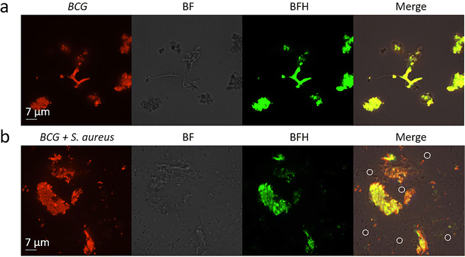
The recognition ability of BFH and BAH NPs to BCG was first investigated by confocal laser scanning microscope (CLSM). The BCG was firstly stained by Texas red dye and then incubated with, PBS, BFH and BAH NPs in 500 μL of 0.9% saline for 4 h, respectively. Then, the BCG solution (10 μL) was suspended in physiological saline, which was further dropped on the center of the confocal Petri dish and observed by CLSM. In PBS group, BCG showed red fluorescence on BCG, indicating the successful staining (Fig. S8, ESI†). However, BFH and BAH NPs treated with BCG showed well-merged fluorescence of red and green signals (Fig. 2a and Fig. S9, ESI†). These results indicated that both BFH and BAH containing WHSGTPH sequence can specifically recognize BCG. However, the BFH group exhibited strong fluorescence with large area, but small area for BAH group. This may be due to fibrillogenesis of BFH NPs into fibrous networks after specifically recognizing and binding to BCG, leading to the agglutination of BCG. On the basis of this observation, the BCG actually can be detected with fluorescence and morphology by the fluorescent BFH peptide. In order to further confirm the specific recognition ability of BFH NPs, the mixture of BCG and S. aureus was incubated with BFH NPs. The green fluorescence of BFH was well merged with red fluorescence BCG to show yellow fluorescence from the mixture of both BCG and S. aureus (Fig. 2b). Meanwhile, there were no obvious fluorescence signals on S. aureus, indicating the specificity of BFH to BCG.The fibrillogenesis of BFH on the surface of BCG
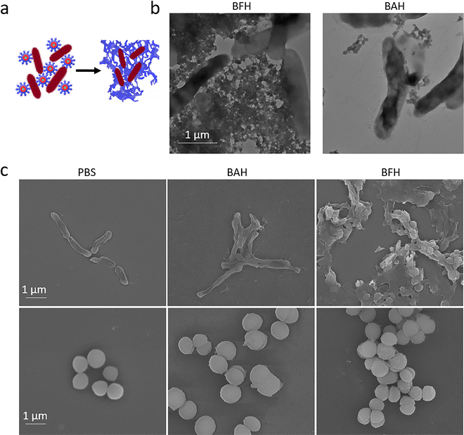
BFH NPs can form NFs in situ after specific recognition of BCG (Fig. 3a). Therefore, the morphological changes of BFH and BAH NPs on the surface of BCG were further observed by bio-TEM. The BFH NPs (the experimental group) and BAH NPs (the control group) (3 × 10⁻⁵ M) were incubated with BCG for 4 h at 37 °C in a constant temperature incubator, respectively. After centrifugation, the BCG was washed with 0.9% saline for TEM measurement. In the experimental group, a large number of fibrous networks appeared around the BCG, validating the BIF ability of BFH. In the control group, scattered nanoparticles were found on the surface of BCG (Fig. 3b). Obviously, due to the conversion from BFH NPs to NFs, the adhesion of BFH on the surface of BCG far exceeded that of BAH, which can explain that BCG treated with BFH NPs had stronger fluorescence intensity than BAH NPs. These results verified that the recognition ability of the targeting peptide in BFH and BAH to BCG, and the importance of KLVFF for the conversion from NPs to NFs after recognition.Scanning electron microscopy (SEM) studies was carried out on BCG incubated with PBS, BAH, and BFH NPs, respectively. PBS group hardly showed BCG aggregation and no fibrous network was found on the surface of BCG. BAH group only exhibited a slight aggregation of BCG, and almost no fibrous networks were formed on the surface of BCG. In contrast, there were not only large amount of BCG agglutination, but also dense fibrous networks entangled on the surface of BCG in BFH group. To further verify the specific binding of BFH to BCG, other bacilli, E. coli, was selected as a control for incubation with BFH. There were no fibrous networks on the surfaces of E. coli, and no agglutination of E. coli from SEM images (Fig. S10, ESI†). At the same time, PBS, BAH and BFH NPs were incubated with S. aureus, and observed by SEM. It was found that there were no entangled fibrous networks with S. aureus for all three groups, indicating no obvious interactions between the targeting peptide WHSGTPH and S. aureus (Fig. 3c).
BFH NPs defend against BCG invasion of macrophages
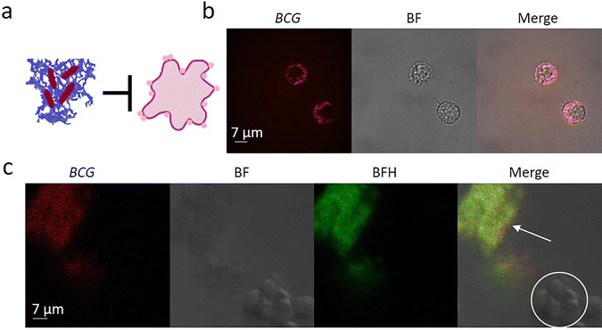
M. tuberculosis is an invasive bacteria that can bind to and invade into host cells (macrophages), causing the formation and spread of tuberculous granulomas. The fibrillogenesis of BFH NPs on the surface of BCG to form NFs was expected to trap and inhibit the invasion of BCG to host cells (Fig. 4a), which was evaluated by the trapping and invading experiment. In the blank group where only BCG and macrophages were incubated without any other treatment, a large amount of BCG was phagocytosed by macrophages (Fig. 4b). In contrast, In the control group, a lot of BCG (merged red and green channel) was observed inside the macrophages, even BCG were pre-treated and recognized by BAH NPs (Fig. S11, ESI†). However, once the BCG was pretreated with BFH NPs, BCG aggregated into larger BCG-BFH clump-like complexes outside the macrophages and there were almost no BCG were observed in macrophages (Fig. 4c). These results suggested that BFH NPs had a significant inhibitory effect on the phagocytosis of BCG by macrophages. We also carried out the toxicity test of BFH NPs against BCG, where BCG in logarithmic growth phase was incubated with BFH NPs for 24 h. It was found that no significant bacterial toxicity at concentrations up to 180 μM (Fig. S12, ESI†).Despite the emerging novel technologies for diagnosis and treatment of tuberculosis in recent years, there's still a great challenge to completely overcome the disease.42 In the initial stage of tuberculosis infection, M. tuberculosis will be engulfed by macrophages and survive inside the phagosome.43 By inhibiting the acidification and maturation of phagolysosomes, it evades killing.44–46 Along with the necrosis of macrophages, M. tuberculosis will be released into extracellular space and induce the granuloma expansion.47,48 To make matters worse, the current anti-tuberculosis drugs can not effectively kill dormant M. tuberculosis,49,50 can not form higher drug concentrations inside macrophages, which makes it more difficult to completely kill M. tuberculosis.51,52 Therefore, a new type of intelligent peptide BFH, was designed, which can target and capture M. tuberculosis, forming nanofibrils on the surface of M. tuberculosis, and inhibiting the bacteria invasion into macrophages. Subsequently, the BFH may prevent the expansion of tuberculosis granulomas and make the extracellular M. tuberculosis more effectively killed by anti-tuberculosis drugs.
Conclusions
In this study, an intelligent peptide BFH with particle unit, fiber unit and recognition unit was designed and synthesized. Due to the strong hydrophobic effect of particle unit BP, the BFH can self-assemble into NPs in polar solvents. In addition, the fluorescent signal caused by the AIE effect of BP provides the possibility to detect the location of BFH. Fiber unit is KLVFF sequence from Aβ that folds a fibrous structure. The recognition unit is WHSGTPH sequence, which can target and bind to M. tuberculosis. It was found that the fluorescence intensity at 525 nm gradually increased with the increase of water content in the mixed solution, indicating the aggregation of BP, which induced the self-assembly of BFH. It was observed by TEM that the BFH were NPs at 0 h, and completely transformed into NFs in 48 h upon BCG incubation.In order to verify its targeting effect of BFH to M. tuberculosis, we co-incubated the BFH NPs with BCG and verified by CLSM. The results showed that strong fluorescence signal completely cover the surface of bacteria, which confirmed the targeting ability of BFH to BCG. To further investigate whether the targeting peptide can induce the transformation of NPs into NFs on the bacterial surface, we co-incubated the BFH NPs with BCG and observed by SEM. The results revealed that BCG was covered and captured by fibrous networks, and agglomeration occurred.
In order to further evaluate the inhibition of BFH on phagocytosis of macrophages on BCG, we co-incubated BFH NPs, BCG and macrophages, and observed the effect of BFH on phagocytosis of BCG by macrophages through CLSM. The results showed that after incubation with BFH, the fluorescence intensity in macrophages was very low, and there were fluorescence signals outside cells. In the control group without BFH, a strong fluorescence signal was observed in macrophages, but no fluorescence signal was observed outside the cells. The above results showed that the BFH can self-assemble into entangled fibrous networks around the bacteria and gather the bacteria to increase their volume, thus inhibiting the phagocytosis of macrophages. We believed that this phenomenon would benefit effective killing of M. tuberculosis by drugs outside macrophages.
The limitation of this study was that only the phenomenon that the BFH inhibiting macrophage phagocytosis of BCG was observed. However, the specific mechanism of action was not clear. The follow-up study was planned to further study whether the above-mentioned phagocytosis inhibition can play a role in killing M. tuberculosis in vitro and in vivo, and to explore the relevant mechanisms.
Experimental
Materials and methods
The starting material BP-COOH was synthesized according to our previous report.41N,N-dimethylformamide (DMF) and dichloromethane need to be treated with molecular sieves before using. Methanol, N′-tetramethylurea hexafluorophosphate (HBTU), 4-methylmorpholine (NMM), trifluoroacetic acid (TFA) and O-(benzotriazol-1-yl) N,N,N′,N′ were purchased from Sigma-Aldrich Chemical Co. All Fmoc amino acids and Wang resins were obtained from GL Biochem (Shanghai) Ltd. The cell lines RAW 264.7 were purchased from the Cell Culture Center of the Institute of Basic Medical Sciences, Chinese Academy of Medical Sciences (Beijing, China). Fetal bovine serum and cell culture medium were purchased from Wisent Inc. (Multicell, Wisent Inc., St. Bruno, Quebec, Canada). BCG was purchased from Chengdu Institute of Biological Products Co., Ltd.![[thin space (1/6-em)]](https://www.rsc.org/images/entities/char_2009.gif) 000 rpm·for 5 minutes, Finally remove the supernatant.
000 rpm·for 5 minutes, Finally remove the supernatant.
![[thin space (1/6-em)]](https://www.rsc.org/images/entities/char_2009.gif) :
:![[thin space (1/6-em)]](https://www.rsc.org/images/entities/char_2009.gif) 1:1 v/v). Then, add HBTU (137.2 mM) and Hobt activated amino acid (137.2 mM) to NMM (5%) anhydrous DMF. Repeat the appeal process until the BP-COOH was successfully coupled. Then, the final product was obtained by cleaving the resin from the resin for 3 h through a mixture of TFA (95%, volume percentage), triisopropylsilane (2.5%, volume percentage) and water (2.5%, volume percentage) in an ice bath. Suction the resin and lysate to obtain the filtrate, and blow with nitrogen to 1/10 of the total volume. An appropriate amount of glacial diethyl ether was added to the remaining filtrate to obtain a precipitated solid. The obtained solid was washed 3 times with glacial diethyl ether and dried in a vacuum oven to obtain the final product. The molecular structures of peptides were confirmed by MALDI-TOF-MS (Bruker Daltonics).
1:1 v/v). Then, add HBTU (137.2 mM) and Hobt activated amino acid (137.2 mM) to NMM (5%) anhydrous DMF. Repeat the appeal process until the BP-COOH was successfully coupled. Then, the final product was obtained by cleaving the resin from the resin for 3 h through a mixture of TFA (95%, volume percentage), triisopropylsilane (2.5%, volume percentage) and water (2.5%, volume percentage) in an ice bath. Suction the resin and lysate to obtain the filtrate, and blow with nitrogen to 1/10 of the total volume. An appropriate amount of glacial diethyl ether was added to the remaining filtrate to obtain a precipitated solid. The obtained solid was washed 3 times with glacial diethyl ether and dried in a vacuum oven to obtain the final product. The molecular structures of peptides were confirmed by MALDI-TOF-MS (Bruker Daltonics).
![[thin space (1/6-em)]](https://www.rsc.org/images/entities/char_2009.gif) 000 rpm for 5 min in a centrifuge and the precipitated bacteria were washed for 3 times with 0.9% saline. The obtained bacteria were used for TEM measurements by a bio-TEM (HT7700 HITACHI TEM).
000 rpm for 5 min in a centrifuge and the precipitated bacteria were washed for 3 times with 0.9% saline. The obtained bacteria were used for TEM measurements by a bio-TEM (HT7700 HITACHI TEM).
![[thin space (1/6-em)]](https://www.rsc.org/images/entities/char_2009.gif) :
:![[thin space (1/6-em)]](https://www.rsc.org/images/entities/char_2009.gif) 3 and placed at 37 °C in a 5% CO2 cell incubator. The medium was changed once in 1–2 days. After 3 generations, the experiment was performed after the cell status was stable.
3 and placed at 37 °C in a 5% CO2 cell incubator. The medium was changed once in 1–2 days. After 3 generations, the experiment was performed after the cell status was stable.
The frozen BCG was quickly thawed in a 37 °C water bath, and stain the BCG in the same way as above. The experimental group was co-incubated with macrophage suspension (500 μL), BCG (106 CFU per mL) and BFH NPs (30 μM). The control group was co-incubated with macrophages (500 μL), BCG (106 CFU per mL) and BAH NPs (30 μM). The blank control group was co-incubated with macrophage suspension (500 μL) and BCG (106 CFU per mL) without any nanomaterials. After 4 h, the suspension (10 μL) was dropped on the center of the confocal Petri dish and covered with a cover glass for CLSM measurement (Ultra-VIEWVox, PerkinElmer).
Conflicts of interest
There are no conflicts to declare.Acknowledgements
This study is supported by the Strategic Priority Research Program of Chinese Academy of Sciences, Grant No. XDB36000000, the National Natural Science Foundation of China (81702174), and Hygiene and Health Development Scientific Research Fostering Plan of Haidian District Beijing, Grant No. HP2021-04-80501.Notes and references
- D. Falzon, S. den Boon, A. Kanchar, M. Zignol, G. B. Migliori and T. Kasaeva, Eur. Respir. J., 2022, 59, 2102753 CrossRef PubMed
.
- C. Jeremiah, E. Petersen, R. Nantanda, B. N. Mungai, G. B. Migliori, F. Amanullah, P. Lungu, F. Ntoumi, N. Kumarasamy, M. Maeurer and A. Zumla, Int. J. Infect. Dis., 2022 DOI:10.1016/j.ijid.2022.03.011
.
- M. G. Moule and J. D. Cirillo, Front. Cell. Infect. Microbiol., 2020, 10, 65 CrossRef PubMed
.
- E. Guirado, L. S. Schlesinger and G. Kaplan, Semin. Immunopathol., 2013, 35, 563–583 CrossRef CAS PubMed
.
- Z. Huang, Q. Luo, Y. Guo, J. Chen, G. Xiong, Y. Peng, J. Ye and J. Li, PLoS One, 2015, 10, e0129744 CrossRef PubMed
.
- M. Silva Miranda, A. Breiman, S. Allain, F. Deknuydt and F. Altare, Clin. Dev. Immunol., 2012, 2012, 139127 CrossRef PubMed
.
- J. M. Davis and L. Ramakrishnan, Cell, 2009, 136, 37–49 CrossRef CAS PubMed
.
- L. Ramakrishnan, Nat. Rev. Immunol., 2012, 12, 352–366 CrossRef CAS PubMed
.
- E. J. Rubin, N. Engl. J. Med., 2009, 360, 2471–2473 CrossRef CAS PubMed
.
- T. D. Bold and J. D. Ernst, Cell, 2009, 136, 17–19 CrossRef CAS PubMed
.
- T. Ulrichs and S. H. Kaufmann, J. Pathol., 2006, 208, 261–269 CrossRef CAS PubMed
.
- K. Borah, Y. Xu and J. McFadden, Front. Immunol., 2021, 12, 762315 CrossRef CAS PubMed
.
- C. L. Cosma, D. R. Sherman and L. Ramakrishnan, Annu. Rev. Microbiol., 2003, 57, 641–676 CrossRef CAS PubMed
.
- K. Rohde, R. M. Yates, G. E. Purdy and D. G. Russell, Immunol. Rev., 2007, 219, 37–54 CrossRef CAS PubMed
.
- C. M. McClean and D. M. Tobin, Pathog. Dis., 2016, 74, ftw068 CrossRef PubMed
.
- H. E. Volkman, T. C. Pozos, J. Zheng, J. M. Davis, J. F. Rawls and L. Ramakrishnan, Science, 2010, 327, 466–469 CrossRef CAS PubMed
.
- J. M. Davis, H. Clay, J. L. Lewis, N. Ghori, P. Herbomel and L. Ramakrishnan, Immunity, 2002, 17, 693–702 CrossRef CAS PubMed
.
- H. E. Volkman, H. Clay, D. Beery, J. C. Chang, D. R. Sherman and L. Ramakrishnan, PLoS Biol., 2004, 2, e367 CrossRef PubMed
.
- P. J. Cardona, Enferm. Infecc. Microbiol. Clin., 2018, 36, 38–46 CrossRef PubMed
.
- L. Ramakrishnan, Adv. Exp. Med. Biol., 2013, 783, 251–266 CrossRef PubMed
.
- H. Clay, H. E. Volkman and L. Ramakrishnan, Immunity, 2008, 29, 283–294 CrossRef CAS PubMed
.
- L. Zhang, Q. Cheng, C. Li, X. Zeng and X. Z. Zhang, Biomaterials, 2020, 248, 120029 CrossRef CAS PubMed
.
- C. Xu, Z. Lu, Y. Luo, Y. Liu, Z. Cao, S. Shen, H. Li, J. Liu, K. Chen, Z. Chen, X. Yang, Z. Gu and J. Wang, Nat. Commun., 2018, 9, 4092 CrossRef PubMed
.
- F. H. Ma, Y. An, J. Wang, Y. Song, Y. Liu and L. Shi, ACS Nano, 2017, 11, 10549–10557 CrossRef CAS PubMed
.
- Y. Zeng, J. Wang, Z. Wang, G. Chen, J. Yu, S. Li, Q. Li, H. Li, D. Wen, Z. Gu and Z. Gu, Nano Today, 2020, 35, 100984 CrossRef CAS
.
- Y. Zhang, K. Cai, C. Li, Q. Guo, Q. Chen, X. He, L. Liu, Y. Zhang, Y. Lu, X. Chen, T. Sun, Y. Huang, J. Cheng and C. Jiang, Nano Lett., 2018, 18, 1908–1915 CrossRef CAS PubMed
.
- P. Zhang, Y. Zhang, X. Ding, W. Shen, M. Li, E. Wagner, C. Xiao and X. Chen, Adv. Mater., 2020, 32, e2000013 CrossRef PubMed
.
- J. M. Lehn, Science, 1993, 260, 1762–1763 CrossRef CAS PubMed
.
- G. V. Oshovsky, D. N. Reinhoudt and W. Verboom, Angew. Chem., Int. Ed., 2007, 46, 2366–2393 CrossRef CAS PubMed
.
- K. Zhang, P. P. Yang, P. P. He, S. F. Wen, X. R. Zou, Y. Fan, Z. M. Chen, H. Cao, Z. Yang, K. Yue, X. Zhang, H. Zhang, L. Wang and H. Wang, ACS Nano, 2020, 14, 7170–7180 CrossRef CAS PubMed
.
- P. P. Yang, Y. J. Li, Y. Cao, L. Zhang, J. Q. Wang, Z. Lai, K. Zhang, D. Shorty, W. Xiao, H. Cao, L. Wang, H. Wang, R. Liu and K. S. Lam, Nat. Commun., 2021, 12, 4494 CrossRef CAS PubMed
.
- J. Q. Fan, Y. J. Li, Z. J. Wei, Y. Fan, X. D. Li, Z. M. Chen, D. Y. Hou, W. Y. Xiao, M. R. Ding, H. Wang and L. Wang, Nano Lett., 2021, 21, 6202–6210 CrossRef CAS PubMed
.
- P.-P. He, X.-D. Li, J.-Q. Fan, Y. Fan, P.-P. Yang, B.-N. Li, Y. Cong, C. Yang, K. Zhang, Z.-Q. Wang, D.-Y. Hou, H. Wang and L. Wang, CCS Chem., 2020, 2, 539–554 CrossRef CAS
.
- J. Fan, Y. Fan, Z. Wei, Y. Li, X. Li, L. Wang and H. Wang, Chin. Chem. Lett., 2020, 31, 1787–1791 CrossRef CAS
.
- Z. Chen, K. Zhang, J. Fan, Y. Fan, C. Yang, W. Tian, Y. Li, W. Li, J. Zhang, H. Wang and L. Wang, Chin. Chem. Lett., 2020, 31, 3107–3112 CrossRef CAS
.
- P. P. He, X. D. Li, L. Wang and H. Wang, Acc. Chem. Res., 2019, 52, 367–378 CrossRef CAS PubMed
.
- G. B. Qi, Y. J. Gao, L. Wang and H. Wang, Adv. Mater., 2018, 30, e1703444 CrossRef PubMed
.
- Y. Fan, X. D. Li, P. P. He, X. X. Hu, K. Zhang, J. Q. Fan, P. P. Yang, H. Y. Zheng, W. Tian, Z. M. Chen, L. Ji, H. Wang and L. Wang, Sci. Adv., 2020, 6, eaaz4767 CrossRef CAS PubMed
.
- H. Yang, L. Qin, Y. Wang, B. Zhang, Z. Liu, H. Ma, J. Lu, X. Huang, D. Shi and Z. Hu, Int. J. Nanomed., 2015, 10, 77–88 Search PubMed
.
- V. Castelletto, P. Ryumin, R. Cramer, I. W. Hamley, M. Taylor, D. Allsop, M. Reza, J. Ruokolainen, T. Arnold, D. Hermida-Merino, C. I. Garcia, M. C. Leal and E. Castaño, Sci. Rep., 2017, 7, 43637 CrossRef CAS PubMed
.
- L. Wang, W. Li, J. Lu, Y.-X. Zhao, G. Fan, J.-P. Zhang and H. Wang, J. Phys. Chem. C, 2013, 117, 26811–26820 CrossRef CAS
.
- C. M. Gill, L. Dolan, L. M. Piggott and A. M. McLaughlin, Breathe, 2022, 18, 210149 CrossRef PubMed
.
- D. Mahamed, M. Boulle, Y. Ganga, C. Mc Arthur, S. Skroch, L. Oom, O. Catinas, K. Pillay, M. Naicker, S. Rampersad, C. Mathonsi, J. Hunter, E. B. Wong, M. Suleman, G. Sreejit, A. S. Pym, G. Lustig and A. Sigal, eLife, 2017, 6, 22028 CrossRef PubMed
.
- I. Vergne, J. Chua, H. H. Lee, M. Lucas, J. Belisle and V. Deretic, Proc. Natl. Acad. Sci. U. S. A., 2005, 102, 4033–4038 CrossRef CAS PubMed
.
- G. Arora, R. Misra and A. Sajid, Curr. Top. Med. Chem., 2017, 17, 2077–2099 CrossRef CAS PubMed
.
- R. Domingo-Gonzalez, O. Prince, A. Cooper and S. A. Khader, Microbiol. Spectr., 2016, 4, TBTB2-0018-2016 Search PubMed
.
- A. J. Pagán and L. Ramakrishnan, Cold Spring Harbor Perspect. Med., 2014, 5, a018499 CrossRef PubMed
.
- P. J. Cardona, Inflammation Allergy: Drug Targets, 2007, 6, 27–39 CrossRef CAS PubMed
.
- M. Gengenbacher and S. H. Kaufmann, FEMS Microbiol. Rev., 2012, 36, 514–532 CrossRef CAS PubMed
.
- R. M. McCune, F. M. Feldmann, H. P. Lambert and W. McDermott, J. Exp. Med., 1966, 123, 445–468 CrossRef CAS PubMed
.
- K. Schaaf, V. Hayley, A. Speer, F. Wolschendorf, M. Niederweis, O. Kutsch and J. Sun, Assay Drug Dev. Technol., 2016, 14, 345–354 CrossRef CAS PubMed
.
- A. Gupta, A. Kaul, A. G. Tsolaki, U. Kishore and S. Bhakta, Immunobiology, 2012, 217, 363–374 CrossRef CAS PubMed
.
Footnotes |
| † Electronic supplementary information (ESI) available. See DOI: https://doi.org/10.1039/d2tb01764d |
| ‡ Gui-Yuan Wang and Bin Lu contribute equally to the study. |
| This journal is © The Royal Society of Chemistry 2023 |

