
Metallic plasmons significantly boosted visible-light photocatalytic hydrogen evolution from water splitting†
Ikram
Ullah
a,
Cong
Ling
a,
Jing-Han
Li
a,
Xiao-Jie
Lu
a,
Chenchuang
Li
a,
Zhengkun
Yang
 b,
Xiao-Jun
Qian
a,
Gang
Wang
*a and
An-Wu
Xu
*a
b,
Xiao-Jun
Qian
a,
Gang
Wang
*a and
An-Wu
Xu
*a
aDivision of Nanomaterials and Chemistry, Hefei National Research Center for Physical Sciences at the Microscale, The First Affiliated Hospital, University of Science and Technology of China, Hefei, Anhui 230026, P. R. China. E-mail: wanggang829@126.com; anwuxu@ustc.edu.cn
bInstitutes of Physical Science and Information Technology, Key Laboratory of Structure and Functional Regulation of Hybrid Materials, Ministry of Education, Anhui Graphene Engineering Laboratory, Anhui University, Hefei, Anhui 230601, P. R. China
First published on 17th November 2022
Abstract
Under visible-light irradiation, graphitic carbon nitride (g-C3N4) is considered a favorable photocatalyst for hydrogen (H2) production from water splitting. However, the poor H2 production and fast recombination rate of charge carriers prevent its practical applications. Therefore, the integration of g-C3H4 with suitable plasmonic materials to develop a nanocomposite photocatalyst is worthwhile for enhancing H2 evolution. Herein, a highly efficient g-C3N4/Ni@N-doped C (termed as CN/Ni@C) plasmonic photocatalyst is developed by the combination of g-C3N4 and nickel supported on nitrogen-doped carbon (Ni@C) for H2 production from water splitting. The results show that photocatalytic performance is enhanced by Ni metallic plasmons over the CN/Ni@C nanocomposite. The optimized CN/Ni@C-1 (1 wt% Ni@C loading) plasmonic heterojunction achieves an efficient H2 evolution rate of 56.67 μmol h−1, which is 4-fold higher than that of bare g-C3N4 (13.55 μmol h−1) with an apparent quantum yield (AQY) of 5.20% under visible-light irradiation (λ ≥ 420 nm). This improved performance is associated with the efficient charge separation, charge transfer, and surface plasmon resonance (SPR) effect of metallic Ni nanoparticles. Additionally, the optimal CN/Ni@C-1 plasmonic heterojunction exhibits excellent photocatalytic stability toward H2 generation. We believe that this study will open the door to constructing and developing other plasmonic material decorated g-C3N4 photocatalysts for potential applications in sustainable and renewable energy.
Introduction
The world's energy crisis is growing at a shocking rate owing to severe environmental problems; therefore, the development of economical and environment-friendly energy sources is essential nowadays.1–3 Hydrogen energy is an attractive source of renewable energy due to its clean, sustainable, and environment-friendly features.4–6 In the past few decades, non-renewable sources have been used to produce H2; however, this depletes fossil fuel supplies and causes serious environmental issues.7 In 1972, Fujishima et al.8 discovered photo-electrocatalytic H2 generation from water splitting over a TiO2 electrode. Recently, researchers have developed various semiconductor-based photocatalysts for H2 evolution, including conjugated polymers,9 nitrides,10 sulfides,11 and oxides;12 because of their low efficiency, poor stability, low charge separation, and high cost, such materials are restricted in practical applications. Thus, great efforts are being made to find promising materials with extraordinary photocatalytic efficiency and stability.At present, g-C3N4 conjugated polymer has been considered a potential visible-light photocatalyst for H2 generation from water splitting. Polymeric g-C3N4 is attractive as compared to conventional semiconductors, owing to its remarkable photocatalytic stability, easy synthesis, narrow bandgap, and capability of visible-light absorption.13–15 Moreover, g-C3N4 is considered as a rich polymer source and readily synthesized by direct calcination of cheap, nitrogen-rich, and reliable compounds, in particular urea,16 thiourea,17 dicyanamide,18,19 and melamine.20 Despite its advantages, bare g-C3N4 appears to be restricted in terms of low photocatalytic efficiency and applicability because of poor light absorption and fast recombination of photoexcited charge carriers. To overcome these shortcomings, researchers have endeavoured to develop new approaches, including protonation, polymerization,21 increasing surface area,22 doping,23 controlling morphology,24 and coupling with other semiconducting materials25 to enhance charge transfer and separation, thus improving the photocatalytic activity of g-C3N4.
The effective fabrication of heterojunctions with plasmonic metals is regarded as an effective strategy to overcome the above-mentioned limitations since it could instantaneously broaden the light absorption of materials and inhibit charge carrier recombination.26,27 Plasmonic photocatalysts with a distinctive surface plasmon resonance (SPR) effect have a unique light absorption ability in the visible range. Additionally, plasmonic metals could be employed as electron-donating agents to improve photocatalytic performance.28 Reported studies proved that under visible-light excitation, metallic plasmons generate hot electrons that could transfer to the attached semiconductor through the Schottky barrier.29–32 Yi et al.33 investigated the influence of SPR on enhancing photocatalytic performance over the constructed Ag/g-C3N4 plasmonic heterostructure. Zhao et al.34 designed a plasmonic Au/g-C3N4 nanocomposite and explored the role of the SPR effect in enhancing H2 evolution. However, these photocatalysts suffer from the shortcoming of the use of noble metals and relatively low photocatalytic activity. Ni is commonly used for different applications, including batteries, coins, and catalysts.35–38 Recently, some studies have been reported to construct Ni-based heterostructured photocatalysts, which exhibited an efficient H2 evolution,39–42 but according to our understanding, metallic Ni plasmons are still less explored for the hydrogen evolution reaction under visible-light. Thus, the incorporation of plasmonic Ni metal into a semiconductor photocatalyst to form a heterostructure is essential to enhance optical absorption, and electron transfer and separation, which in turn increases the performance of the photocatalyst.
Herein, we design and fabricate g-C3N4/Ni@N-doped C (termed as CN/Ni@C) nanocomposites as proficient visible-light photocatalysts for H2 generation from water splitting. The as-synthesized CN/Ni@C nanocomposites show a remarkable photocatalytic H2 evolution rate with improved photostability due to strong interfacial interaction between g-C3N4 and plasmonic Ni@C NPs. The successful incorporation of plasmonic Ni@C onto g-C3N4 nanosheets favours the formation of an internal built-in electric field, which in turn boosts charge transport and reduces electron–hole pair recombination, which is beneficial for boosting H2 generation. A series of CN/Ni@C-x samples (x is the weight content of Ni@C) were synthesized, displaying a higher H2 production rate than bare g-C3N4. This work could encourage the development of other heterostructured plasmonic photocatalysts for high performance.
Experimental section
Materials
Urea (CH4N2O, ≥99%), triethanolamine (C6H15NO3, TEOA, ≥78%), lactic acid (C3H6O3, ≥85%), ascorbic acid (C6H8O6, ≥95%), methanol (CH3OH, ≥99.5%), ethanol (C2H5OH, ≥99.7%), chloroplatinic acid hexahydrate (H2PtCl6·6H2O, 37.5% Pt), dicyanamide (C2H4N4, DCDA, ≥98%), ammonium sulfate (N2H8SO4, ≥99%), nickel powder (Ni, ≥99.5%), sodium sulfate (Na2SO4, ≥99%), and Nafion solution (C7HF13O5S·C2F4, 5%) were obtained from Sinopharm Chemical Reagent Co. Ltd. All materials were used without additional treatment.Preparation of g-C3N4 nanosheets
In a conventional synthetic route, g-C3N4 was fabricated by calcining urea at 550 °C for 4 h. The light-yellow powder was cooled down to ambient temperature, and then labelled as g-C3N4.Preparation of Ni@C nanoparticles
First, DCDA powder (20 g) and ammonium sulfate (0.08 g) were added in deionized water (20 mL), followed by stirring in an oil bath at 85 °C until the solvent was removed. After that, the resultant white powder (16 g) and nickel powder (0.14 g) were ground in an agate mortar and then annealed at 900 °C for 4 h in an N2 flow. The resultant material was named Ni@C.Preparation of g-C3N4/Ni@C (CN/Ni@C-x) nanocomposites
The g-C3N4/Ni@C plasmonic photocatalysts were prepared by grinding specified amounts of g-C3N4 and Ni@C in a mortar for 40 min. Then, the mixture was heated at 500 °C for 2 h in an N2 flow. The resultant samples were marked as CN/Ni@C-x, (x = 0.5, 1, 2, and 3 wt% of Ni@C).Results and discussion
Bare g-C3N4 was synthesized by calcination of urea at 550 °C for 4 h, while metallic Ni supported on nitrogen-doped carbon (Ni@C) was prepared via a sulfur-diffusion method; for construction of CN/Ni@C plasmonic photocatalysts, Ni@C was well-mixed with g-C3N4 in an agate mortar and annealed at 500 °C for 2 h under a N2 flow (see the Experimental section, Fig. S1†). The structure and crystallinity of Ni@C, g-C3N4, and CN/Ni@C-1 samples were investigated by X-ray diffraction (XRD) measurements. As displayed in Fig. 1a, one broad peak at 27.7° is attributed to the (002) crystal plane induced by interlayers of conjugated aromatic structures, while the weak peak which appeared at 13.3° can be assigned as the (100) plane, which is associated with the compositional pattern of g-C3N4.43,44 From the XRD pattern of Ni@C, it is clearly seen that metallic Ni0 was formed. The XRD pattern of the CN/Ni@C-1 sample displays similar peaks to that of g-C3N4, which suggests that the incorporation of Ni@C did not alter the structure of g-C3N4. The FTIR spectra of Ni@C, g-C3N4, and CN/Ni@C-x samples are illustrated in Fig. 1b. It is clearly seen that CN/Ni@C-x nano-heterostructures have similar bands to those of g-C3N4 with no shift. The bands located at 3000 to 3500 cm−1 are attributed to the –NH and –OH stretching vibrations, respectively, owing to unconverted amino groups and water physisorption.45 The extensive bands starting from 1220 to 1645 cm−1 are attributed to the conventional extending forms of CN heterocycles.46 Furthermore, the band which appeared at around 813 cm−1 is associated with the stretching vibration of s-triazine modes.47 Consequently, the incorporation of Ni@C has no obvious influence on the molecular arrangement of g-C3N4 nanosheets. Fig. 1c shows UV-vis diffuse reflectance spectra (DRS) of g-C3N4 and CN/Ni@C-x photocatalysts. In comparison to bare g-C3N4, the absorption band edges of the CN/Ni@C-x nanocomposites show small blue shifts. Moreover, CN/Ni@C heterostructured photocatalysts have increased light-absorption in the visible range as compared to g-C3N4, as a result of metallic Ni@C plasmons. This result confirms the successful incorporation of Ni@C onto the g-C3N4 surface. The UV-DRS spectra of Ni@C NPs is depicted in Fig. S2.† Furthermore, the electron paramagnetic resonance (EPR) test was conducted to evaluate the role of Ni@C in charge transport and the inherent electronic configuration of the samples. As depicted in Fig. 1d, the samples present an isotropic signal (g = 2.0034), which is ascribed to the delocalized conducting electrons.48 The EPR intensity of the CN/Ni@C-1 nanocomposite is enhanced as compared to that of g-C3N4, thus providing the maximum concentration of conducting electrons and electronic conductivity of CN/Ni@C-1. It is concluded that incorporating plasmonic Ni@C NPs onto g-C3N4 could help the formation of unpaired electrons and radicals, thereby optimizing the electronic band structure for effective charge separation and transfer. Consequently, more photoinduced conducting electrons are helpful for enhanced photocatalytic hydrogen evolution.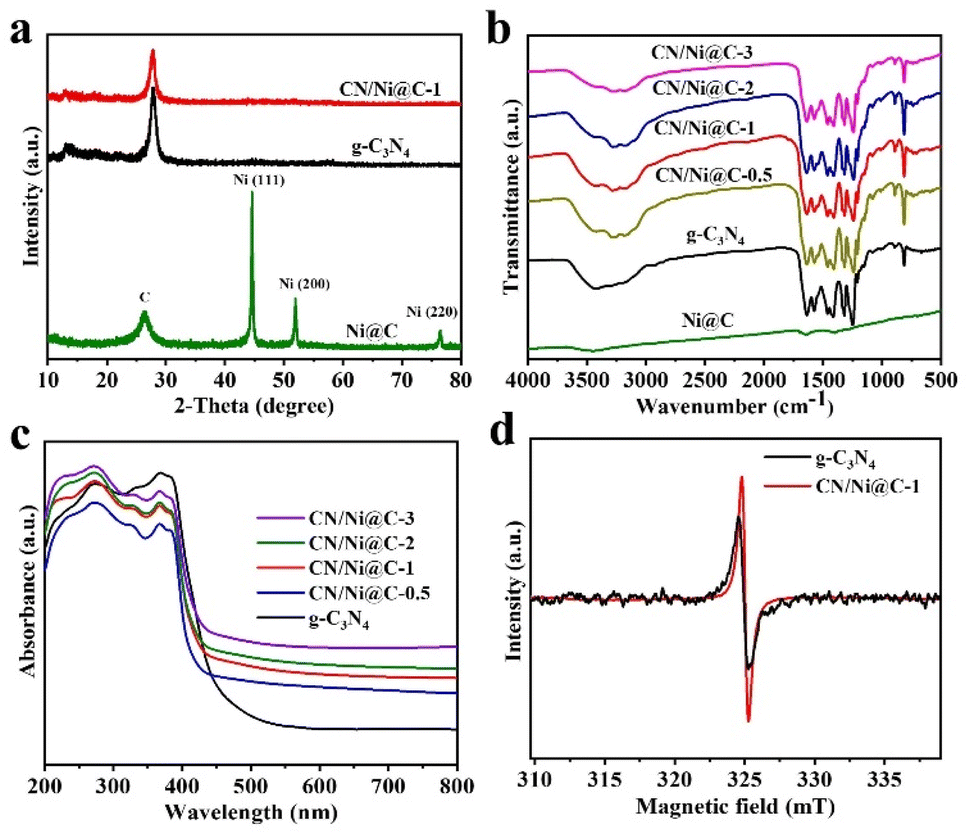 | ||
| Fig. 1 (a) XRD patterns of Ni@C, g-C3N4, and CN/Ni@C-1 photocatalysts. (b) FTIR spectra of Ni@C, g-C3N4, and CN/Ni@C-x samples (x represents the weight content of Ni@C in the samples). (c) UV-vis DRS of g-C3N4 and CN/Ni@C-x samples. (d) EPR plot of g-C3N4 and CN/Ni@C-1 photocatalysts. | ||
The morphology and structure of the g-C3N4, Ni@C, and CN/Ni@C-1 samples were examined by transmission electron microscopy (TEM). As presented in Fig. 2a–c, Ni@C shows a nanotube like structure, and g-C3N4 shows a traditional layered sheet-like structure. While the CN/Ni@C-1 nanocomposite photocatalyst exhibits similar morphology to g-C3N4, this result obviously reveals that the incorporation of plasmonic Ni@C did not change the morphology of g-C3N4. The scanning electronic microscopy (SEM) images of Ni@C, g-C3N4, and CN/Ni@C-1 are displayed in Fig. S3,† which is in agreement with the TEM results. In addition, high-angle annular dark-field scanning transmission microscopy (HAADF-STEM) and its corresponding EDS elemental mapping images (Fig. 2d–g) show that C, N, and Ni elements are present in CN/Ni@C-1, confirming the formation of CN/Ni@C-1 heterostructures.
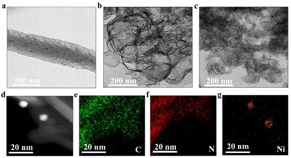 | ||
| Fig. 2 TEM images of (a) Ni@C NPs, (b) g-C3N4 nanosheets, and (c) the CN/Ni@C-1 nanocomposite. (d–g) HAADF-STEM image and its corresponding EDS elemental mapping images of the CN/Ni@C-1 photocatalyst. | ||
The X-ray photoelectron spectroscopy (XPS) measurements were conducted to investigate the chemical state and composition of the as-synthesized samples. XPS survey spectra prove the presence of C, N, and Ni elements in the optimal CN/Ni@C-1 heterojunction (Fig. 3a). The C 1s spectrum can be deconvoluted into three peaks that occur at 284.4, 285.3, and 288.3 eV (Fig. 3b). The peak at 284.4 eV is ascribed to sp2-hybridized carbon contamination, such as C![[double bond, length as m-dash]](https://www.rsc.org/images/entities/char_e001.gif) C, whereas the peaks at 285.3 and 288.3 eV are readily assigned to covalently bonded carbon located within N–CN and C–NH2 in the carbon nitride interlayers.49 The N 1s spectra can be split into four peaks that appeared at 398.8, 400.2, 401.3, and 404.5 eV (Fig. 3c). The peaks positioned at 398.8, 400.2, and 401.3 eV are attributed to sp2-hybridized confined nitrogen in the core of C-atoms from the conjugated ring (CN–C) and tertiary nitrogen in the C3–N groups, respectively.50 The weak peak at 404.5 eV is attributed to the existence of π–π localization of the C–N heterocycles.51Fig. 3d illustrates the Ni 2p spectra of the CN/Ni@C-1 sample displaying two main peaks at 854.7 (Ni 2p3/2) and 872.4 eV (Ni 2p1/2). This result indicates the existence of Ni2+ in the CN/Ni@C-1 sample, which is ascribed to the surface oxidation of metallic Ni0 upon exposure to air. Consequently, the XPS results confirm that plasmonic Ni@C was successfully introduced into g-C3N4.
C, whereas the peaks at 285.3 and 288.3 eV are readily assigned to covalently bonded carbon located within N–CN and C–NH2 in the carbon nitride interlayers.49 The N 1s spectra can be split into four peaks that appeared at 398.8, 400.2, 401.3, and 404.5 eV (Fig. 3c). The peaks positioned at 398.8, 400.2, and 401.3 eV are attributed to sp2-hybridized confined nitrogen in the core of C-atoms from the conjugated ring (CN–C) and tertiary nitrogen in the C3–N groups, respectively.50 The weak peak at 404.5 eV is attributed to the existence of π–π localization of the C–N heterocycles.51Fig. 3d illustrates the Ni 2p spectra of the CN/Ni@C-1 sample displaying two main peaks at 854.7 (Ni 2p3/2) and 872.4 eV (Ni 2p1/2). This result indicates the existence of Ni2+ in the CN/Ni@C-1 sample, which is ascribed to the surface oxidation of metallic Ni0 upon exposure to air. Consequently, the XPS results confirm that plasmonic Ni@C was successfully introduced into g-C3N4.
 | ||
| Fig. 3 (a) XPS survey spectra of Ni@C, g-C3N4, and CN/Ni@C-1 photocatalysts. (b) High-resolution XPS spectra of C 1s, (c) N 1s, and (d) Ni 2p of the CN/Ni@C-1 photocatalyst. | ||
Photoluminescence (PL) measurements were carried out to evaluate the efficiency of charge separation and recombination. The PL spectra of g-C3N4 and CN/Ni@C-1 exhibit wide emission peaks around 450 nm (Fig. 4a). Specifically, the decreased PL intensity of the CN/Ni@C-1 sample as compared to that of g-C3N4, suggests that efficient inhibition of photoinduced e−/h+ recombination is found for the CN/Ni@C-1 heterojunction, which is beneficial in promoting H2 production. The time-resolved photoluminescence (TRPL) spectra were conducted to evaluate the lifetime of photoinduced charge carriers. As illustrated in Fig. 4b, the average lifetimes for g-C3N4 and CN/Ni@C-1 are 2.27 ± 0.39 ns and 1.69 ± 0.27 ns, respectively. This reduction in the charge carrier lifetime implies the efficient transport and separation of photoinduced e−/h+ pairs.52 Furthermore, transient photocurrent tests were performed in a conventional three-electrode arrangement with several on/off visible-light irradiation cycles to investigate charge transfer over g-C3N4 and CN/Ni@C-1 samples (Fig. 4c). The transient photocurrent response of the CN/Ni@C-1 sample is higher than that of g-C3N4, demonstrating that in the presence of plasmonic Ni@C NPs, the separation and transfer of photoinduced charge carriers is highly accelerated,53 which agrees well with the PL and TRPL results. Fig. 4d represents the interfacial charge transfer resistance measured by electrochemical impedance spectroscopy (EIS) Nyquist analysis. Clearly, the arc diameter of the CN/Ni@C photocatalyst is relatively narrower than that of g-C3N4, demonstrating that the introduction of plasmonic Ni@C NPs can reduce charge transfer resistance and enhance charge separation.54 As such, the photoelectrochemical results confirm that plasmonic Ni@C anchored on g-C3N4 could prevent the recombination of photoinduced e−/h+ pairs and in turn improve H2 generation.
 | ||
| Fig. 4 (a) PL spectra of g-C3N4 and CN/Ni@C-1 samples. (b) TRPL spectra of g-C3N4 and CN/Ni@C-1 photocatalysts. (c) Transient photocurrent response curves of g-C3N4 and CN/Ni@C-1 samples under on/off visible-light irradiation cycles. (d) EIS Nyquist curves of pristine g-C3N4 and CN/Ni@NC-1 samples under visible-light irradiation. | ||
We carried out comparative photocatalytic H2 evolution experiments over the obtained photocatalysts under visible-light irradiation (Fig. 5). TEOA is found to be a more efficient sacrificial agent as compared to other sacrificial reagents after evaluating the H2 evolution rate over the optimal CN/Ni@C-1 sample (see Fig. S4†). Pristine g-C3N4 without the Pt cocatalyst achieves a minimum H2 generation rate of 0.53 μmol h−1 owing to its quick charge recombination (Fig. 5a). In the presence of 1 wt% Pt cocatalyst, the H2 evolution rate is increased to 13.55 μmol h−1 over g-C3N4. To confirm the role of plasmonic Ni@C, a comparison test was conducted on CN/Ni@C-1 without 1 wt% Pt, which shows 2.07 μmol h−1 H2 evolution rate. Moreover, the H2 evolution rate increased significantly to 56.67 μmol h−1 over the CN/Ni@C-1 plasmonic photocatalyst with 1 wt% Pt, which is 4 times higher than that of g-C3N4. As such, it is concluded that plasmonic Ni@C loaded on g-C3N4 to construct heterojunctions significantly improves visible-light absorption, charge transport and separation, thus leading to an enhanced H2 evolution rate. We next studied the influence of the weight contents of Ni@C in CN/Ni@C-x samples on photocatalytic activity. The hydrogen evolution rate of 0.45 μmol h−1 is achieved over the pure Ni@C sample (Fig. 5b). Furthermore, the H2 evolution rate increased with an increase of Ni@C contents in the CN/Ni@C photocatalysts, and then declined when the content of Ni@C further increased. The optimized H2 evolution rate of 56.67 μmol h−1 is achieved over the CN/Ni@C-1 sample, which is 4 times to that of bare g-C3N4 (13.55 μmol h−1), and higher than that of g-C3N4 photocatalysts fabricated earlier (see Table S1†). As the weight content of Ni@C increased up to 3 wt%, the H2 evolution rate reduced to 27.76 μmol h−1. A possible reason for this might be that excess Ni@C could contribute to the shielding effect, which blocks active centers and inhibits visible-light from the surface of g-C3N4.55 It is concluded that a proper amount of plasmonic Ni@C is vital to boost the performance of CN/Ni@C plasmonic photocatalysts. Additionally, Brunauer–Emmett–Teller (BET) surface area of CN/Ni@C-1 (91 m2 g−1) is higher than that of g-C3N4 (74 m2 g−1) (Fig. S5a†), and the pore size distribution curve reveals that CN/Ni@C-1 possesses more mesopores than g-C3N4 (Fig. S5b†); this result suggests that the incorporation of plasmonic Ni@C onto g-C3N4 leads to an increase in surface areas. Furthermore, the AQY of CN/Ni@C-1 was measured via an extensive range of monochromatic light irradiation wavelength (λ = 420, 450, 500, 550 and 600 nm ± 5 nm). As depicted in Fig. 5c, the AQY decreased with increasing wavelength, which well agrees with the UV-vis absorption spectrum of CN/Ni@C-1, and it is noted that an AQY of 5.20% is obtained at λ ≥ 420 nm. The stability and recycling are the most crucial factors to estimate the possibility of photocatalysts for practical photocatalytic applications. The recycling tests were carried out to evaluate the stability of the CN/Ni@C-1 sample. As presented in Fig. 5d, after 5 consecutive cycles (each cycle of 4 hours) under the same reaction conditions, no distinguishable decline is noticed in photocatalytic H2 evolution. Besides, no noticeable alteration is detected in the XRD patterns of the CN/Ni@C-1 sample before and after the photocatalytic reaction (Fig. S6†). This result proves that the CN/Ni@C-1 plasmonic photocatalyst has outstanding photocatalytic stability and reusability without structural deformation.
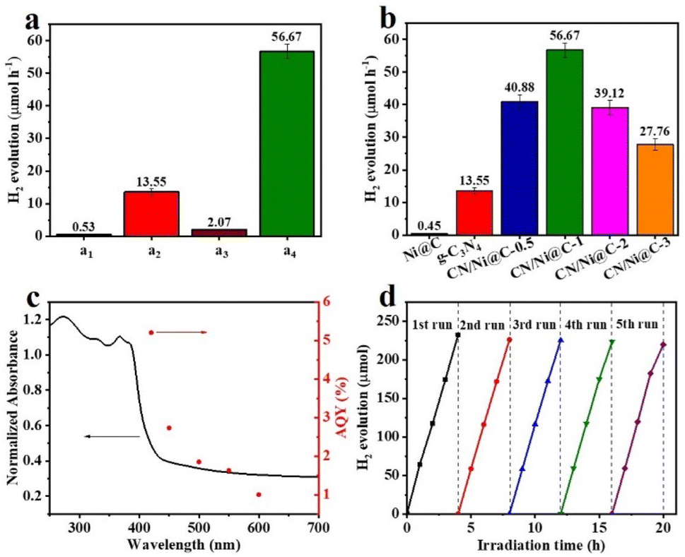 | ||
| Fig. 5 (a) Control experiments of H2 evolution over various synthesized photocatalysts (a1, g-C3N4 in the absence of Pt, a2, g-C3N4 in the presence of Pt, a3, CN/Ni@C-1 in the absence of Pt, and a4, CN/Ni@C-1 in the presence of Pt). (b) Photocatalytic H2 evolution over the Ni@C, g-C3N4, and CN/Ni@C-x samples. (c) The wavelength-dependent AQY of H2 evolution over the CN/Ni@C-1 sample. (d) The stability test of the CN/Ni@C-1 photocatalyst. | ||
Taken together, the improved H2 evolution from water splitting over CN/Ni@C-x photocatalysts is indeed steered by the construction of the heterostructures from g-C3N4 and plasmonic Ni@C NPs which leads to highly efficient charge carrier generation, separation and migration. A possible mechanism is proposed for g-C3N4/Ni@C plasmonic photocatalysts to understand the phenomena of photocatalytic H2 generation from water splitting. The valence band (VB) and conduction band (CB) of g-C3N4 are located at −6.1 eV and −3.4 eV vs. the absolute vacuum scale, respectively.56 The measured optical bandgap of g-C3N4 is 2.75 eV (Fig. S7†). No band gap is detected for Ni@C, indicating its metallic characteristics. While the Fermi level of metallic Ni is positioned at −4.59 eV vs. vacuum level.57 As such, plasmonic Ni is promoted to excited states via 3d electron excitation due to its strong surface plasmon absorption under visible-light irradiation (Fig. 6). The excited Ni nanoparticles produced a lot of surface plasmons and these surface plasmons decay quickly and generate hot electrons, which jump to the above Fermi level of Ni, and plasmonic hot electrons are transported to the CB of g-C3N4 because of the surface plasmon resonance (SPR) effect, then further transferred to Pt active sites for H2 evolution (reductive product). Simultaneously, to retain charge equilibrium, photoinduced holes in g-C3N4 are quenched by TEOA to generate TEOA+ (oxidative product). Therefore, continuously photoinduced hot electrons are transported from plasmonic Ni NPs, and effective separation and transport of photoexcited e−/h+ leads to a significantly improved H2 evolution rate.
 | ||
| Fig. 6 Schematic presentation of the energy level, charge separation and transfer over g-C3N4/Ni@C plasmonic photocatalysts under visible-light irradiation for H2 evolution from water splitting. | ||
Conclusions
In summary, CN/Ni@C-x plasmonic photocatalysts have been successfully developed and fabricated. The optimal CN/Ni@C-1 plasmonic heterojunction displays an optimized photocatalytic H2 evolution rate of 56.67 μmol h−1 with an AQY of 5.20% under visible-light irradiation (λ ≥ 420 nm). The PL, TRPL, transient photocurrent response, and EIS analysis of the CN/Ni@C-1 sample show higher charge separation efficiency as compared to bare g-C3N4, and a possible mechanism of plasmon-enhanced photocatalytic performance is proposed. Moreover, the as-synthesized CN/Ni@C-1 nano-heterostructure exhibits good photocatalytic stability under visible-light irradiation for 20 h. The boosted photocatalytic activity is associated with efficient charge separation, charge transport, and the SPR effect of Ni NPs. Overall, our study could provide a creative strategy for developing other metallic plasmon-modified graphitic carbon nitride photocatalytic systems to fulfil the renewable energy demands.Conflicts of interest
The authors confirm no conflict of interest.Acknowledgements
The authors appreciatively acknowledge the special financial support from the National Natural Science Foundation of China (22271266, 21771171, and 82102905), the Scientific Research Grant of the Hefei National Synchrotron Radiation Laboratory (UN2017LHJJ), and the Fundamental Research Funds for the Central Universities (YD2340002001).Notes and references
- A. G. Olabi and M. A. Abdelkareem, Renewable Sustainable Energy Rev., 2022, 158, 112111 CrossRef CAS.
- B. Nastasi, N. Markovska, T. Puksec, N. Duić and A. Foley, Renewable Sustainable Energy Rev., 2022, 157, 112071 CrossRef.
- Y. Zhang, H. Liu, F. Gao, X. Tan, Y. Cai, B. Hu, Q. Huang, M. Fang and X. Wang, EnergyChem, 2022, 4, 100078 CrossRef CAS.
- M. D. Scovell, Int. J. Hydrogen Energy, 2022, 47, 10441–10459 CrossRef CAS.
- A. Bahadoran, Q. Liu, S. Ramakrishna, B. Sadeghi, M. M. De Castro and P. D. Cavaliere, Energies, 2022, 15, 3222 CrossRef CAS.
- W. Huang, J. Dai and L. Xiong, Sustain. Energy Technol. Assess., 2022, 52, 102059 Search PubMed.
- M. Amin, H. H. Shah, A. G. Fareed, W. U. Khan, E. Chung, A. Zia, Z. U. R. Farooqi and C. Lee, Int. J. Hydrogen Energy, 2022, 47, 33112–33134 CrossRef CAS.
- A. Fujishima and K. Honda, Nature, 1972, 238, 37–38 CrossRef CAS PubMed.
- Z. Li, H. Fang, Z. Chen, W. Zou, C. Zhao and X. Yang, Appl. Catal., B, 2022, 312, 121374 CrossRef CAS.
- Q. Liu, J. Huang, H. Tang, X. Yu and J. Shen, J. Mater. Sci. Technol., 2020, 56, 196–205 CrossRef.
- H. Lv, H. Wu, X. Wu, J. Zheng and Y. Liu, Appl. Surf. Sci., 2022, 593, 153448 Search PubMed.
- D.-H. Wang, L. Jia, X.-L. Wu, L.-Q. Lu and A.-W. Xu, Nanoscale, 2012, 4, 576–584 RSC.
- R. Sharma, M. Almáši, S. P. Nehra, V. S. Rao, P. Panchal, D. R. Paul, I. P. Jain and A. Sharma, Renewable Sustainable Energy Rev., 2022, 168, 112776 Search PubMed.
- C. Cheng, C.-L. Dong, J. Shi, L. Mao, Y.-C. Huang, X. Kang, S. Zong and S. Shen, J. Mater. Sci. Technol., 2022, 98, 160–168 CrossRef.
- H. Y. Yuan, J. Y. Bai, B. Xu, X. Y. Li, S. Y. Xiao, P. F. Liu, X. L. Wang and H. G. Yang, Chem. Commun., 2021, 57, 3042–3045 RSC.
- P. Niu, H. Li, Y. Ma and T. Zhai, J. Phys. Chem. C, 2018, 122, 20717–20726 CrossRef CAS.
- Y. Hong, E. Liu, J. Shi, X. Lin, L. Sheng, M. Zhang, L. Wang and J. Chen, Int. J. Hydrogen Energy, 2019, 44, 7194–7204 CrossRef CAS.
- M. R. Gholipour, F. Béland and T.-O. Do, ACS Sustain. Chem. Eng., 2017, 5, 213–220 CrossRef CAS.
- C. Hu, W.-L. Chiu, C.-Y. Wang and V.-H. Nguyen, J. Taiwan Inst. Chem. Eng., 2021, 129, 128–134 Search PubMed.
- N. D. Shcherban, V. V. Shvalagin, G. V. Korzhak, P. S. Yaremov, M. A. Skoryk, S. A. Sergiienko and S. Y. Kuchmiy, J. Mol. Struct., 2022, 1250, 131741 CrossRef CAS.
- L. Wang, Y. Hong, E. Liu, Z. Wang, J. Chen, S. Yang, J. Wang, X. Lin and J. Shi, Int. J. Hydrogen Energy, 2020, 45, 6425–6436 Search PubMed.
- C. Han, P. Su, B. Tan, X. Ma, H. Lv, C. Huang, P. Wang, Z. Tong, G. Li, Y. Huang and Z. Liu, J. Colloid Interface Sci., 2021, 581, 159–166 Search PubMed.
- K. Liu, J. Ma, X. Yang, Z. Liu, X. Li, J. Zhang, R. Cui and R. Sun, Chem. Eng. J., 2022, 437, 135232 CrossRef CAS.
- Z. Chen, F. Guo, H. Sun, Y. Shi and W. Shi, J. Colloid Interface Sci., 2022, 607, 1391–1401 Search PubMed.
- L. Ding, F. Qi, Y. Li, J. Lin, Y. Su, Y. Song, L. Wang, H. Sun and C. Tong, J. Colloid Interface Sci., 2022, 614, 92–101 CrossRef CAS PubMed.
- F. Ding, T. Ming, H. Zhang, Y. Gao, V. Dragutan, Y. Sun, I. Dragutan and Z. Xu, R. Chem. Mater., 2022, 1, 1–7 Search PubMed.
- Q. Zhang, D. T. Gangadharan, Y. Liu, Z. Xu, M. Chaker and D. Ma, J. Materiomics, 2017, 3, 33–50 Search PubMed.
- Y. Zhang, S. He, W. Guo, Y. Hu, J. Huang, J. R. Mulcahy and W. D. Wei, Chem. Rev., 2018, 118, 2927–2954 CrossRef CAS.
- C. Jia, X. Li, N. Xin, Y. Gong, J. Guan, L. Meng, S. Meng and X. Guo, Adv. Energy Mater., 2016, 6, 1600431 Search PubMed.
- M. Valenti, M. P. Jonsson, G. Biskos, A. Schmidt-Ott and W. A. Smith, J. Mater. Chem. A, 2016, 4, 17891–17912 RSC.
- S. Khanam and S. K. Rout, ACS Omega, 2022, 7, 25466–25475 CrossRef CAS PubMed.
- B. Ma, J. Bi, J. Lv, C. Kong, P. Yan, X. Zhao, X. Zhang, T. Yang and Z. Yang, Chem. Eng. J., 2021, 405, 126709 Search PubMed.
- J. Yi, X. She, Y. Song, H. Xu, P. Zhang, Z. Mo, L. Liu, D. Du and H. Li, RSC Adv., 2016, 6, 112420–112428 RSC.
- Z. Mo, H. Xu, Z. Chen, X. She, Y. Song, P. Yan, Y. Xu, Y. Lei, S. Yuan and H. Li, Chin. J. Catal., 2018, 39, 760–770 CrossRef CAS.
- A. K. Shukla, S. Venugopalan and B. Hariprakash, J. Power Sources, 2001, 100, 125–148 CrossRef CAS.
- X. Shi, S. Posysaev, M. Huttula, V. Pankratov, J. Hoszowska, J.-C. Dousse, F. Zeeshan, Y. Niu, A. Zakharov, T. Li, O. Miroshnichenko, M. Zhang, X. Wang, Z. Huang, S. Saukko, D. L. González, S. van Dijken, M. Alatalo and W. Cao, Small, 2018, 14, 1704526 Search PubMed.
- G. S. Pawar, A. Elikkottil, B. Pesala, A. A. Tahir and T. K. Mallick, Int. J. Hydrogen Energy, 2019, 44, 578–586 Search PubMed.
- L. Pei, T. Li, Y. Yuan, T. Yang, J. Zhong, Z. Ji, S. Yan and Z. Zou, Chem. Commun., 2019, 55, 11754–11757 Search PubMed.
- J. Wen, J. Xie, H. Zhang, A. Zhang, Y. Liu, X. Chen and X. Li, ACS Appl. Mater. Interfaces, 2017, 9, 14031–14042 CrossRef CAS PubMed.
- Z. Lin, C. Du, B. Yan, C. Wang and G. Yang, Nat. Commun., 2018, 9, 4036 Search PubMed.
- X. Liu, G. Verma, Z. Chen, B. Hu, Q. Huang, H. Yang, S. Ma and X. Wang, Innovation, 2022, 3, 100281 CAS.
- C. Cheng, J. Zhang, R. Zeng, F. Xing and C. Huang, Appl. Catal., B, 2022, 310, 121321 Search PubMed.
- B.-R. Wulan, S.-S. Yi, S.-J. Li, Y.-X. Duan, J.-M. Yan and Q. Jiang, Appl. Catal., B, 2018, 231, 43–50 Search PubMed.
- M. Wang, H. Xu, C. Huang, Z. Cui, M. Li, B. Song, G. Shao, H. Wang, H. Lu and R. Zhang, Inorg. Chem. Commun., 2021, 129, 108645 Search PubMed.
- S. Zhao, J. Wu, Y. Xu, Z. Wang, Y. Han and X. Zhang, Int. J. Hydrogen Energy, 2020, 45, 20851–20858 CrossRef CAS.
- X. Wu, X. Wang, F. Wang and H. Yu, Appl. Catal., B, 2019, 247, 70–77 Search PubMed.
- C. Wang, C. Yang, J. Qin, S. Rajendran and X. Zhang, Mater. Chem. Phys., 2022, 275, 125299 Search PubMed.
- T. Zhao, T.-Y. Cheang, H.-B. Chong, C. Ling, X.-J. Lu, C.-C. Li, X.-X. Fang, L.-B. Ma, G. Wang and A.-W. Xu, Catal. Sci. Technol., 2021, 11, 4776–4782 Search PubMed.
- S. Yılmaz, E. G. Acar, G. Yanalak, E. Aslan, M. Kılıç, İ. Hatay Patır and Ö. Metin, Appl. Surf. Sci., 2022, 593, 153398 Search PubMed.
- X. Zhang, C. Xi, Y. Yue, P. Deng, L. Zhang and Y. Hou, Int. J. Hydrogen Energy, 2022, 47, 1624–1632 CrossRef CAS.
- L. Luo, Z. Gong, J. Ma, K. Wang, H. Zhu, K. Li, L. Xiong, X. Guo and J. Tang, Appl. Catal., B, 2021, 284, 119742 CrossRef CAS.
- R. Boppella, W. Yang, J. Tan, H.-C. Kwon, J. Park and J. Moon, Appl. Catal., B, 2019, 242, 422–430 CrossRef CAS.
- J.-S. Zhang and W.-D. Zhang, J. Colloid Interface Sci., 2020, 560, 11–20 CrossRef CAS PubMed.
- X. Yan, H. An, Z. Chen and G. Yang, Chin. J. Chem. Eng., 2022, 43, 31–39 CrossRef.
- F. Sarwar, M. Tahir and H. Alias, Mater. Sci. Semicond. Process., 2022, 137, 106187 Search PubMed.
- X. Wang, K. Maeda, A. Thomas, K. Takanabe, G. Xin, J. M. Carlsson, K. Domen and M. Antonietti, Nat. Mater., 2009, 8, 76–80 Search PubMed.
- B. Geng, Y. He, F. Yan, C. Zhu, X. Zhang, X. Zhang and Y. Chen, Mater. Today Nano, 2022, 18, 100176 Search PubMed.
Footnote |
| † Electronic supplementary information (ESI) available: Characterization studies, photoelectrochemical tests, and photocatalytic H2 evolution measurements. Schematic illustration of the as-synthesized samples. UV-DRS specturm of Ni@C sample. SEM images of Ni@C, g-C3N4, and CN/Ni@C-1 photcatalysts. Photocatalytic H2 evolution rate over CN/Ni@C-1 nanocomposite under various sacrificial agents. Comparison table of the photocatalytic H2 evolution rate of our study with previous reports. Nitrogen adsorption–desorption isotherms and pore size distribution of g-C3N4 and CN/Ni@C-1 samples. XRD spectra of CN/Ni@C-1 before and after the recycling experiment. Plot of energy band gap of g-C3N4. See DOI: https://doi.org/10.1039/d2se01523d |
| This journal is © The Royal Society of Chemistry 2023 |