
 Open Access Article
 This Open Access Article is licensed under a Creative Commons Attribution-Non Commercial 3.0 Unported Licence
Development of antioxidant-rich edible active films and coatings incorporated with de-oiled ethanolic green algae extract: a candidate for prolonging the shelf life of fresh produce†
Kona Mondal,
Sayan Kumar Bhattacharjee,
Chethana Mudenur,
Tabli Ghosh,
Vaibhav V. Goud and
Vimal Katiyar*
and
Vimal Katiyar*
Department of Chemical Engineering, Indian Institute of Technology Guwahati (IITG), Assam-781039, India. E-mail: vkatiyar@iitg.ac.in
First published on 3rd May 2022
Abstract
The concept of sustainability and the substitution of non-biodegradable packaging using biodegradable packaging has attracted gigantic interest. The objective of the present study was to revalorize the biowaste “de-oiled green algae biomass (DAB)” of Dunaliella tertiolecta using a green approach and the development of biodegradable chitosan (CS)-based edible active biocomposite films and coatings for prolonging the shelf life of fresh produce. Ultrasound-assisted green extraction was conducted using food-grade solvent ethanol for obtaining the bio-actives, namely “crude algae ethanolic extract (CAEE)” from DAB. The edible films (CS/CAEE) and coating solutions were developed by incorporating CAEE with varying concentrations (0 to 28%). The CAEE was subjected to MALDI-TOF-MS, NMR, and other biochemical analyses, and was found to be rich in DPPH antioxidant activity (∼40%). The CS/CAEE films were fabricated using a solvent casting method and characterized by several biochemical and physicochemical (FESEM, TGA, FTIR, XRD, WVP, UTM, and rheological) characterization techniques. The addition of CAEE into the CS matrix reduced the maximum film transparency (∼20%), water vapor permeability (∼60%); improved the crystallinity (∼24%), tensile strength (∼25%), and antioxidant activity (∼27%); and exhibited UV-Vis blocking properties as compared to the control film. Besides, the developed coating solutions and CAEE showed biocompatibility with BHK-21 fibroblast cells and antimicrobial activity against common food pathogens. The developed coating solution was applied on green chilli using a dipping method and stored at ambient temperature (25 ± 2 °C, 50–70 % RH) for 10 days. The shelf life of chillies was extended without altering the quality as compared to uncoated green chillies. Therefore, the formulated coating could be applicable for prolonging the shelf life of fresh produce.
Introduction
Active and edible packaging is an emerging concept that aims to deliver quality and safe food along with a prolonged shelf life while at the same time mitigating environmental hazards caused by non-biodegradable food-packaging waste. Besides providing barrier properties, this innovative packaging also acts as a preservation method. The improved functionality of this packaging material is due to the incorporation of certain active substances, that are not available in conventional packaging.1,2 In general, the active packaging system consists of antimicrobial agents, antioxidants, and several other beneficial compounds.3 In recent times, a mass of the scientific community has focused their research predominantly on the development of biodegradable active edible packaging utilizing various natural bioresources as a functional preservative with due concern for food safety and security for consumers, overcoming the environmental drawbacks of current packaging, and the preservation of fossil fuel resources.4,5In this context, chitosan, a biodegradable, nontoxic, bio-based substance with good film-forming properties and biocompatible polymers, obtained from the deacetylation of chitin, found in crustaceans and insects,6 has received much attention for the development of active and edible food-packaging materials.7 Chitosan is a linear polysaccharide, associated with the β-(1-4) glycosidic bond between D-glucosamine and N-acetyl-D-glucosamine (NADG). Interestingly, it is one of the potential candidates for developing active packaging materials among other polysaccharides due to its inherent antimicrobial and antioxidant activity based on its concentration and molecular weight.2 Chitosan-based films and coatings have demonstrated wide antimicrobial properties against Gram-positive, Gram-negative, yeast, and molds, due to the presence of certain cations that lead to surface damage in the microbial cell via interacting with the anions present in it.8 Besides, chitosan can restore essential oils and bioactive compounds into their natural structure, which diversify its applications in controlled-drug delivery and food-coating systems.9,10 Nevertheless, there has been a limited use of chitosan-based films in the area of food packaging to date due to its inherent poor barrier and mechanical properties, which makes it hydrophilic and brittle, which are undesirable properties for a food-packaging material where high strength, control of moisture, and gas transfer are essential.11 To overcome these limitations biofillers, such as oil, lipids, fatty acids, waxes, proteins, polysaccharides, and various plant extracts, have been added to chitosan for obtaining improved physicochemical and structural properties.12 However, the addition of oils and lipids adversely affect the mechanical properties of chitosan by altering the three-dimensional (3D) network structure.13 Further, the incorporation of proteins and polysaccharides into chitosan films sometimes provide incompatibility for certain uses; thereby restricting their use. In this regard, Ferreira et al. (2009)14 reported that whey protein-rich surfaces are more hydrophobic than chitosan; however, enhancing the protein amount decreased the mechanical strength and elongation. Furthermore, the concept of developing selected biopolymer-based multi-layered films and composites as biodegradable packaging materials has attracted a lot of attention due to the improved mechanical and other properties possible; albeit the higher cost limit the use of this concept.15,16 In this context, a promising strategy has been put forward to address all the limitations and to prompt the development of novel sustainable packaging materials via utilizing the concept of green technology, where various biomaterials from different Agri-based waste may be utilized as a key factor for fabricating eco-friendly packaging materials.17,18 In this regard, various plant-based extracts, such as banana peel extract,19 pomegranate peel extract,9 mutra leaf extract,20 extract of pine nut shell, peanut shell, jujube leaf,21 and others, have been used as biofillers in the chitosan matrix, and the associated literature on these have reported improvements in the antioxidant and other functional properties of the developed biocomposites. Recently, one study reported that the addition of spice extract into chitosan film enhanced different characteristic properties of the film and extended the shelf life of a food material.22 Besides, Lorenzo et al. (2018) reported the non-toxicity of the plant extract at a higher dose level compared to doses taken through daily diets.23
Dunaliella tertiolecta is a green microalga that has been mostly studied for bio-energy production.24 However, it also has potential use in the pharmaceutical and food industries as a healthy food supplement due to its bioactive compounds.25 Predominantly, Dunaliella algae biomass has been utilized for biodiesel and bioethanol production.26 Moreover, after extraction of the oil, the de-oiled residue or leftover part is recognized as de-oiled green algae biomass (DAB), which has commonly been treated as cattle feed or dumped in coastal regions, whereby it is responsible for causing negative environmental impacts. However, fresh algae biomass has gained much attention in various sectors compared to the residual part, with very few studies focused on de-oiled ABR. Interestingly, this waste is rich in several bioactive compounds, such as carbohydrates, proteins, and carotenoids.27 In this context, Deshmukh et al. (2021) reported on the development of chitosan-based biocomposites for food packaging, whereby Chlorella de-fatted biomass powder was added as a biofiller material.4 In addition, cellulose nanocrystals have been isolated from Dunaliella DAB and utilized as one of the potential biofillers for the development of poly(lactic acid)- and poly(ε-caprolactone)-based biodegradable biocomposites, as reported in our previous study.28,29
The present study focuses on the ultrasound-assisted green technology-based liquid extraction of bioactive compounds from Dunaliella DAB and the development of chitosan (CS)-based active edible biocomposite films and coatings. The ultrasound-assisted crude algae ethanolic extract (CAEE) was used in varying concentrations as a biofiller/food additive in the chitosan matrix to obtain active edible packaging materials with several improved physicochemical and functional properties. Further, the developed filmogenic solution was applied to a real food system as a primary packaging material in the form of an active edible coating, with an aim to extend the shelf life. Moreover, several physicochemical and functional properties of the developed films were studied. Further, the coating effects were confirmed by conducting physicochemical studies of coated green chilli. Interestingly, to the best of our knowledge, this is the first study on the development of an active edible food-packaging material additivated with Dunaliella tertiolecta green algae biomass extract. Further, more detailed experiments, analyses, and observations of the developed edible films and coating applications on various fresh produce have been documented elsewhere (Indian patent file no. 202131013653).
Experimental
Extraction of the de-oiled crude algae ethanolic extract
The green extraction of the de-oiled algae residue powder (provided by TERI, India) of Dunaliella tertiolecta is depicted in Fig. 1. Here, 50 gm of the standardized particles of the de-oiled algae biomass residue (passing through a 40 mesh sieve) was added into 500 mL ethanol (Finner, India) and kept under shaking overnight, before being subjected to ultrasound-assisted extraction. An ultrasonicator probe (Biologics, Inc., USA) was used for the extract preparation and sonication was conducted for 2–3 min, at room temperature, 20 kHz at 99% efficiency. Thereafter, the suspension was subjected to centrifugation at 5000 rpm for 5 min to separate the solid part from solution, and finally, filtration was conducted using Whatman filter paper no. 3 for obtaining the clear transparent green-colored crude algae ethanolic extract (CAEE) without any particles. Finally, the obtained extract was kept at −20 °C in the dark until further use.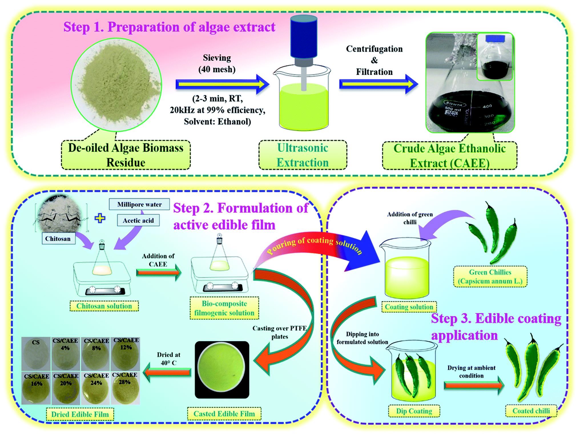 | ||
| Fig. 1 Extraction of the biofiller CAEE and fabrication flowsheet of the active edible films and coating. | ||
Fabrication of biocomposites active edible films and coatings
The films were prepared by a solution casting technique, as described in Fig. 1. The concentrations of chitosan (CS, medium molecular weight, Sigma Aldrich, (USA)) (1 g/100 mL filmogenic solution) and acetic acid (Finner, India) (0.3%) were kept constant. Afterwards, the incorporation of CAEE to the CS solution was followed with varying the amount of CAEE at concentrations of 0, 4, 8, 12, 16, 20, 24, and 28 mg g−1 of CS. The solution was magnetically stirred for 24 h at room temperature. The developed filmogenic solution was kept under a vacuum drier for stabilizing the air bubbles generated from the solution for 2 h. Thereafter, the solution was spread over Teflon plates (140 cm dia) and dried at 40 °C for 24 h inside a hot air oven. A digital micrometer (Indi 6156, India) was used to measure the average film thickness by taking 10 random measurements on the film surface. Finally, the formulated filmogenic solution was applied as an active edible coating material (Fig. 1).Characterization
| Total Chl = 8.02 × A(663) + 20.2 × A(645) |
| Chl ‘a’ = 12.7 × A(663) − 2.69 × A(645) |
| Chl ‘b’ = 22.9 × A(645) − 4.68 × A(663) |
The yield of chlorophyll from the CAEE using the green solvent via ultrasonic extraction was calculated with the below equation:

![[thin space (1/6-em)]](https://www.rsc.org/images/entities/char_2009.gif) :6 (matrix:CAEE), which meant 1 μL of matrix solution was added to 6 μL of analyte solution (CAEE). MALDI-TOF-MS analysis was performed on a Bruker Autoflex Speed spectrometer at an accelerating voltage of 19 kV (per spectrum around 2000 laser shots). The sample with the matrix solution was placed over a stainless-steel plate and dried before being subjected to the analysis. The spectrum reproducibility was checked by taking measurements at five different spots on the sample.
:6 (matrix:CAEE), which meant 1 μL of matrix solution was added to 6 μL of analyte solution (CAEE). MALDI-TOF-MS analysis was performed on a Bruker Autoflex Speed spectrometer at an accelerating voltage of 19 kV (per spectrum around 2000 laser shots). The sample with the matrix solution was placed over a stainless-steel plate and dried before being subjected to the analysis. The spectrum reproducibility was checked by taking measurements at five different spots on the sample. :F–C reagent = 10:1), followed by the addition of 2 mL of sodium carbonate (Himedia, India) (7.5%, diluted in distilled water) into the mixture after 5 min. The absorbance of the solution was measured at 740 nm after an incubation period of 2 h in the dark. The result was determined using the standard gallic acid (Himedia, India) curve and reported in terms of μg of gallic acid per mL of extract.
where WVTR is the test result obtained from the instrument after completion of a cycle in (g m−2 day), L indicates the thickness of the film, and ΔP is the pressure difference at 37.8 °C saturated vapor pressure condition. The WVP is reported in this work in kg m m−2 Pa−1 s−1. All the tests were performed in triplicate.

| η = m(γ˙)n−1 |
Different rheological parameters were analyzed:
•  the storage modulus at the critical strain.
• γL, the critical strain.
• tanδ, the ratio of loss to the storage modulus, which determines the liquid or solid behavior of the solutions.
• The difference between the moduli G′′–G′ at a fixed strain value, which depicts the reinforcing effect of the extract incorporation of the solutions in their viscous and elastic behaviors.
Cytotoxicity study
Shelf-life study of green chilli (Capsicum annum L.)
Physiological parameters of the coated green chilli

Results and discussion
Biochemical analysis of the biofiller CAEE
The ultrasound-assisted extraction from de-oiled algae biomass residue is schematically described in Fig. 1. The CAEE was rich in antioxidants with 40 ± 1.41% DPPH radical-scavenging activity, a total phenolic content of 515.72 ± 4.67 μg g−1, total flavonoid content of 151.75 ± 2.05%, and total chlorophyll content of 23.81 ± 1.1 mg mL−1, respectively. The yield of chlorophyll was 238.14 mg mL−1, which indicated ethanol acted as a superior chlorophyll extracting solvent.30 In addition, the CAEE contained a higher percentage of chlorophyll ‘a’ (15.63 ± 0.16) compared to chlorophyll ‘b’ (8.19 ± 0.09). Moreover, the obtained results indicated that the CAEE was rich in antioxidants and polyphenols. The color coordinates L, and a* and b* values of CAEE were 72.62 ± 0.04, −14.1 ± 0.01, and 59.29 ± 0.05, respectively. The negative value of a* was attributed to the green color of the extract, which was due to the presence of the pigment chlorophyll, whereas the higher positive value of b* indicated a yellow color, which could be due to the presence of carotenoids, such as β-carotene and others, which is the most available pigment found in green algae.31MALDI-TOF-MS analysis of CAEE
Matrix-assisted laser desorption/ionization mass spectroscopy is one of the methods for the structural identification of organic matter of any biological sample. A series of masses m/z of CAEE were observed from the MALDI-TOF-MS spectrum and are depicted in Fig. 2. The obtained peaks at the lower m/z range of 89.22, 105.32, 117.25, and 181.42 corresponded to proteins alanine, serine, valine, and tyrosine, respectively.32 Besides, the masses m/z at 183.98, 211.66, 232.74, 253.82, 258.21, 284.25, 324.94, 340.89, 344.73, 359.21, 372.41, 409.25, and 425.01 corresponded to protonated hexose sugar, sulfated hexose sugar, magnesium ion (Mg+) attached with a hexose sugar, uronic acid, two hexose sugar, two pentose sugar with a Mg+ ion, di-saccharide (heptose and pentose), deprotonated di-saccharide (hexose and heptose) associated with a sodium ion, and sugars linked with other ions, like Na, Mg, P, S, etc. The mentioned metals present in the de-oiled algae biomass were also detected by ICPMS analysis, as reported in our previous study.28 The observed peak at m/z 662.19 might be related to the tetra-saccharide of two sugars, i.e., both pentose and hexose. The mass deltas between the peaks were observed consecutively at 1–2, 14, 18, and 28 Da, corresponding to the degrees of saturation and ring structures, polymeric chains ((CH2)n), loss of water molecule and deprived CO bond from the esters of lactone.32 Further, the m/z values in the range between 500 to 600 were predominantly responsible for the carotenoids. The MALDI spectra were highlighted and intensified for masses m/z at 536.58, 538.73, 549.67, 568.57, and 593.86, representing the presence of carotenoids, such as β-carotene along with its derivatives, zeaxanthin and deprotonated astaxanthin, which is similar to the reported literature by Fraser et al. (2007).33 The identified peaks at m/z 871.69 and 888.74 corresponded to chlorophyll a or pheophytin and astaxanthin esters.33 | ||
| Fig. 2 MALDI-TOF-MS spectra of crude algae ethanolic extract (CAEE). | ||
Nuclear magnetic resonance (NMR) analysis of CAEE
![[double bond, length as m-dash]](https://www.rsc.org/images/entities/char_e001.gif) C–CH3)n, vinyl CH3, CH2, and iso dimethyl attached to a cyclic ring, respectively.25 In this context, the presence of β-carotene and astaxanthin pigments was also confirmed due to these signals. The observed chemical shift at 7.13 ppm in the doublet form in both the NMR spectra was related to the olefinic protons of fucoxanthin.31 Further, the 1H NMR spectra predicted the presence of proteins, such as alanine and tyrosine, in CAEE due to the appearance of chemical shifts at 3.62, 1.47, 2.30, 2.34, and 7.14. This finding corroborated the obtained mass results from the MALDI-TOF-MS spectrum. In this context, a similar observation was reported by Iglesias et al. (2019).31
C–CH3)n, vinyl CH3, CH2, and iso dimethyl attached to a cyclic ring, respectively.25 In this context, the presence of β-carotene and astaxanthin pigments was also confirmed due to these signals. The observed chemical shift at 7.13 ppm in the doublet form in both the NMR spectra was related to the olefinic protons of fucoxanthin.31 Further, the 1H NMR spectra predicted the presence of proteins, such as alanine and tyrosine, in CAEE due to the appearance of chemical shifts at 3.62, 1.47, 2.30, 2.34, and 7.14. This finding corroborated the obtained mass results from the MALDI-TOF-MS spectrum. In this context, a similar observation was reported by Iglesias et al. (2019).31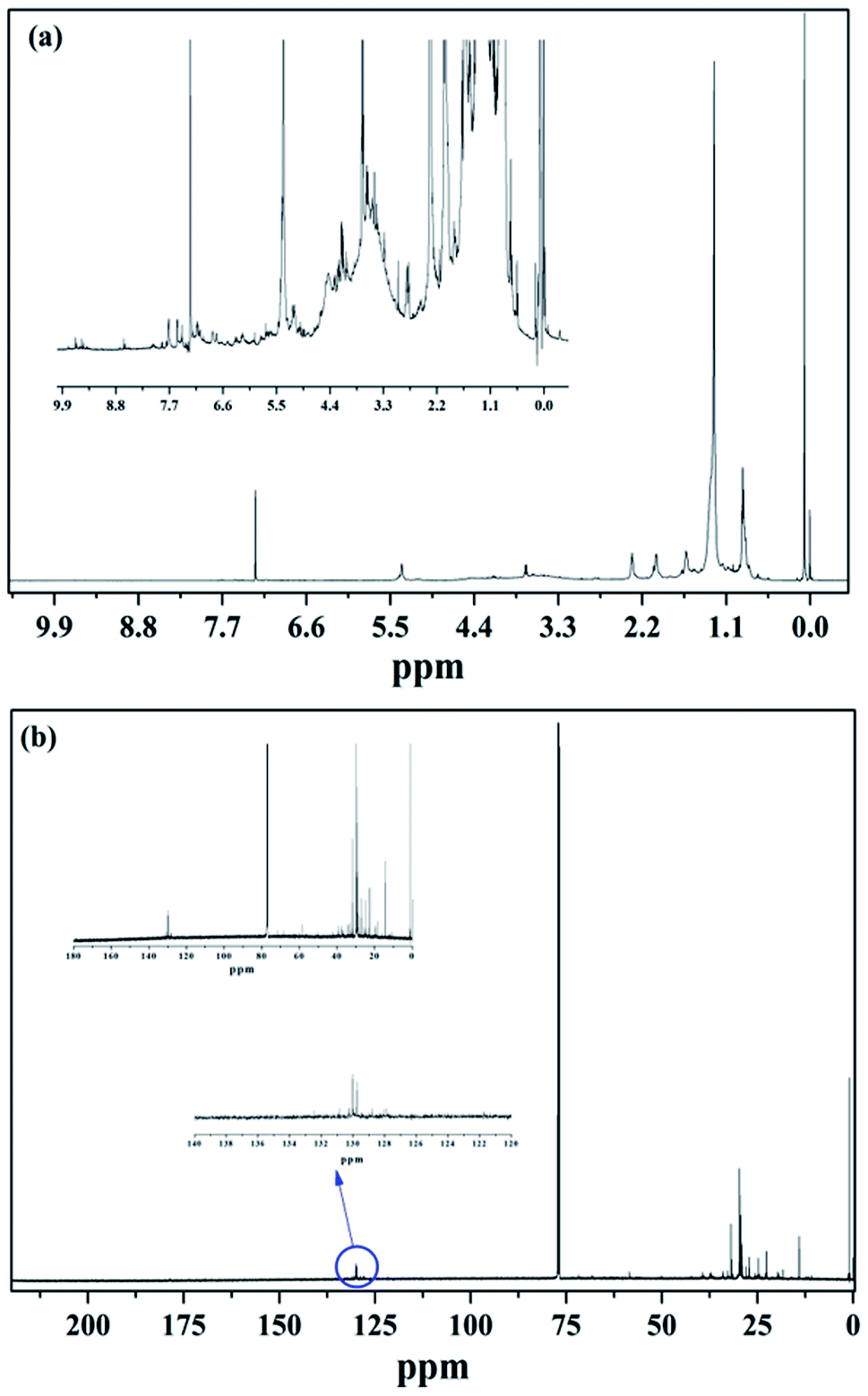 | ||
| Fig. 3 (a) 1H NMR spectra of CAEE in CDCl3 solvent (b) 13C NMR spectra of CAEE in CDCl3 solvent. | ||
Film characterization
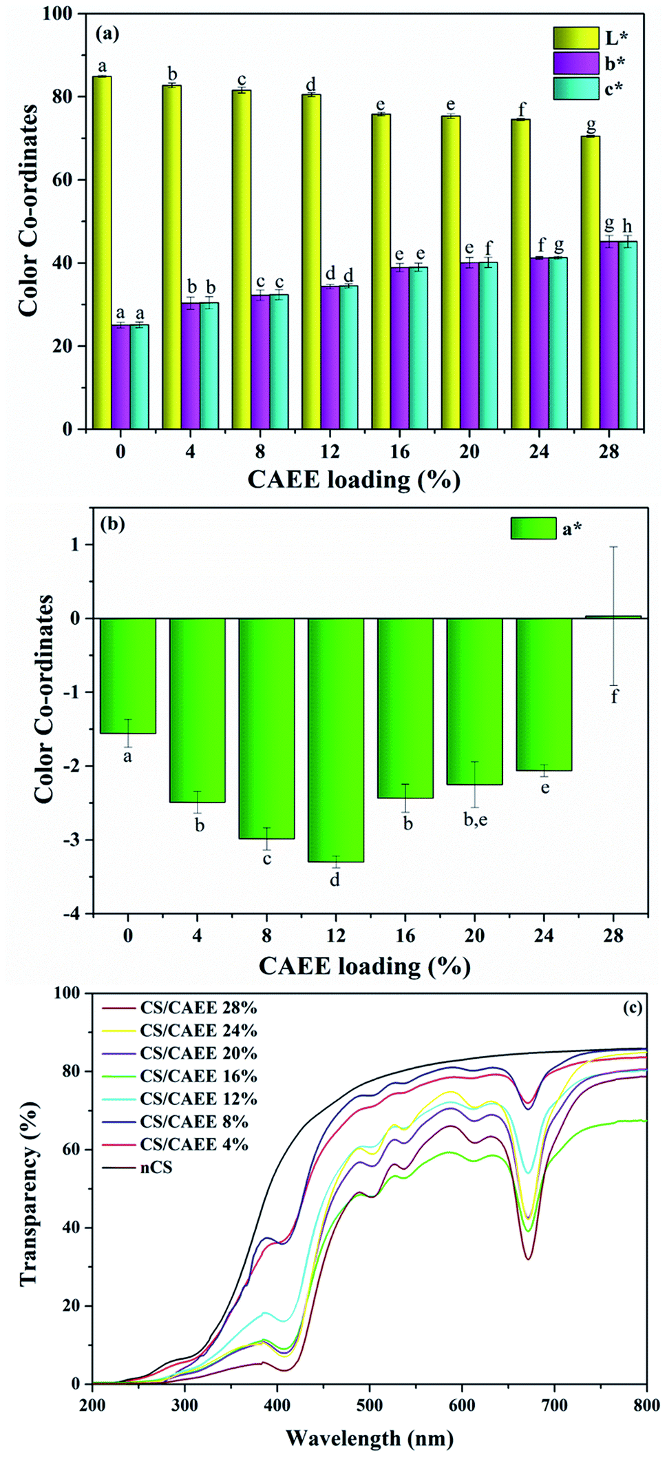 | ||
| Fig. 4 Color coordinate values of (a) L*, b*, and c* (b) a*, and (c) UV-Vis light transparency of the CAEE incorporated edible active films (biocomposites) along with the control film. | ||
 | ||
| Fig. 5 (a) Thermogravimetric, and (b) derivative thermogram of CAEE incorporated edible active films (bio-composites) along with control film, (c) FTIR spectrum of de-oil algae residue powder and CAEE, and (d) FTIR spectrum of developed bio-composite films with control. | ||
A two-step degradation was recorded in each of the samples (Table SI†). The first stage of degradation was recorded around 50–100 °C (about 5% decomposition for each film), which could be attributed to the loosely bound water evaporation from the polymeric structures. The second step of degradation was observed between 270–320 °C and it could correspond to the decomposition of CS molecules and CAEE.19 The predominant mass loss was recorded in the second degradation stage at around 270–305 °C and it was because of the denaturation of the CS-polymeric organization and biofiller. In the second stage, the biocomposite films showed a slight reduction in mass loss as compared with the nCS film, which could be due to the formation of strong and heavy interactions via hydrogen bonding between CS molecules with the phenolic compounds of CAEE.19 The temperatures at 5% (T5%) and 10% (T10%) mass loss are reported in Table SI,† where it can be seen that the biocomposite showed higher temperatures compared to the nCS film in both cases. Furthermore, the maximum temperature of the first degradation for the control film was ∼75.5 °C (Table SI†) whereas, the incorporation of CAEE increased the temperature to 106.8 °C and 113.8 °C in the biocomposites, which indicated an improved thermal stability of the biocomposites compared to the nCS film. Similar types of observation have been reported previously, where the incorporation of sweet potato extract, banana peel extract, and others tailored the thermal stability in developed biocomposites.19,38
O) group and –CS group the stretching vibrational.40 The other lower region peaks were assigned to C–N and –CS groups. The FTIR spectrum of CAEE (Fig. 5c) exhibited a broad absorption peak at ∼3337 cm−1 attributed to –OH stretching. However, the observed absorption bands in the de-oiled algae biomass residue powder and the extract were different. A strong absorption peak was observed at 2970 cm−1, corresponding to C–H symmetric stretching vibrations of the polyphenols. The presence of bands at 2884 and 1656 cm−1 might be attributed to ethanol and amide I, or CO stretching of proteins or carboxylic acid, respectively. The presence of bands at 1276, 1327, and 1380 cm−1 could be related to CH2 vibration, amide III, and symmetric CH3(CO) vibration of 1,8-cineole, respectively. Further, the possibility of the presence of polysaccharides could be attributed to the occurrence of bands at 1087 and 1045 cm−1. The observed band at 879 cm−1 corresponded to the –CH2 stretching vibration of 1,8-cineole or ethanol. Fig. 5d depicts the spectra for the nCS and developed biocomposite edible films. The nCS displayed a wide absorption peak centered at 3288 cm−1, which could be associated with stretching vibrations of N–H and O–H. The occurrence of peaks at 2922 and 1642 cm−1 might be attributed to C–H asymmetric stretching due to methylene groups and the CO stretching vibration of the residual amide bond. The absorption bands at 1538 and 1404 cm−1 corresponded to amide II and amide III, while the peak at 1152 cm−1 was attributed to C–H and 1064 cm−1 was ascribed to C–O stretching vibration.41Moreover, while the incorporation of CAEE into the CS matrix exhibited variations in intensity, the frequency was unalerted.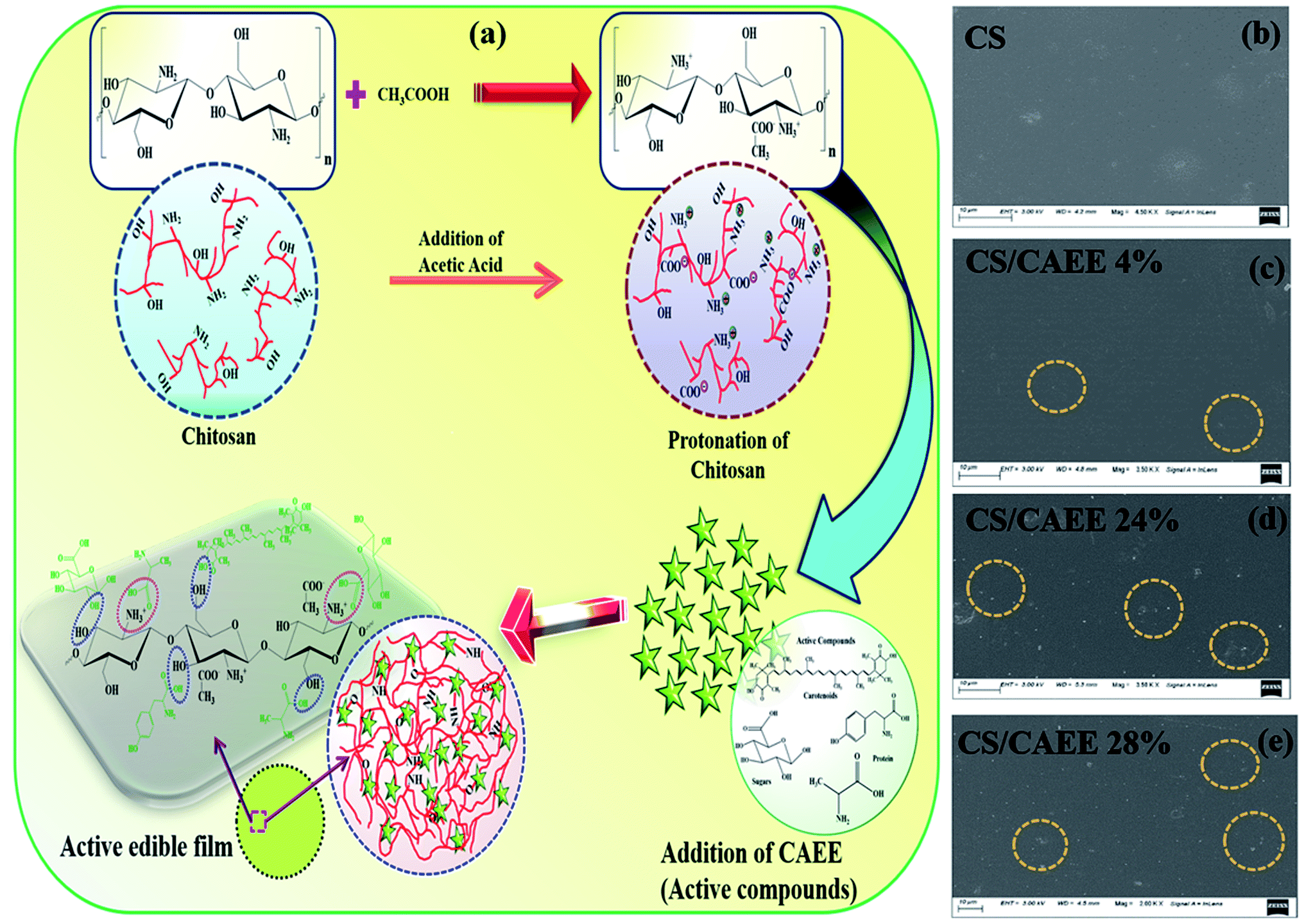 | ||
| Fig. 6 (a) Probable mechanism of the CS/CAEE biocomposite film formation, and (b–e) surface morphology of the developed biocomposite films compared with the control. | ||
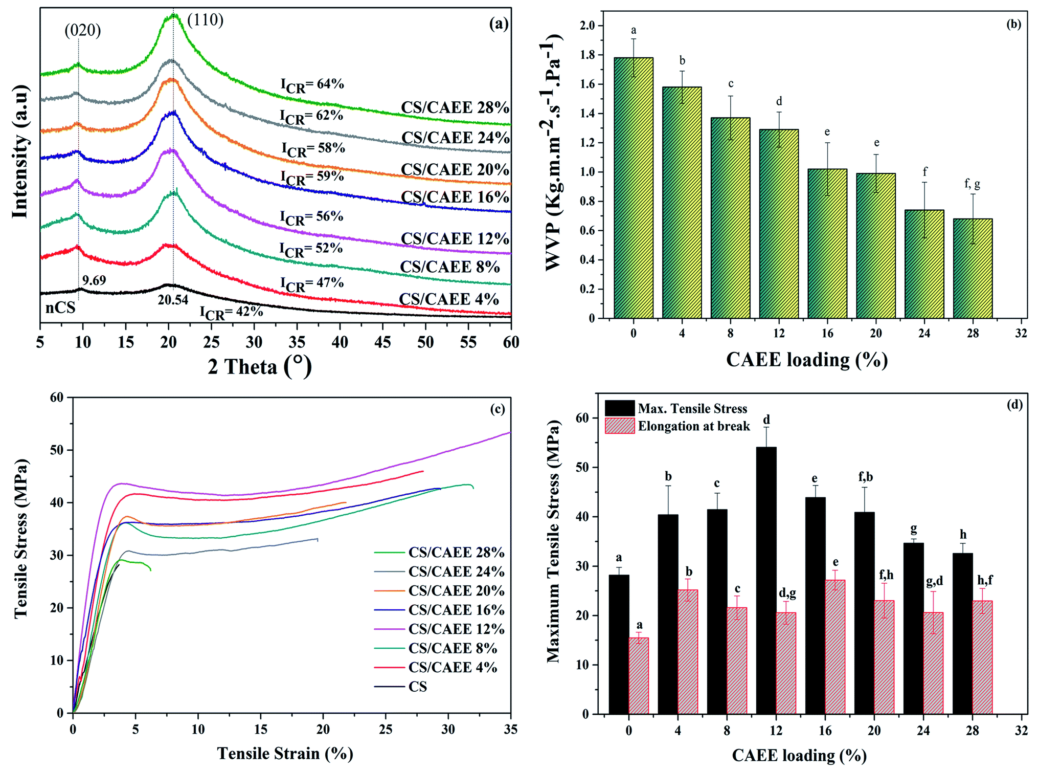 | ||
| Fig. 7 (a) XRD patterns, (b) water vapor permeability results, (c) stress–strain curves, and (d) maximum tensile stresses and elongations at break of the developed biocomposite films compared with the control. | ||
Rheological behavior
 | ||
| Fig. 8 (a) Strain sweep parameters of the G′ and G′′ moduli at f = 1 Hz, (b) emphasizing the LVR, (c) frequency sweep parameters of the G′ and G′′ moduli, and (d) flow curves of the developed biocomposite films together with the control. | ||
Overall, the elasticity (G′) decreased over time with increasing the shear strain for all the samples, including the control. However, the addition of CAEE significantly improved the elastic behavior compared to the control (nCS), which was more significant at higher strain. Further, a significant increase in  values with increasing the filler concentration from 4 to 20% was observed; whereas at a higher concentration of CAEE (28%), the
  dropped down, but not below the control value, as described in Table 1, at a fixed shear strain of 1%. Besides, at lower shear strain, a similar trend was visualized for all the samples, except for the higher loading of CAEE (28%), where a slight reduction of G′ was observed. Additionally, the non-linearity of the G′ and G′′ moduli under applied strain indicated the end limit of the LVR region.9 Moreover, the higher G′′ value of the CAEE-incorporated polymeric solution compared to the control solution indicated its viscous nature. The G′′ values showed a significant improvement from 0.97 (nCS) to 2.0 (CAEE 20%) Pa with increasing the CAEE concentration from 4 to 28%; however at 28% CAEE addition, G′′ decreased sharply, but it was still higher than the control. One of the important parameters of oscillatory rheology is the loss factor or loss tangent (tanδ = G′′/G′), which indicates the characteristics of the material; whereby tanδ > 1 indicates a viscous nature of the sample as G′′ > G′, while the sample is elastic when tanδ < 1 (G′ > G′′), and the sample is in an intermediate phase of a highly concentrated polymer solution and a real gel when tanδ > 0.1, but where the sample is not a true gel.46 The tanδ value of all the filmogenic solutions in the present study was greater than 1, which predicted a concentrated and viscous nature of the polymeric solution. The incorporation of CAEE into the polymeric matrix decreased the tanδ value for the 4% and 12% CAEE (4.14 and 3.61) with increasing the filler loading compared to the control (4.49), which was expected as the addition of these specified concentrations of CAEE improved the elasticity of the CS-based filmogenic solution. However, while the addition of 20% and 28% CAEE significantly increased the tanδ value, the
  value was significantly lower at this higher concentration (28%). The discussed parameters from the sweep strain measurements, supported by the G′′–G′ values at a specified shear strain of 1%, showed that the incorporation of CAEE increased the viscous property of the biocomposite filmogenic solutions. The strain sweep measurements showed the influence of the added filler CAEE on the rheological properties of the chitosan-based biocomposite solutions. The observed significant effects might be correlated to the major interaction between the phenolics groups of CAEE and the cationic groups (NH3+) of chitosan via hydrogen, covalent, and hydrophobic bonding. The effects were even more with increasing the biofiller content up to 20%. In some studies, no great effects on the rheological behavior were found after the addition of extracts like grape seed, and jabuticaba peel extract in the polymeric matrix due to the use of a small range of extracts.47 However, other studies showed the influence of the incorporated extract on the rheological properties of the chitosan matrix,46 like the results obtained from the present study.
 G′′ modulus at the LVR limit
 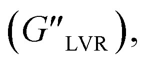 loss factor value (tanδ) and the difference between G′′ and G′ moduli at 1% strain
| Sample | γL (%) |
|
|
tanδ |
G′′–G′ (Pa) | Power-law model (n) | R2 |
|---|---|---|---|---|---|---|---|
| nCS | 31.6 | 0.21 | 0.97 | 4.49 | 0.75 | 0.32 | 0.996 |
| CS/CAEE 4% | 46.4 | 0.28 | 1.18 | 4.14 | 0.90 | 0.31 | 0.991 |
| CS/CAEE 12% | 68.1 | 0.43 | 1.56 | 3.61 | 1.13 | 0.37 | 0.990 |
| CS/CAEE 20% | 99.9 | 0.42 | 2.00 | 4.72 | 1.58 | 0.40 | 0.996 |
| CS/CAEE 28% | 147 | 0.17 | 1.22 | 6.88 | 1.05 | 0.23 | 0.994 |

 δ values (tanδ > 1) (see Table 2). The crossover frequency parameters, obtained from frequency sweep curves, are listed in Table 2. Between the nCS and CS/CAEE 4%, a shift in the crossover frequency (ωcrossover) was observed from 1 to 63.1 rad s−1 and an increased value of
  from 0.207 to 7.367 was also observed. No crossover point was noticed for the CS/CAEE 20% and CS/CAEE 28% samples.
 at the crossover point
| Sample | ωcrossover (rad s−1) |
|
|---|---|---|
| nCS | 1 | 0.207 |
| CS/CAEE 4% | 63.1 | 7.367 |
| CS/CAEE 12% | 0.6 | 0.205 |
| CS/CAEE 20% | — | — |
| CS/CAEE 28% | — | — |

Further, the incorporation of CAEE reduced the viscosity in the biocomposites compared to in nCS from 717.66 to 242.98 mPa s (Fig. 8d). The obtained results indicated that the addition of CAEE weakened the intramolecular polymeric bonds of chitosan and acetic acid. The overall viscosity was decreased with increasing the CAEE concentration; however, an improvement in viscosity was noticed in the case of the 12% and 20% loadings as compared to the 4% CAEE. However, this increment was overcome by the significant reduction of the viscosity with the highest CAEE (28%) concentration as compared to the control. As mentioned earlier, CAEE contains various phenolic compounds, such as carotenoids, pheophytin, chlorophyll, β-carotene, and astaxanthin, which loosen the bond interaction among the polymeric chains, as polyphenols are rich in hydroxyl groups (–OH), which could create space between the polymeric chains, thereby increasing the mobility of the chains by providing free space.48 In this context, similar observations of a reduction in viscosity with the addition of polyphenols, such as pomegranate seed extract,9 grape seed extract,46 mutra leaf extract,48 apple skin,49 and green tea extract,50 into CS-based film-forming solutions have been reported in the literature. Besides, the flow behavior index (n) obtained by plotting the viscosity as a function of the shear rate and using the power-law model indicates a pseudoplastic behavior when ‘n’ is between 0 to 1, with values beyond 1 following a Newtonian behavior.9 The ‘n’ values in this study ranged from 0.32–0.40 as shown in Table 1, indicating the pseudoplastic behavior of all the filmogenic solutions, which was corroborated by the observed steady flow behavior. Further, the increase in CAEE concentration in the polymeric solution led to increasing ‘n’ values, except at higher concentrations (28%), where a slight reduction was noticed. However, the addition of CAEE did not alter the pseudoplastic behavior of the filmogenic solutions.
 | ||
| Fig. 9 (a) Antioxidant activity, (b) total phenolic content, (c) antimicrobial activity, and (d) biocompatibility (MTT assay) of the developed biocomposite films compared with the control. | ||
The total phenolic content (TPC) of the CS/CAEE biocomposite films was determined by the Folin–Ciocalteu phenol reagent method. The TPCs of the films are shown in Fig. 9b. The control film exhibited a TPC of 97.96 μg gallic acid/g film. Moreover, an increasing TPC was observed in the biocomposite with increasing the CAEE loading. The highest TPC obtained in the biocomposites was 423.50 μg gallic acid/g film, which was ∼38 times greater than that of the control. Therefore, it can be postulated that the presence of phenolic compounds was responsible for the desirable DPPH antioxidant activity in the films and which showed an increasing trend with enhancing the biofiller (CAEE) concentration, corroborating the results obtained for the DPPH antioxidant activity.54
Coating efficiency effect on the physiological characteristics of green chilli
 | ||
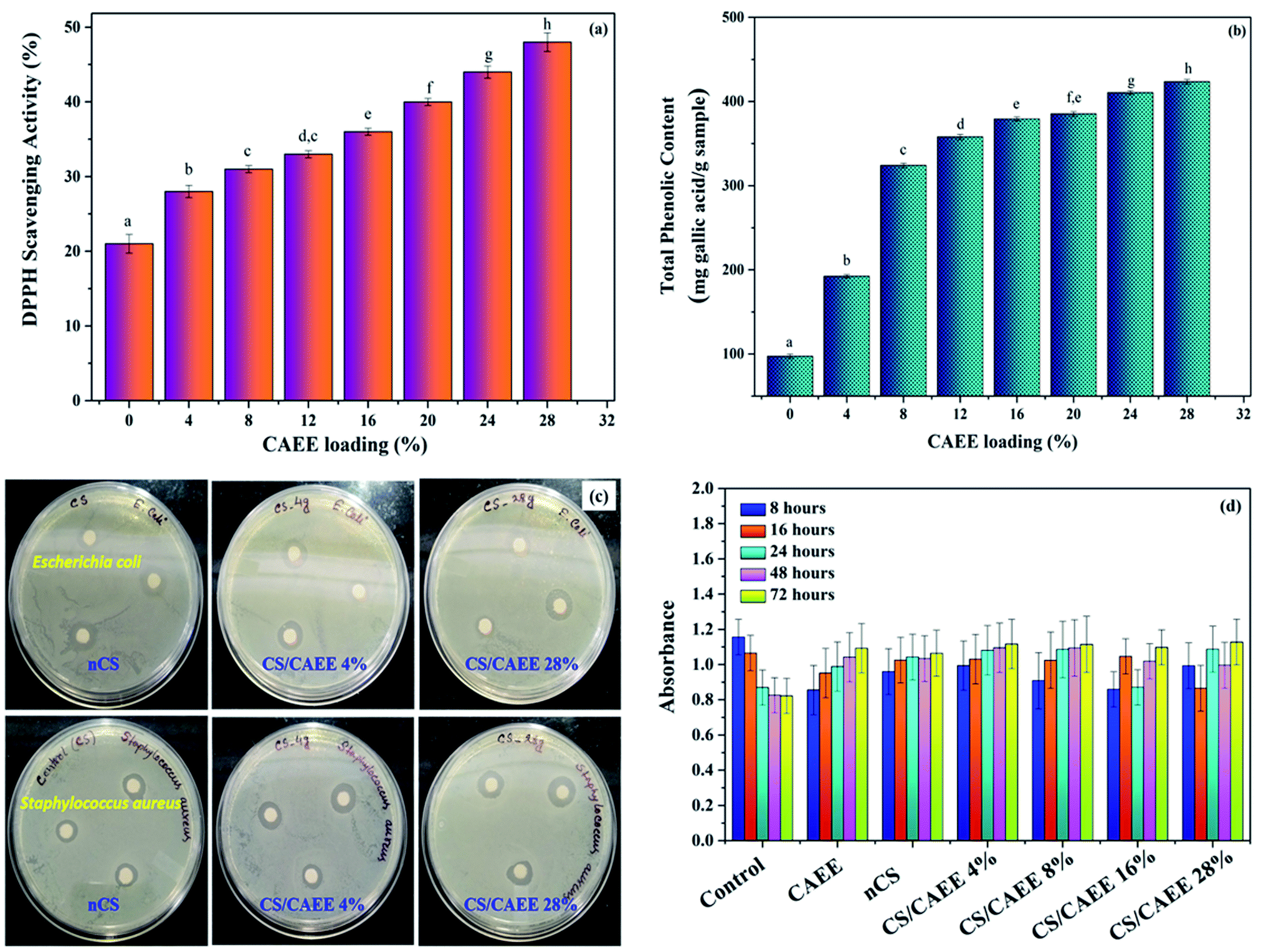
| Fig. 10 (a) Physiological weight loss, (b) visual observation, (c) polarized optical microscopic observation, and (d) firmness of stored coated and control green chilli during ambient storage. | ||
It is crystal clear that the control sample exhibited surface dryness and a wilting effect during storage under ambient conditions, whereas the coated samples appeared to be fresh. However, a higher concentration of CAEE (28%) was less effective compared to a lower concentration, which might be due to the agglomeration of CAEE particles inside the polymer matrix. Further, the microstructure of the stored samples was captured under a polarized optical microscope (POM), where surface roughness was observed in the control compared to the coated samples (Fig. 10c). Although, some particles were also visualized over the CS/CAEE-coated samples due to the addition of CAEE.
Conclusion
The present study involved the successful fabrication of chitosan-based edible active biocomposites incorporated with CAEE as a biofiller as a secondary packaging material and application of a formulated filmogenic solution as a coating material on a real food system (fresh produce). The CAEE was obtained by the ultrasound-assisted sustainable extraction of waste de-oil green algae biomass (Dunaliella tertiolecta). The extract, CAEE, was rich in antioxidants and various bioactive compounds, predominantly carotenoids, proteins, and polysaccharides. The properties of the fabricated active edible films with varying CAEE contents were analyzed and compared with the control film. The fabricated edible films displayed a superior antioxidant activity, total phenolic content, water vapor barrier property, thermal stability, and mechanical strength. An excellent UV-Vis light-blocking property was observed in the developed edible active film due to the strong interaction of polyphenols with the polymer matrix. The rheological property of the filmogenic solutions exhibited a stable shear thinning (viscous liquid) behavior. The cell line study suggested the developed coating material was biocompatible and nontoxic. Finally, green chillies were coated with the filmogenic solution as a primary packaging and stored under ambient conditions, and a prolonged shelf life of the coated green chilli was demonstrated compared to the control. Besides, coated chilli packed edible pouches made by the developed edible films as secondary packaging material preserved the chillies for a longer period. The promising outcomes from this study suggest that the fabricated edible films could be used as edible active pouches and at the same time the developed filmogenic solution could also be used as a coating material for extending the shelf life of fresh produce.Author contributions
Kona Mondal: data curation, formal analysis, investigation, original draft writing, review, and editing. Sayan Bhattacharjee: experimental, and original draft. Chethana Mudenur: experimental. Tabli Ghosh: experimental. Vaibhav V. Goud: supervision, and original draft. Vimal Katiyar: conceptualization; funding acquisition, project administration, resources, supervision, validation, visualization, review, and editing.Conflicts of interest
The authors declare no conflicts of interest.Acknowledgements
The authors are sincerely thankful to the Centre of Excellence for Sustainable Polymers (CoE-SusPol), Centre for Sustainable Polymers (CSP), and CIF (Center Instrument Facility) at the Indian Institute of Technology Guwahati (IITG) for providing all the possible research and experimental facilities during this work. We are also thankful to TERI-DBT (The Energy and Resource Institute) CoE on Bio-fuels and Bio-commodities (xCLESPNxDBT00860xxVK010, DBT, GOI) for providing raw materials for this work.Notes and references
- S. Remya, C. O. Mohan, J. Bindu, G. K. Sivaraman, G. Venkateshwarlu and C. N. Ravishankar, J. Food Sci. Technol., 2016, 53, 685–693 CrossRef CAS PubMed.
- U. Siripatrawan and B. R. Harte, Food Hydrocolloids, 2010, 24, 770–775 CrossRef CAS.
- D. Moreira, B. Gullón, P. Gullón, A. Gomes and F. Tavaria, Food Funct., 2016, 7, 3273–3282 RSC.
- A. R. Deshmukh, H. Aloui, C. Khomlaem, A. Negi, J. H. Yun, H. S. Kim and B. S. Kim, Food Chem., 2021, 337, 127777 CrossRef CAS PubMed.
- J. Deng, E. Q. Zhu, G. Xu, N. Naik, V. Murugadoss, M. G. Ma and Z. Shi, Green Chem., 2021, 24, 480–492 RSC.
- M. S. Nair, A. Saxena and C. Kaur, Food Bioprocess Technol., 2018, 11, 1317–1327 CrossRef CAS.
- K. Mondal, T. Ghosh, P. Bhagabati, and V. Katiyar, Sustainable Nanostructured Materials in Food Packaging, Dynamics of Advanced Sustainable Nanomaterials and their Related Nanocomposites at the Bio-Nano Interface, Elsevier, 2019, pp. 171–213 Search PubMed.
- P. K. Dutta, S. Tripathi, G. K. Mehrotra and J. Dutta, Food Chem., 2009, 114, 1173–1182 CrossRef CAS.
- M. R. Bertolo, V. C. Martins, M. M. Horn, L. B. Brenelli and A. M. Plepis, Carbohydr. Polym., 2020, 228, 115386 CrossRef CAS PubMed.
- T. Ghosh, Y. Teramoto and V. Katiyar, J. Agric. Food Chem., 2019, 67, 4289–4299 CrossRef CAS PubMed.
- S. M. Ojagh, M. Rezaei, S. H. Razavi and S. M. H. Hosseini, Food Chem., 2010, 122, 161–166 CrossRef CAS.
- F. Nowzari, B. Shábanpour and S. M. Ojagh, Food Chem., 2013, 141, 1667–1672 CrossRef CAS PubMed.
- T. C. Dos Santos, N. Rescignano, L. Boff, F. H. Reginatto, C. M. O. Simões, A. M. de Campos and C. U. Mijangos, Carbohydr. Polym., 2017, 173, 638–644 CrossRef PubMed.
- C. O. Ferreira, C. A. Nunes, I. Delgadillo and J. A. Lopes-da-Silva, Food Res. Int., 2009, 42, 807–813 CrossRef CAS.
- N. K. Kalita, M. K. Nagar, C. Mudenur, A. Kalamdhad and V. Katiyar, Polym. Test., 2019, 76, 522–536 CrossRef CAS.
- W. Lan, R. Zhang, S. Ahmed, W. Qin and Y. Liu, LWT, 2019, 113, 108297 CrossRef CAS.
- U. Bhardwaj, P. Dhar, A. Kumar and V. Katiyar, Am. Chem. Soc., 2014, 275–314 CAS.
- H. Akretche, G. Pierre, R. Moussaoui, P. Michaud and C. Delattre, Green Chem., 2019, 21, 3065–3073 RSC.
- W. Zhang, X. Li and W. Jiang, Int. J. Biol. Macromol., 2020, 154, 1205–1214 CrossRef CAS PubMed.
- A. Silva-Weiss, V. Bifani, M. Ihl, P. J. A. Sobral and M. C. Gómez-Guillén, Food Hydrocolloids, 2013, 31, 458–466 CrossRef CAS.
- X. Zhang, H. Lian, J. Shi, W. Meng and Y. Peng, Int. J. Biol. Macromol., 2020, 148, 1242–1250 CrossRef CAS PubMed.
- T. Liu, L. Liu, X. Gong, F. Chi and Z. Ma, LWT, 2021, 135, 110181 CrossRef CAS.
- J. M. Lorenzo, P. E. Munekata, A. S. Sant'Ana, R. B. Carvalho, F. J. Barba, F. Toldrá and M. A. Trindade, Trends Food Sci. Technol., 2018, 77, 1–10 CrossRef CAS.
- S. Zou, Y. Wu, M. Yang, C. Li and J. Tong, Energy Fuel., 2009, 23, 3753–3758 CrossRef CAS.
- A. S. Sarpal, C. M. Teixeira, S. Mesquita, I. C. Costa and R. M. S. Paulo, Adv. Appl. Microbiol., 2018, 11, 555812 Search PubMed.
- A. S. Sarpal, C. M. Teixeira, P. R. M. Silva, T. V. da Costa Monteiro, J. I. da Silva, V. S. da Cunha and R. J. Daroda, Appl. Microbiol. Biotechnol., 2016, 100, 2471–2485 CrossRef CAS PubMed.
- P. M. Foley, E. S. Beach and J. B. Zimmerman, Green Chem., 2011, 13, 1399–1405 RSC.
- K. Mondal, S. Sakurai, Y. Okahisa, V. V. Goud and V. Katiyar, Carbohydr. Polym., 2021, 261, 117881 CrossRef CAS PubMed.
- K. Mondal, P. Bhagabati, V. V. Goud, S. Sakurai and V. Katiyar, Int. J. Biol. Macromol., 2021, 191, 521–530 CrossRef CAS PubMed.
- A. Hosikian, S. Lim, R. Halim and M. K. Danquah, Int. J. Chem. Eng., 2010, 2010 Search PubMed.
- M. J. Iglesias, R. Soengas, I. Probert, E. Guilloud, P. Gourvil, M. Mehiri and F. L. Ortiz, Phytochemistry, 2019, 164, 192–205 CrossRef PubMed.
- R. Nicolau, M. Leloup, D. Lachassagne, E. Pinault and G. Feuillade-Cathalifaud, Talanta, 2015, 136, 102–107 CrossRef CAS PubMed.
- P. D. Fraser, E. M. Enfissi, M. Goodfellow, T. Eguchi and P. M. Bramley, Plant J., 2007, 49, 552–564 CrossRef CAS PubMed.
- A. Mishra, K. Kavita and B. Jha, Carbohydr. Polym., 2011, 83, 852–857 CrossRef CAS.
- K. Tsukida, K. Saiki and M. Sugiura, J. Nutr. Sci. Vitaminol., 1981, 27, 551–561 CrossRef CAS PubMed.
- L. Wang, Y. Dong, H. Men, J. Tong and J. Zhou, Food Hydrocolloids, 2013, 32, 35–41 CrossRef.
- X. Zhang, Y. Liu, H. Yong, Y. Qin, J. Liu and J. Liu, Food Hydrocolloids, 2019, 94, 80–92 CrossRef CAS.
- H. Yong, X. Wang, R. Bai, Z. Miao, X. Zhang and J. Liu, Food Hydrocolloids, 2019, 90, 216–224 CrossRef CAS.
- A. R. Deshmukh, J. W. Jeong, S. J. Lee, G. U. Park and B. S. Kim, ACS Sustainable Chem. Eng., 2019, 7, 17114–17125 CrossRef CAS.
- A. R. Deshmukh, H. Aloui and B. S. Kim, J. Cleaner Prod., 2020, 270, 122339 CrossRef CAS.
- M. Bajić, T. Ročnik, A. Oberlintner, F. Scognamiglio, U. Novak and B. Likozar, Food Packag. Shelf Life, 2019, 21, 100365 CrossRef.
- S. U. Kadam, S. K. Pankaj, B. K. Tiwari, P. J. Cullen and C. P. O'Donnell, Food Packag. Shelf Life, 2015, 6, 68–74 CrossRef.
- P. Hernández-Muñoz, A. López-Rubio, J. M. Lagarón and R. Gavara, Biomacromolecules, 2004, 5, 415–421 CrossRef PubMed.
- A. Gupta, A. K. Pal, E. M. Woo and V. Katiyar, Sci. Rep., 2018, 8, 1–13 Search PubMed.
- E. Chiellini, P. Cinelli, V. I. Ilieva and M. Martera, Biomacromolecules, 2008, 9, 1007–1013 CrossRef CAS PubMed.
- M. Romanelli Vicente Bertolo, V. da Conceicao Amaro Martins, A. M. de GuzziPlepis and S. Bogusz Jr, J. Appl. Polym. Sci., 2021, 138, 50052 CrossRef.
- M. Á. V. Rodrigues, M. R. V. Bertolo, C. A. Marangon, V. D. C. A. Martins and A. M. de GuzziPlepis, Int. J. Biol. Macromol., 2020, 160, 769–779 CrossRef CAS PubMed.
- A. Silva-Weiss, V. Bifani, M. Ihl, P. J. A. Sobral and M. C. Gómez-Guillén, J. Food Eng., 2014, 140, 28–38 CrossRef CAS.
- W. X. Du, C. W. Olsen, R. J. Avena-Bustillos, M. Friedman and T. H. McHugh, J. Food Sci., 2011, 76, M149–M155 CrossRef CAS PubMed.
- Y. Peng, Q. Wang, J. Shi, Y. Chen and X. Zhang, Food Sci. Technol., 2019, 40, 162–170 CrossRef.
- M. Kaya, P. Ravikumar, S. Ilk, M. Mujtaba, L. Akyuz, J. Labidi and S. K. Erkul, Innovative Food Sci. Emerging Technol., 2018, 45, 287–297 CrossRef CAS.
- K. I. Priyadarsini, D. K. Maity, G. H. Naik, M. S. Kumar, M. K. Unnikrishnan, J. G. Satav and H. Mohan, Free Radicals Biol. Med., 2003, 35, 475–484 CrossRef CAS PubMed.
- E. Talón, K. T. Trifkovic, V. A. Nedovic, B. M. Bugarski, M. Vargas, A. Chiralt and C. González-Martínez, Carbohydr. Polym., 2017, 157, 1153–1161 CrossRef PubMed.
- M. Jouki, F. T. Yazdi, S. A. Mortazavi and A. Koocheki, Food Hydrocolloids, 2014, 36, 9–19 CrossRef CAS.
- S. Ben-Yehoshua, and V. Rodov, Transpiration and Water Stress, Postharvest Physiology and Pathology of Vegetables, CRC Press, 2002, pp. 143–197 Search PubMed.
- O. P. Chauhan, C. Nanjappa, N. Ashok, N. Ravi, N. Roopa and P. S. Raju, J. Food Sci. Technol., 2015, 52, 1200–1205 CrossRef CAS PubMed.
- K. Chitravathi, O. P. Chauhan and P. S. Raju, Postharvest Biol. Technol., 2014, 92, 146–148 CrossRef CAS.
- K. Chitravathi, O. P. Chauhan, P. S. Raju and N. Madhukar, Food Bioprocess Technol., 2015, 8, 1386–1392 CrossRef CAS.
- K. Chitravathi, O. P. Chauhan and P. S. Raju, J. Food Sci. Technol., 2016, 53, 3320–3328 CrossRef CAS PubMed.
- Y. Xing, X. Li, Q. Xu, J. Yun, Y. Lu and Y. Tang, Food Chem., 2011, 124, 1443–1450 CrossRef CAS.
- M. I. Minguez-Mosquera and D. Hornero-Mendez, J. Agric. Food Chem., 1994, 42, 1555–1560 CrossRef CAS.
- R. J. Cogdell, T. D. Howard, R. Bittl, E. Schlodder, I. Geisenheimer and W. Lubitz, Philos. Trans. R. Soc. London, Ser. B, 2000, 355, 1345–1349 CrossRef CAS PubMed.
- P. S. Tanada-Palmu and R. F. G. Carlos, Postharvest Biol. Technol., 2005, 36.2, 199–208 CrossRef.
- K. Godlewska, I. Michalak, Ł. Tuhy and K. Chojnacka, BioMed Res. Int., 2017, 2017, 7248634 Search PubMed.
- S. Dudonné, P. Poupard, P. Coutiere, M. Woillez, T. Richard, J. M. Mérillon and X. Vitrac, J. Agric. Food Chem., 2011, 59, 4527–4536 CrossRef PubMed.
- C. M. Bitencourt, C. S. Fávaro-Trindade, P. D. A. Sobral and R. A. Carvalho, Food Hydrocolloids, 2014, 40, 145–152 CrossRef CAS.
- Y. Fang, M. A. Tung, I. J. Britt, S. Yada and D. G. Dalgleish, J. Food Sci., 2002, 67, 188–193 CrossRef CAS.
- N. Limchoowong, P. Sricharoen, S. Techawongstien and S. Chanthai, Food Chem., 2016, 200, 223–229 CrossRef CAS PubMed.
Footnote |
| † Electronic supplementary information (ESI) available: Supporting figures and tables. See https://doi.org/10.1039/d2ra00949h |
| This journal is © The Royal Society of Chemistry 2022 |