 Open Access Article
Creative Commons Attribution 3.0 Unported Licence
Surface-mediated assembly, polymerization and degradation of thiophene-based monomers†
G.
Galeotti‡
 ab,
F.
De Marchi‡
 a,
T.
Taerum
c,
L. V.
Besteiro
 ad,
M.
El Garah
a,
J.
Lipton-Duffin
 e,
M.
Ebrahimi§
 *a,
D. F.
Perepichka
 *c and
F.
Rosei
 *ad
*ad
aCentre Energie, Matériaux et Télécommunications, Institut National de la Recherche Scientifique, 1650 Boulevard Lionel-Boulet, Varennes, Québec, Canada J3X 1S2. E-mail: maryam.ebrahimi@emt.inrs.ca; rosei@emt.inrs.ca
bIstituto di Struttura della Materia, CNR, Via Fosso del Cavaliere 100, 00133 Roma, Italy
cDepartment of Chemistry, McGill University, 801 Sherbrooke Street West, Montreal, Quebec, Canada H3A 0B8. E-mail: dmitrii.perepichka@mcgill.ca
dInstitute of Fundamental and Frontier Science, University of Electronic Science and Technology of China, Chengdu 610054, PR China
eInstitute for Future Environments, Queensland University of Technology (QUT), 2 George Street, Brisbane, 4001 QLD, Australia
First published on 16th April 2019
Abstract
Ullmann coupling of halogenated aromatics is widely used in on-surface synthesis of two-dimensional (2D) polymers and graphene nanoribbons. It stands out among other reactions for regioselectively connecting aromatic monomers into 1D and 2D π-conjugated polymers, whose final structure and properties are determined by the initial building blocks. Thanks to their exceptional electronic properties, thiophene-containing monomers are frequently used for the synthesis of various conjugated materials. On the other hand, their use in on-surface polymerization is hampered by the possibility of ring opening when adsorbed on metal surfaces. In the present work, we mapped the temperature regime for these two competing reactions by investigating the adsorption of a thiophene-based prochiral molecule using scanning tunneling microscopy, X-ray photoelectron spectroscopy and density functional theory calculations. We followed the formation of organometallic (OM) networks, their evolution into covalent structures and the competition between C–C coupling and thiophene ring opening. The effect of surface reactivity was explored by comparing the adsorption on three (111) coinage metal substrates, namely Au, Ag and Cu. While outlining strategies to minimize the ring opening reaction, we found that the surface temperature during deposition is of paramount importance for the preparation of 2D OM networks, greatly enhancing the overall ordering of the product by depositing on hot Ag surface. Notably, the same protocol permits the creation of OM structures on the air-stable Au surface, thereby allowing the synthesis and application of 2D OM networks outside the ultra-high vacuum environment.
Introduction
Two-dimensional (2D) materials have been extensively studied due to their unprecedented properties arising from reduced dimensionality.1 Thanks to their tunable band gap, π-conjugated 2D polymers2,3 constitute an emerging class of 2D materials with potential applications in nanoelectronics and optoelectronics.4–6 However, their synthesis is challenging due to the difficulty in controlling the dimensionality during conventional solution-based polymerization, while chemical vapor deposition methods7 require an extremely high chemoselectivity considering the molecular complexity of the monomers.8,9One promising approach to synthesize 2D polymers is based on the self-assembly of reactive molecules on atomically flat surfaces,10 followed by polymerization.11 Several reactions have been explored for this purpose, including Sonogashira coupling of haloaromatic precursors with terminal alkynes,12 oxidative terminal alkyne (Glaser–Hay) coupling,13 photo/electro-initiated polymerization of diacetylene derivatives,11 decarboxylative coupling14 and electro-oxidative polymerization of thiophenes.15 Among these, on-surface Ullmann coupling is the most broadly applicable and frequently used reaction for the synthesis of 2D π-conjugated polymers.16–20 The reaction is catalyzed by coinage metal (Cu, Ag, and Au) surfaces and involves the dissociation of a carbon–halogen bond resulting in an organometallic (OM) phase21,22 which is followed by carbon–carbon coupling.23 The main reasons for the widespread use of Ullmann coupling are its efficiency and wide scope. By changing the structure of the monomer and the position of the halogen along the molecular backbone, it is possible to control the polymer's dimensionality and topology.19
The required polymerization temperature is dictated by the chosen substrate and halogen, and a multistep hierarchical growth process can be engineered by including different types of halogens.24,25 The substrate affects both the thermodynamics of the system and the reaction kinetics, driving the reaction along different pathways, and changing the relative stability of its intermediates (e.g., destabilizing the OM structures).26,27
A variety of building blocks have been used in the on-surface synthesis of π-conjugated 2D polymers. The structure of the monomers and their network connectivity control electron delocalization, band gap and charge carrier mobility.28–30 Incorporating heteroatoms into the monomer's backbone is a well-established approach to modify the polymer's properties, and a useful tool in tuning the doped carbon materials towards applications in catalysis, energy and semiconducting devices.31 In this context, thiophene-containing monomers are particularly promising for the synthesis of π-conjugated 2D polymers, due to their synthetic feasibility, structural diversity, highly efficient π-conjugation, and already well established applications of solution-processable (1D) polythiophenes in semiconducting devices.32,33
Nevertheless, on-surface polymerization of thiophene monomers via Ullmann coupling is challenging. In fact, the sulphur–metal interaction is found to be too strong on nickel and copper surfaces, resulting in partial or complete scission of C–S bonds.18,34–36 On the other hand, it was recently reported that 2,5-dichloro(3,4-ethylenedioxythiophene) could be polymerized into linear PEDOT without ring openings on the Ag surface (but not on Cu or Au).37 Thus, the extent of desulfurization and its dependence on the substrate, the molecular structure and the reaction temperature need to be understood in order to access well-defined sulfur-doped carbon nanomaterials.
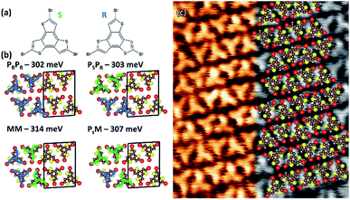
Here we present a comparative study of self-assembly and two-step reactivity of a tridentate tribromoterthienobenzene (TBTTB, Fig. 1a) on Au(111), Ag(111) and Cu(111), using scanning tunneling microscopy (STM) and X-ray photoelectron spectroscopy (XPS), supported by density functional theory (DFT) calculations. The terthienobenzene (TTB) core38 of this monomer presents a higher S/C ratio in comparison to other previously studied monomers,18,39,40 and has been used as a building block in several semiconducting materials,41 including star-shaped oligomers,42 linear conjugated polymers,43 and 3D microporous polymers.44–46
By depositing the molecules over a substrate kept at room temperature (RT) we obtain 2D self-assembled molecular networks on Au, and OM structures on Ag and Cu. Our results demonstrate that while annealing at elevated temperatures is necessary to trigger Ullmann coupling, it also causes the adsorbates to undergo competing ring opening reactions, due to the high affinity of sulphur with coinage metals and relatively low C–S bond dissociation energy (3.24 eV for C–S vs. 3.05 eV for C–Br).47 We map out the temperature dependence of these two competing reactions on the three investigated surfaces, and show that the activation barrier for the ring opening exceeds the barrier for dehalogenation for all three surfaces.26,48
By depositing directly on a heated substrate, we show that it is possible to improve the overall ordering of the 2D OM network on Ag and to obtain similar hexagonal networks on Au. While common for Ag and Cu surfaces,27,49 Ullmann coupling on Au rarely yields stable OM intermediates. Although short 1D Au-bridged OM chains have been observed in recent studies,50–53 the formation of 2D OM networks on gold has not been previously reported.
Results and discussion
TBTTB on Au(111)
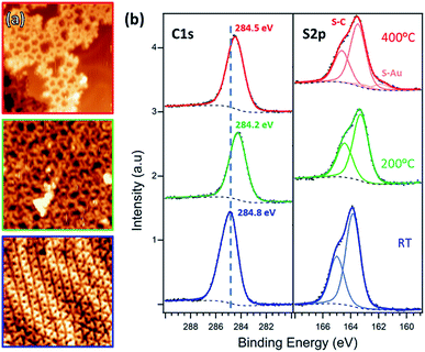
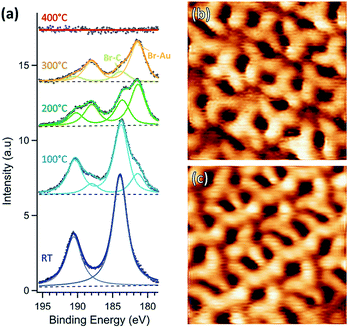
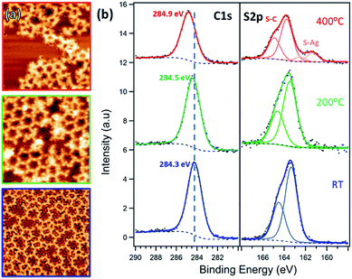
After deposition on Au at RT, the molecules organize into a close-packed 2D row structure (Fig. 1c). The unit cell, with dimensions of (1.73 ± 0.05) × (2.45 ± 0.05) nm2 and an angle of 87 ± 3° between two vectors, contains four molecules in two rows of alternating R/S enantiomers (the identification of the enantiomers is described in the ESI, Section 1†). To confirm this assignment, we performed DFT gas phase calculations (PBE-GGA with D3 dispersion correction), calculating all the possible combinations of enantiopure (Ps or PR) and racemic (M) rows (Fig. 1b and S1†). The total cohesive energies indicate that the racemic networks are slightly more stable (ΔE ≈ 0.05 eV per unit cell) than the homo-enantiomeric assemblies. As such, the simulations point toward the MM combination as the most stable assembly, as inferred from STM images. The herringbone reconstruction of the Au(111) surface is maintained (Fig. 2a, blue frame), consistent with weak molecule–substrate interactions.50,54 XPS analysis shows a Br 3p doublet at binding energies (BEs) of 183.7 eV (Br 3p3/2) and 190.2 eV (Br 3p1/2) and a S 2p doublet at BEs of 163.9 eV (S 2p3/2) and 165.1 eV (S 2p1/2), as expected for Br–C and S–C bonds, confirming that the molecules are intact (Fig. 2b and 3a).18Annealing the chiral phase on Au(111) reduces the level of order of the molecular network. Starting from 100 °C, additional arrangements are observed to coexist with the intact RT phase (Fig. 3b, c and S2†). XPS shows the emergence of a new Br 3p3/2 peak at BE of 181.4 eV (Br–Au), due to partial Br–C dissociation (Fig. 3a). We infer that the new arrangements are produced by the presence of partially debrominated monomers and the dissociated Br atoms that locally perturb the self-assembled structures (Fig. 3b and c). As the temperature increases, the Br–Au peak grows at the expense of the Br–C signal, while Br atoms progressively desorb leaving the surface bromine-free at 400 °C (Fig. 3c).
After annealing for 30 minutes at 200 °C, this gradual dehalogenation results in the formation of disordered OM networks (Fig. 2a, green frame and S3†), with the C and S peaks shifting toward lower BEs (S 2p3/2 at 163.5 eV, C 1s at 284.2 eV). These changes in the core levels can be ascribed to the increased electron density on the emitter atoms after binding to electropositive Au atoms.17,55
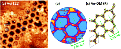
OM networks were further investigated by direct deposition on a surface held at 200 °C. When compared to the structures obtained by the annealing of the RT-deposited sample, XPS data show no chemical difference between the two cases (Fig. S4†). However, the STM images reveal striking differences, with small domains of well-ordered morphologies appearing in the heated surface (Fig. 4a).
When dosed on a heated surface (200 °C), the dehalogenated molecules do not react with their immediate neighbours but possess sufficient thermal energy and time to diffuse to optimal positions, permitting the formation of a low-density hexagonal structure. This observation is consistent with the previous work56 for the Ullmann reaction of dibromo-m-terphenyl on Cu, where the formation of macrocycles was favoured by low deposition rates.
DFT calculations were performed to simulate the observed OM hexagonal phase. The center-to-center distance measured in STM images (1.95 ± 0.10 nm, Fig. 4b) is in agreement with a calculated OM structure (1.93 nm, Fig. 4c), and is considerably larger than expected for a polymer structure (1.48 nm, Fig. S5†). The STM images also exhibit bright spots between every vertex (Fig. 4), attributed to a bridging Au atom.
Post-annealing at various temperatures up to 400 °C (or direct deposition at 300 °C) does not further improve the order of the OM phase, but instead produces a disordered phase (Fig. 2a, red frame, and S6†). The XPS spectra show a shift toward higher BE, i.e. 163.3 eV for S 2p3/2 and 284.5 eV for C 1s, consistent with the expected depletion of C–Au and formation of C–C bonds.17 However, the simultaneous appearance of the Au-related S 2p3/2 peak at 161.8 eV suggests that a fraction of the thiophene rings are open. By comparing the size of the two S 2p components, we estimate that a 10% of the thiophenes present broken C–S bonds. Moreover, there is also an overall decrease of both C 1s and S 2p peak areas as the annealing temperature increases (ESI, Section 2†).
TBTTB on Ag(111)
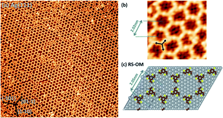
Deposition of TBTTB on silver at RT immediately yields an OM network (Fig. 5a, blue frame) composed of both open and closed polygons, with 4 to 8 vertices and irregular shapes. The order of the molecular phase increases with annealing until it forms a hexagonal closed structure above 200 °C (Fig. 5a, green frame). This suggests that the dynamic nature of the C–Ag bond is an essential feature for the network's self-assembly and “self-repair”,57,58 whereas this was not observed on gold, possibly because of the higher bond dissociation energy of the C–Au bond (1.39 eV for C–Ag vs. 1.99 eV for C–Au).47 This ordering does not occur in experiments starting at saturated coverage with subsequent annealing, presumably because molecular diffusion is hindered at higher densities. STM images at submolecular resolution reveal a random distribution of the R and S enantiomers in the porous OM networks (Fig. S10†). This difference with the close-packed self-assembled molecular networks on Au(111) is consistent with the variation in chiral expression between close-packed and porous networks observed for the related TTB-tricarboxylic acid.59 The Ag OM phase (Fig. 5a, green frame and Fig. 6) is commensurate with the Ag(111) substrate with an overlayer matrix of containing two TBTTB molecules in the unit cell (Fig. S11†).
The experimental pore-to-pore distance for a hexagonal network is measured to be 2.02 ± 0.10 nm, in agreement with the calculated 2.05 nm distance of OM structure (Fig. 6b and c), calculated by first optimizing the TBTTB adsorption position on the Ag(111) surface and then building the OM structure, also including the adsorbed Br atoms (ESI Section 4†).
Annealing above 300 °C destroys the hexagonal structure, yielding a disordered network of distorted polygons (Fig. 5a, red frame) similar to the phase observed on gold. The 1.20 ± 0.10 nm vertex-to-vertex distance of the OM phase is reduced to 0.90 ± 0.10 nm after annealing to 400 °C (Fig. S15†), consistent with the transformation of the C–Ag to C–C bonds (Fig. S5†). XPS shows that the C 1s peak shifts towards higher BE, as expected for the conversion from OM to polymer phase. The appearance of a second S 2p doublet at lower binding energies (BE) is attributed to the thiophene ring opening,18via breaking a C–S–C bond and formation of C–S–Ag bonds. This suggests that the polymerization reaction competes with desulphurization, which explains the lack of order in the polymer phase. From an analysis of the S 2p peak components we can observe that at complete polymerization, i.e. 400 °C, 18% of the C–S bonds are broken (ESI, Section 2†). Once again, we observe a decrease in the C 1s and S 2p peak intensities during annealing (between 200 °C and 400 °C), confirming that in addition to desulphurization, the molecular fragments desorb from the surface.
To improve the domain size of the OM phase for such applications, we deposited TBTTB on a Ag(111) surface kept at 200 °C, which resulted in larger (>30 nm) hexagonally packed domains (Fig. 6a). XPS shows no difference between deposition at 200 °C and RT deposition with post-annealing to the same temperature (Fig. S4†). These hexagonal domains obtained via hot-surface deposition still contain multiple line defects but exhibit much higher degree of order compared to the OM networks obtained at RT.
We performed a statistical analysis of the OM lattice layout, by a minimum spanning tree (MST) network that connects the centres of mass of each closed pore (Fig. 7a and b and S16†) following the procedure previously followed by Ourdjini et al.60 By comparing the MST's statistically aggregated values (details in ESI, Section 6†) with the calculated trajectory61 that joins the perfect hexagonal lattice with the random arrangement point, it is evident that when TBTTB was dosed on a hot surface much more ordered structures were produced (Fig. 7c).
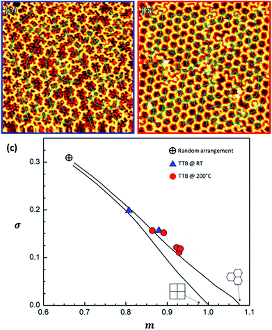 | ||
| Fig. 7 STM images of a 20 × 20 nm RT OM structure (a) and a 30 × 30 hot-dosed OM layer (b), with the MST network superimposed. (c) Plot confronting the area-normalized average of the distribution of edge lengths in the MST, m, vs. its standard deviation, σ, as obtained from the STM images of TBTTB/Ag at RT (in blue, It = 0.35 nA; Vt = 0.52 V) and hot-dosed (in red, It = −0.49 nA; Vt = −0.11 V); the lines show the distortion trajectory57 for squared or hexagonal lattice; additional information in the ESI, Section 6.† | ||
The OM grown on Ag is more extended than the OM obtained on Au(111). This is similar to the observations by Bieri et al.,62 who reported that high diffusivity of molecules and a low coupling probability are essential for achieving long-range ordered structures.
TBTTB on Cu(111)
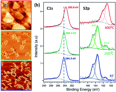
Deposition of TBTTB on copper at RT results in branched OM chains on the surface (Fig. 8a, blue frame). XPS analysis shows that the molecules are fully dehalogenated, and the C 1s peak position at 284.3 eV suggests the formation of the C–Cu bonds.17 The observed OM chains are similar to those reported by Bieri et al.62 for cyclohexa-m-phenylene on Cu(111). 2D networks were observed on gold and silver instead of the copper chain structure, presumably due to the faster diffusion of the molecules on the former substrates.29Annealing up to 200 °C increases the size of the molecular domains and the number of closed structures, but no long-range order is achieved (Fig. 8a, green frame). Further annealing above 200 °C does not improve the order. Instead, the STM contrast suggests fragmentation of the molecules (Fig. 8a, red frame).
This is confirmed by the shift of S 2p3/2 peaks from 163.34 to 161.08 eV, characteristic of the copper-bonded sulphur, which is present even at RT, where 10% of the thiophene rings are broken (ESI, Section 2†). This percentage increases with annealing temperature, and at 400 °C only S–Au is observed, meaning that all of the thiophene rings are open. At this temperature the sulphur atoms appear to be completely removed from the molecule, both decorating the step edges and forming a distinct 2 × 2 overlayer (Fig. 8a, red frame).63 No long-range ordered structure was observed upon dosing TBTTB on a hot Cu(111) surface.
Surface comparison
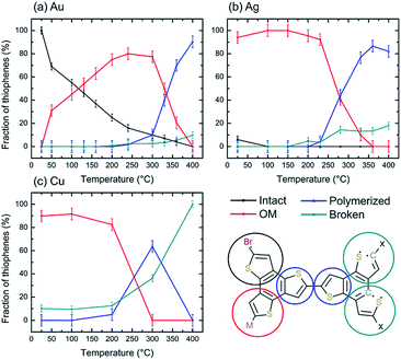
The choice between the three coinage metals strongly affects the final result of TBTTB deposition. At RT we observe intact molecules on Au, partially dehalogenated on Ag and fully dehalogenated on Cu. This trend fits with the halogen affinity of the three surfaces: bond dissociation energies (BDEs) for Cu–Br, Ag–Br and Au–Br bonds are 3.43, 2.90 and 2.21 eV respectively.47 The reported BDE values for Cu–S, Ag–S and Au–S bonds are 2.85, 2.25 and 2.63 eV respectively.47 While the largest BDE of Cu–S bond is in line with the fastest thiophene ring opening on this surface, the second highest BDE belongs to Au–S bond, which does not explain the lowest reactivity observed on this surface. The difference between Ag(111) and Au(111) – the two apparently similar surfaces (with almost the same nearest-neighbor distance, 2.88 and 2.89 Å respectively), is well known and has been reported for the chemically related self-assembled monolayers of alkanethiols.64The high chemical ‘nobility’ of Au(111) compared to all other metals is almost universally accepted, and is generally attributed to the high cohesive energy of the Au 5d states.65 Although thermal annealing does lead to organometallic and polymeric domains on all three substrates, the nature of the surface is still decisive in determining the purity/phase composition, as well as the molecular structure of the polymer. This is evident from the comparison in Fig. 9, which shows the fraction of monomer in each state: intact, OM, polymer, broken. While for Ag it is possible to obtain a full OM layer stable in a wide temperature range (RT-200 °C), for Au these structures coexist with both intact and polymeric moieties. This is further reflected by the lack of long-range order in the TBTTB/Au annealed phase.
For the C–C coupling polymerization, the Au substrate was the only one resulting in reasonable reaction efficiency (90% at 400 °C), but still the defects associated with the thiophene ring opening prevented the formation of ordered 2D polymers networks.
Conclusions and perspectives
The interactions between molecules and surfaces play a key role in steering many chemical phenomena. Our investigation of TBTTB deciphers the role of these interactions in four interweaved processes: (i) molecular self-assembly, (ii) carbon–halogen bond cleavage forming OM structures, (iii) carbon–carbon bond formation producing covalent polymers and (iv) carbon–sulfur bond cleavage which open the thiophene rings in the polymer. Deposition of the TBTTB monomer at RT resulted in non-covalent molecular networks on Au(111), OM structures on Ag(111) and partially broken molecules on Cu(111).We demonstrate that varying the substrate temperature during the deposition drives the growth process toward different final products. By depositing TBTTB on a hot surface, we formed an ordered 2D OM network on Au and a highly extended version of the same network on Ag. Further heating of the OM phase (>200 °C on Cu, >250 °C on Ag, >300 °C on Au) triggers C–C coupling of the TTB core, but the polymerization cannot be completed because of concomitant C–S bond cleavage. The maximum efficiency of the C–C coupling peaks at ca. 90% for Au at 400 °C (Fig. 9, ESI Section 2†). Comparison with other thiophene-containing monomers suggests that the probability of side-reactions correlates with the S/C ratio, as expected from the statistical probability of desulphurization. Thus, in the case of tetrathienoanthracene (S/C = 0.182) small polymer domains could be prepared on Ag,18 while for TTB (S/C = 0.25), even on the least reactive Au(111) surface, desulfurization is observed simultaneously with the polymerization. In addition, the non-fused thiophene rings might be more resistant to C–S scission due to a more significant aromatic stabilization, as observed for 3,4-(ethylenedioxy)thiophene on Ag.37
Despite their lower prominence in the field of soluble (1D) conjugated polymers, O– and N– containing building blocks (furan, pyrrole, pyridine, etc.) appear to present better alternatives for the design of surface-templated semiconducting materials, although ring opening reactions are still a problem when using highly reactive surfaces such as Cu.66
On the other hand, this study marks the formation of long-range ordered OM phases, covering the whole surface. Such networks can be prepared at lower annealing temperature, where no desorption and ring opening take place. The reaction efficiency on Ag is close to 100%, allowing to obtain extended domains larger than 20 × 20 nm2 – a size comparable with the typical feature sizes of state-of-the-art silicon technology. Due to their high degree of order and promising electronic properties, such OM structures may find different applications in molecular recognition, 2D nanopatterning and non-linear optics.67,68 The realization of these networks on the air-stable Au surface is a promising result, which opens new possibilities for their characterization outside the ultra-high vacuum (UHV), as well as a starting point for the development of OM-based devices.
Experimental
All the experiments were performed within UHV chambers with base pressures below 2 × 10⁻¹⁰ mbar. TBTTB was synthesized as described in previous work46 and deposited onto the samples using Knudsen-type effusion cells at temperatures between 100–120 °C. The deposition was performed on samples held at various temperatures: RT, 150, 200 and 250 °C. The 111 single crystals of Au, Ag and Cu (MaTecK GmbH) were cleaned by repeated cycles of Ar+ sputtering and annealed at 450–500 °C. STM images were acquired in constant-current mode using a SPECS Aarhus 150 STM and an Omicron VT-STM. STM images were analyzed using the free WSxM software,69 and were treated for plane subtraction, line-by-line flattening, contrast, and corrected based on the lattice parameters of the metal substrate.XPS experiments were performed using an electron spectrometer (SPHERA II U5 analyzer-Oxford Instruments Omicron Nanoscience), coupled to the UHV chamber that also hosts the Omicron VT-STM. The electron spectrometer consists of a hemispherical analyzer and a five-channeltron detector. All XPS analyses were performed in normal emission, using a twin anode Mg/Al X-ray source (DAR 400), supplying non-monochromatic Mg Kα radiation at 1254.6 eV photon energy. The BE scale of the XPS spectra was calibrated to the Ag 3d 5/2, Au 4f 7/2 and Cu 2p 3/2 photoelectron core levels at 368.25 eV, 84.00 eV, and 932.67 eV, respectively. Spectral peak fitting was based on residual minimization with Voigt line shapes and Shirley backgrounds, unless stated otherwise, and was performed using the Casa XPS software.70
Theoretical calculations were performed with the Vienna Ab-initio Simulation Package (VASP).71,72 DFT calculations were made using the Pedrew–Burke–Ernzerhof73 generalized-gradient approximation (PBE-GGA) for exchange–correlation potential, the projector augmented wave (PAW) method,74,75 and a plane-wave basis set with an energy cut off of 450 eV. The zero-damping DFT-D3 method of Grimme,76 was used for dispersion correction. The lattice constant of silver was first optimized (4.1362 Å) using 146-irreducible k-points (13 × 13 × 1 k-mesh), which was then used to construct the subsequent Ag(111) supercells with an energy convergence criterion of 5 meV using 61-irreducible k-points (11 × 11 × 1 k-mesh). The experimentally determined unit-cell of the adsorbed molecules on the Ag(111) surface and the epitaxy of the molecules with respect to the surface (see ESI, Section 4†) were used to construct a 5-layer slab with a vacuum gap of 15 Å. The bottom bi-layer was kept frozen and all the other surface atoms and the adsorbed molecules were fully relaxed until the net force on each atom was less than 0.05 eV Å−1. The Ag(111) slab constructed for calculating the OM phase contained 240 Ag atoms, arranged in five layers, with dimensions a = 20.47 Å, b = 20.47 Å, c = 30.00 Å, and an angle of 60° between a and b vectors (which corresponds to a (7 × 7) unit cell, as described in Section 4.1 of ESI†). All calculations for the adsorption of the molecules or their moieties on the surface were performed by sampling the gamma point of the Brillouin zone, and without using spin-polarization. However, in the case that adatoms were included on the surface (slab containing 243 Ag atoms), the convergence and energy were also tested with spin-polarization applied. The adsorption of individual TBTTB molecules on the Ag(111) surface was simulated to identify the most energetically stable adsorption site and orientation, which was subsequently used in the OM and polymer structure calculations. To simulate the molecular self-assembly, the OM network and the polymer obtained on the Au surface, because of the complexity of herringbone surface reconstruction, the DFT calculations were instead performed in the gas-phase. Images of the simulated structures were generated using VESTA software.77
Conflicts of interest
The authors state that there are no conflicts to declare.Acknowledgements
The authors acknowledge funding from the Natural Sciences and Engineering Research Council of Canada (NSERC) and the Fonds Québécois de la Recherche sur la Nature et les Technologies (FQNRT) through a Team Grant. F. R. is grateful to the Canada Research Chairs Program for funding and partial salary support. F. R. is also grateful to the Government of China for a Chang Jiang scholar short term award and to Sichuan Province for a 1000 talent short term award. G. G. is grateful to the Italian Ministero degli Affari Esteri e della Cooperazione Internazionale (MAECI) and to Giorgio Contini for the support. We thank Dr Chaoying Fu for NMR analysis of the TBTTB. Computations were performed mostly on the TCS supercomputer at the SciNet HPC Consortium, funded by the Canada Foundation for Innovation under Compute Canada, the Government of Ontario, Ontario Research Fund – Research Excellence, and the University of Toronto, and in part on Graham, Cedar and Niagara clusters of the Compute Canada (http://www.computecanada.ca). The simulations were also enabled by facilities of the Shared Hierarchical Academic Research Computing Network (SHARCNET: http://www.sharcnet.ca), as well as WestGrid (http://www.westgrid.ca).Notes and references
- F. Rosei, J. Phys.: Condens. Matter, 2004, 16, S1373 CrossRef CAS.
- M. Ebrahimi and F. Rosei, Nature, 2017, 542, 423–424 CrossRef CAS PubMed.
- W. Liu, X. Luo, Y. Bao, Y. P. Liu, G.-H. Ning, I. Abdelwahab, L. Li, C. T. Nai, Z. G. Hu, D. Zhao, B. Liu, S. Y. Quek and K. P. Loh, Nat. Chem., 2017, 9, 563–570 CrossRef CAS.
- K. Moth-Poulsen and T. Bjornholm, Nat. Nanotechnol., 2009, 4, 551–556 CrossRef CAS PubMed.
- M. Osada and T. Sasaki, Adv. Mater., 2012, 24, 210–228 CrossRef CAS PubMed.
- A. K. Geim, Science, 2009, 324, 1530–1534 CrossRef CAS PubMed.
- R. Muñoz and C. Gómez-Aleixandre, Chem. Vap. Deposition, 2013, 19, 297–322 CrossRef.
- J. Sakamoto, J. van Heijst, O. Lukin and A. D. Schlüter, Angew. Chem., Int. Ed., 2009, 48, 1030–1069 CrossRef CAS PubMed.
- G. Franc and A. Gourdon, Phys. Chem. Chem. Phys., 2011, 13, 14283–14292 RSC.
- F. Rosei, M. Schunack, Y. Naitoh, P. Jiang, A. Gourdon, E. Laegsgaard, I. Stensgaard, C. Joachim and F. Besenbacher, Prog. Surf. Sci., 2003, 71, 95–146 CrossRef CAS.
- A. Miura, S. De Feyter, M. M. S. Abdel-Mottaleb, A. Gesquière, P. C. M. Grim, G. Moessner, M. Sieffert, M. Klapper, K. Müllen and F. C. De Schryver, Langmuir, 2003, 19, 6474–6482 CrossRef CAS.
- D. Zhong, J.-H. Franke, S. K. Podiyanachari, T. Blömker, H. Zhang, G. Kehr, G. Erker, H. Fuchs and L. Chi, Science, 2011, 334, 213–216 CrossRef CAS PubMed.
- Y. Q. Zhang, N. Kepcija, M. Kleinschrodt, K. Diller, S. Fischer, A. C. Papageorgiou, F. Allegretti, J. Bjork, S. Klyatskaya, F. Klappenberger, M. Ruben and J. V. Barth, Nat. Commun., 2012, 3, 1286 CrossRef PubMed.
- H.-Y. Gao, P. A. Held, M. Knor, C. Mück-Lichtenfeld, J. Neugebauer, A. Studer and H. Fuchs, J. Am. Chem. Soc., 2014, 136, 9658–9663 CrossRef CAS PubMed.
- H. Sakaguchi, H. Matsumura, H. Gong and A. M. Abouelwafa, Science, 2005, 310, 1002–1006 CrossRef CAS PubMed.
- J. Lipton-Duffin, O. Ivasenko, D. Perepichka and F. Rosei, Small, 2009, 5, 592–597 CrossRef CAS PubMed.
- M. Di Giovannantonio, M. El Garah, J. Lipton-Duffin, V. Meunier, L. Cardenas, Y. Fagot Revurat, A. Cossaro, A. Verdini, D. F. Perepichka, F. Rosei and G. Contini, ACS Nano, 2013, 7, 8190–8198 CrossRef CAS PubMed.
- R. Gutzler, L. Cardenas, J. Lipton-Duffin, M. El Garah, L. E. Dinca, C. E. Szakacs, C. Fu, M. Gallagher, M. Vondracek, M. Rybachuk, D. F. Perepichka and F. Rosei, Nanoscale, 2014, 6, 2660–2668 RSC.
- L. Grill, M. Dyer, L. Lafferentz, M. Persson, M. V. Peters and S. Hecht, Nat. Nanotechnol., 2007, 2, 687–691 CrossRef CAS PubMed.
- M. El Garah, J. M. MacLeod and F. Rosei, Surf. Sci., 2013, 613, 6–14 CrossRef CAS.
- G. McCarty and P. Weiss, J. Phys. Chem. B, 2002, 106, 8005–8008 CrossRef CAS.
- G. S. McCarty and P. S. Weiss, J. Am. Chem. Soc., 2004, 126, 16772–16776 CrossRef CAS PubMed.
- M. Di Giovannantonio, M. Tomellini, J. Lipton-Duffin, G. Galeotti, M. Ebrahimi, A. Cossaro, A. Verdini, N. Kharche, V. Meunier, G. Vasseur, Y. Fagot-Revurat, D. F. Perepichka, F. Rosei and G. Contini, J. Am. Chem. Soc., 2016, 138, 16696–16702 CrossRef CAS PubMed.
- L. Lafferentz, V. Eberhardt, C. Dri, C. Africh, G. Comelli, F. Esch, S. Hecht and L. Grill, Nat. Chem., 2012, 4, 215–220 CrossRef CAS PubMed.
- G. Galeotti, M. Di Giovannantonio, J. Lipton-Duffin, M. Ebrahimi, S. Tebi, A. Verdini, L. Floreano, Y. Fagot-Revurat, D. Perepichka, F. Rosei and G. Contini, Faraday Discuss., 2017, 204, 453–469 RSC.
- J. Björk, F. Hanke and S. Stafström, J. Am. Chem. Soc., 2013, 135, 5768–5775 CrossRef PubMed.
- L. Dong, S. Wang, W. Wang, C. Chen, T. Lin, J. Adisoejoso and N. Lin, in On-Surface Synthesis, Springer, 2016, pp. 23–42 Search PubMed.
- R. Gutzler and D. F. Perepichka, J. Am. Chem. Soc., 2013, 135, 16585–16594 CrossRef CAS PubMed.
- R. Gutzler, Phys. Chem. Chem. Phys., 2016, 18, 29092–29100 RSC.
- G. Vasseur, Y. Fagot-Revurat, M. Sicot, B. Kierren, L. Moreau, D. Malterre, L. Cardenas, G. Galeotti, J. Lipton-Duffin, F. Rosei, M. Di Giovannantonio, G. Contini, P. Le Fèvre, F. Bertran, L. Liang, V. Meunier and D. F. Perepichka, Nat. Commun., 2016, 7, 10235 CrossRef CAS PubMed.
- W. Yong, W. Xinchen and A. Markus, Angew. Chem., Int. Ed., 2012, 51, 68–89 CrossRef.
- C. Nacci, F. Ample, D. Bleger, S. Hecht, C. Joachim and L. Grill, Nat. Commun., 2015, 6, 7397 CrossRef PubMed.
- I. Perepichka and D. Perepichka, Handbook of Thiophene-Based Materials: Applications in Organic Electronics and Photonics, 2 Volumes Set, Wiley, 2009 Search PubMed.
- L. Cardenas, R. Gutzler, J. Lipton-Duffin, C. Fu, J. L. Brusso, L. E. Dinca, M. Vondracek, Y. Fagot-Revurat, D. Malterre, F. Rosei and D. F. Perepichka, Chem. Sci., 2013, 4, 3263–3268 RSC.
- L. E. Dinca, C. Fu, J. M. MacLeod, J. Lipton-Duffin, J. L. Brusso, C. E. Szakacs, D. Ma, D. F. Perepichka and F. Rosei, ACS Nano, 2013, 7, 1652–1657 CrossRef CAS PubMed.
- L. E. Dinca, J. M. MacLeod, J. Lipton-Duffin, C. Fu, D. Ma, D. F. Perepichka and F. Rosei, J. Phys. Chem. C, 2015, 119, 22432–22438 CrossRef CAS.
- I. Di Bernardo, P. Hines, M. Abyazisani, N. Motta, J. MacLeod and J. Lipton-Duffin, Chem. Commun., 2018, 54, 3723–3726 RSC.
- X. Guo, S. Wang, V. Enkelmann, M. Baumgarten and K. Müllen, Org. Lett., 2011, 13, 6062–6065 CrossRef CAS PubMed.
- J. A. Lipton-Duffin, J. A. Miwa, M. Kondratenko, F. Cicoira, B. G. Sumpter, V. Meunier, D. F. Perepichka and F. Rosei, Proc. Natl. Acad. Sci. U. S. A., 2010, 107, 11200–11204 CrossRef CAS PubMed.
- G. D. Nguyen, F. M. Toma, T. Cao, Z. Pedramrazi, C. Chen, D. J. Rizzo, T. Joshi, C. Bronner, Y.-C. Chen, M. Favaro, S. G. Louie, F. R. Fischer and M. F. Crommie, J. Phys. Chem. C, 2016, 120, 2684–2687 CrossRef CAS.
- J. M. MacLeod, J. Lipton-Duffin, C. Fu, T. Taerum, D. F. Perepichka and F. Rosei, ACS Nano, 2017, 11, 8901–8909 CrossRef CAS PubMed.
- R. De Bettignies, Y. Nicolas, P. Blanchard, E. Levillain, J.-M. Nunzi and J. Roncali, Adv. Mater., 2003, 15, 1939–1943 CrossRef CAS.
- C. B. Nielsen, J. M. Fraser, B. C. Schroeder, J. Du, A. J. P. White, W. Zhang and I. McCulloch, Org. Lett., 2011, 13, 2414–2417 CrossRef CAS PubMed.
- G. Cheng, H. Ning, C. Youchun, Q. Leiqiang, X. Hong, Z. Shitong, L. Fenghong, M. Yuguang and J. Donglin, Angew. Chem., Int. Ed., 2015, 54, 13594–13598 CrossRef PubMed.
- A. Ringk, A. Lignie, Y. Hou, H. N. Alshareef and P. M. Beaujuge, ACS Appl. Mater. Interfaces, 2016, 8, 12091–12100 CrossRef CAS PubMed.
- T. Taerum, O. Lukoyanova, R. G. Wylie and D. F. Perepichka, Org. Lett., 2009, 11, 3230–3233 CrossRef CAS PubMed.
- Y.-R. Luo, Comprehensive handbook of chemical bond energies, CRC press, 2007 Search PubMed.
- L. Dong, P. N. Liu and N. Lin, Acc. Chem. Res., 2015, 48, 2765–2774 CrossRef CAS PubMed.
- M. Lackinger, Chem. Commun., 2017, 53, 7872–7885 RSC.
- T. A. Pham, F. Song, M.-T. Nguyen, Z. Li, F. Studener and M. Stöhr, Chem.–Eur. J., 2016, 22, 5937–5944 CrossRef CAS PubMed.
- A. Saywell, W. Greń, G. Franc, A. Gourdon, X. Bouju and L. Grill, J. Phys. Chem. C, 2014, 118, 1719–1728 CrossRef CAS.
- M. Liu, M. Liu, L. She, Z. Zha, J. Pan, S. Li, T. Li, Y. He, Z. Cai, J. Wang, Y. Zheng, X. Qiu and D. Zhong, Nat. Commun., 2017, 8, 14924 CrossRef CAS PubMed.
- Q. Sun, L. Cai, H. Ma, C. Yuan and W. Xu, ACS Nano, 2016, 10, 7023–7030 CrossRef CAS PubMed.
- T. A. Pham, F. Song and M. Stohr, Phys. Chem. Chem. Phys., 2014, 16, 8881–8885 RSC.
- M. Chen, J. Xiao, H.-P. Steinrück, S. Wang, W. Wang, N. Lin, W. Hieringer and J. M. Gottfried, J. Phys. Chem. C, 2014, 118, 6820–6830 CrossRef CAS.
- Q. Fan, T. Wang, J. Dai, J. Kuttner, G. Hilt, J. M. Gottfried and J. Zhu, ACS Nano, 2017, 11, 5070–5079 CrossRef CAS.
- J. Eichhorn, T. Strunskus, A. Rastgoo-Lahrood, D. Samanta, M. Schmittel and M. Lackinger, Chem. Commun., 2014, 50, 7680–7682 RSC.
- J. Eichhorn, D. Nieckarz, O. Ochs, D. Samanta, M. Schmittel, P. J. Szabelski and M. Lackinger, ACS Nano, 2014, 8, 7880–7889 CrossRef CAS PubMed.
- J. M. MacLeod, O. Ivasenko, C. Fu, T. Taerum, F. Rosei and D. F. Perepichka, J. Am. Chem. Soc., 2009, 131, 16844–16850 CrossRef CAS PubMed.
- O. Ourdjini, R. Pawlak, M. Abel, S. Clair, L. Chen, N. Bergeon, M. Sassi, V. Oison, J.-M. Debierre, R. Coratger and L. Porte, Phys. Rev. B: Condens. Matter Mater. Phys., 2011, 84, 125421 CrossRef.
- C. Dussert, G. Rasigni, M. Rasigni, J. Palmari and A. Llebaria, Phys. Rev. B: Condens. Matter Mater. Phys., 1986, 34, 3528–3531 CrossRef.
- M. Bieri, M. T. Nguyen, O. Groning, J. M. Cai, M. Treier, K. Ait-Mansour, P. Ruffieux, C. A. Pignedoli, D. Passerone, M. Kastler, K. Mullen and R. Fasel, J. Am. Chem. Soc., 2010, 132, 16669–16676 CrossRef CAS PubMed.
- H. Walen, D.-J. Liu, J. Oh, H. Lim, J. W. Evans, Y. Kim and P. A. Thiel, J. Chem. Phys., 2015, 142, 194711 CrossRef PubMed.
- A. Ulman, Chem. Rev., 1996, 96, 1533–1554 CrossRef CAS PubMed.
- B. Hammer and J. K. Norskov, Nature, 1995, 376, 238–240 CrossRef CAS.
- M. Abyazisani, J. Bradford, N. Motta, J. Lipton-Duffin and J. MacLeod, J. Phys. Chem. C, 2018, 122, 17836–17845 CrossRef CAS.
- H. Furukawa, K. E. Cordova, M. O'Keeffe and O. M. Yaghi, Science, 2013, 341 Search PubMed.
- H.-C. Zhou, J. R. Long and O. M. Yaghi, Chem. Rev., 2012, 112, 673–674 CrossRef CAS PubMed.
- I. Horcas, R. Fernandez, J. Gomez-Rodriguez, J. Colchero, J. Gómez-Herrero and A. Baro, Rev. Sci. Instrum., 2007, 78, 013705 CrossRef CAS PubMed.
- N. Fairley, CasaXPS Manual 2.3. 15: Introduction to XPS and AES, Casa Software, 2009 Search PubMed.
- G. Kresse and J. Hafner, Phys. Rev. B: Condens. Matter Mater. Phys., 1993, 47, 558–561 CrossRef CAS.
- G. Kresse and J. Furthmüller, Phys. Rev. B: Condens. Matter Mater. Phys., 1996, 54, 11169–11186 CrossRef CAS.
- J. P. Perdew, M. Ernzerhof and K. Burke, J. Chem. Phys., 1996, 105, 9982–9985 CrossRef CAS.
- P. E. Blöchl, Phys. Rev. B: Condens. Matter Mater. Phys., 1994, 50, 17953–17979 CrossRef.
- G. Kresse and D. Joubert, Phys. Rev. B: Condens. Matter Mater. Phys., 1999, 59, 1758–1775 CrossRef CAS.
- S. Grimme, J. Antony, S. Ehrlich and H. Krieg, J. Chem. Phys., 2010, 132, 154104 CrossRef PubMed.
- K. Momma and F. Izumi, J. Appl. Crystallogr., 2011, 44, 1272–1276 CrossRef CAS.
Footnotes |
| † Electronic supplementary information (ESI) available: Additional STM, XPS and DFT results, higher size version of Fig. 2, 5, 8, and a discussion of chirality. See DOI: 10.1039/c8sc05267k. |
| ‡ Authors share contribution. |
| § Current address: Physics Department E20, Technical University of Munich, James-Franck-Str.1, D-85748 Garching, Germany. |
| This journal is © The Royal Society of Chemistry 2019 |

