
One-step co-assembly method to fabricate photosensitive peptide nanoparticles for two-photon photodynamic therapy†
Jieling
Li
,
Anhe
Wang
,
Peng
Ren
,
Xuehai
Yan
 * and
Shuo
Bai
*
* and
Shuo
Bai
*
State Key Laboratory of Biochemical Engineering, Institute of Process Engineering, Chinese Academy of Sciences, No. 1 North 2nd Street, Zhongguancun, 100190 Beijing, China. E-mail: yanxh@ipe.ac.cn; baishuo@ipe.ac.cn
First published on 31st January 2019
Abstract
Peptide-based nanoparticles were employed to load and disperse hydrophobic porphyrins in a one-step co-assembly method in aqueous media. The isolated porphyrins doped within nanoparticles showed enhanced two-photon absorption ability and could effectively generate 1O2 to induce the apoptosis of cancer cells, which holds great prospects in two-photon PDT.
Differing from the traditional one-photon photodynamic therapy that generally requires a laser within the UV-visible range to activate photosensitizers (PSs) and induce the death of cancer cells,1,2 two-photon photodynamic therapy (TP-PDT) has recently emerged as a novel cancer-therapy modality employing two low energy photons of near-infrared light for the excitation of PSs, which is beneficial for deeper tissue penetration and lower photo-bleaching of PSs.3,4 The nonlinear photon absorption allows for the activation of PSs at the focal point of the laser beam, resulting in better spatial control of PS activation and lower photo-induced damage of adjacent healthy tissues.5 So far, TP-PDT has been successfully employed for precise cancer treatment6–9 and the exploitation of PSs with high two-photon absorption ability is emphasized for enhanced TP-PDT efficiency.
Porphyrin derivatives have been studied as photosensitizers for PDT due to their relatively efficient 1O2 productivity and their chemical diversity with a broad assortment of compounds to be used in PDT.10–12 Besides the one-photon excitation of porphyrins,13–15 the two-photon absorption property of porphyrins has also attracted great attention and exploiting porphyrin-based two-photon PSs is of great significance in the development of TP-PDT.16 Efforts to improve porphyrin-based TP-PDT have long focused on increasing the two-photon absorption (TPA) cross sections of the porphyrin,5,17–20 which usually requires a complex porphyrin modification process. However, most of the porphyrins are too hydrophobic to dissolve in aqueous media and tend to aggregate to induce self-quenching resulting in limitations for effective two-photon activated PDT. To achieve high TP-PDT efficiency, developing effective porphyrin delivery systems avoiding porphyrin aggregation and self-quenching still remains a big challenge.
The employment of nanoparticles as porphyrin carriers has been proven to be a promising solution to solve the problem due to the encapsulation of porphyrins into nanoparticles helping to isolate porphyrins in monomeric form, retain their photodynamic activity, improve their solubility and maximize the 1O2 quantum yield.21 A series of nanoparticles such as quantum dots,22 gold nanoparticles,23 and polymeric nanomaterials,24 have recently been reported as porphyrin carriers to enhance their two-photon absorption ability for TP-PDT application. Yet these nanoparticles suffer from the disadvantages of low biodegradability, low long-term biosafety and poor loading efficiency.
Peptide self-assembly is a facile method for the fabrication of nanocarriers with various morphologies.25–30 In particular, their properties of excellent biocompatibility, non-immunogenicity and rapid clearance from the body have distinguished peptide-based nanocarriers from other carriers in the application of nanomedicine.31–33 Notably, peptide nanoparticles have recently been reported to be good candidate nanocarriers to load hydrophobic PSs for both photothermal therapy (PTT) and one-photon PDT applications.34–36 In our previous study, we reported the use of peptide nanoparticles as photosensitizer carriers for traditional one-photon photodynamic therapy. These nanoparticles were reported to be of high drug loading efficiency and were responsive to various stimuli for drug release, demonstrating the great benefits of peptide-based carriers in tumor treatment.15 On the basis of this previous work, we here firstly prepared peptide self-assembled nanoparticles as carriers of hydrophobic porphyrins by a one-step method to exploit the two-photon absorption property of porphyrins for TP-PDT applications. Short peptide fluorenylmethoxycarbonyl-leucine-leucine-leucine-OMe (Fmoc-L3-OMe) could co-assemble with hydrophobic porphyrin derivative meso-tetra(p-hydroxyphenyl)porphine (m-THPP) under the driving forces of π–π stacking and hydrophobic interaction (Scheme 1).
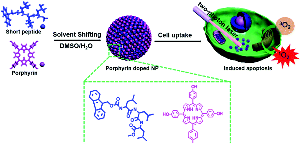 | ||
| Scheme 1 Scheme of the fabrication of porphyrin-doped peptide nanoparticles and their application in TP-PDT and the molecular structures of peptide and porphyrin used in this work. | ||
By doping into peptide nanoparticles, the solubility of hydrophobic porphyrin in aqueous media was easily solved, and porphyrin molecules could be isolated from each other to avoid self-aggregation and self-quenching, maintaining their intrinsic fluorescent property and reducing the overdose of porphyrin induced side effects. Without employing complex modification methods of porphyrin, biocompatible peptide-porphyrin co-assembled nanoparticles with intracellular tracing and enhanced two-photon absorption properties for improved TP-PDT efficiency could serve as novel photosensitizing agents and hold great prospects in cancer treatment through two-photon photodynamic therapy.
The morphology of m-THPP doped peptide nanoparticles was characterized with SEM, TEM and CLSM as shown in Fig. 1. The SEM image showed that the nanoparticles had an average diameter of ca. 170 nm, which was in good consistency with those of the TEM and DLS results. The DLS results also showed that these nanoparticles had a very narrow size distribution (PDI 0.118). This indicated that the nanoparticles were rigid and solid particles with good monodispersity, which was good for their potential use in nanomedicine. The m-THPP doped peptide nanoparticles also exhibited great red fluorescence signal, suggesting the intracellular tracing function of these nanoparticles.
 | ||
| Fig. 1 Morphology characterization of the nanoparticles: (A) SEM image; (B) TEM image and (C) CLSM images and (D) DLS result; (E) photos of nanoparticles and pure m-THPP in water. | ||
Fig. 1E showed the dispersion photos of pure m-THPP and m-THPP doped peptide nanoparticles in water. Owing to the strong hydrophobicity, pure m-THPP could hardly dissolve in aqueous media. Though with the help of an amphipathic organic solvent such as DMSO, we could obtain a uniform m-THPP dispersion, it was unstable and could easily aggregate to form a precipitate. However, when co-assembled with Fmoc-L3-OMe, uniform m-THPP/peptide co-assembled nanoparticles were obtained and could stay stable for one week at least. The stability of the nanoparticles was mainly maintained through electrostatic repulsion, as described in our previous study.35
To verify that the isolation of porphyrins by peptides could enhance the two-photon absorption ability of m-THPP, CLSM images of the nanoparticles under the irradiation of a two-photon laser (850 nm) were first recorded. As seen in Fig. 2A, the nanoparticles could emit a strong fluorescence signal when excited under the irradiation of an 850 nm two-photon laser, suggesting the two-photon absorption and emission property of the m-THPP in the nanoparticles and light irradiation didn’t change the morphology of the assembled nanoparticles (Fig. S1, ESI†). However, for the particles formed by pure m-THPP, owing to the severe aggregation, no fluorescent signal could be observed (Fig. S2, ESI†).
 | ||
| Fig. 2 (A) CLSM images of nanoparticles excited by a two-photon laser of 850 nm: (a) light field; (b) and (c) fluorescence images; (B) two-photon fluorescence spectra of nanoparticles (curve a) and pure m-THPP (curve b); (C) two-photon fluorescence spectra of nanoparticle samples with different m-THPH molar ratio; (D) nanoparticle samples with different m-THPP concentration in DMSO. | ||
Next, the two-photon fluorescence spectra of m-THPP in nanoparticles were measured in water under a two-photon laser of a wavelength of 850 nm, with pure m-THPP as a comparison (Fig. 2B). Owing to the severe self-aggregation, almost no fluorescence signal could be detected for pure m-THPP in water (curve b in Fig. 2B), but m-THPP doped in the peptide nanoparticles could exhibit a strong fluorescence signal (curve a in Fig. 2B). The above results preliminary proved that the isolation of m-THPP by peptides could avoid self-aggregation and self-quenching of m-THPP.
To further confirm that the isolation of m-THPP could enhance the two-photon absorption ability, we then gradually increased the molar ratio of m-THPP within the nanoparticles (table in Fig. 2D) to test the fluorescence intensity changes of m-THPP under two-photon laser excitation. As can be seen in Fig. 2C, the fluorescence intensity of the nanoparticles showed a tendency to decrease with the increase of m-THPP molar ratio. This was probably because with the increase of m-THPP ratio, the peptide became relatively insufficient to completely isolate m-THPP molecules from each other such that m-THPP began to self-aggregate, inducing fluorescence quenching and the decrease of fluorescence intensity. The above results indicated that the doping of m-THPP into the peptide-based nanoparticles could enhance the two-photon absorption and emission ability of m-THPP, which was good for these nanoparticles to be used as photosensitizers for two-photon photodynamic therapy.
For endocytosis, MCF-7 cancer cells were firstly co-incubated with the m-THPP/peptide nanoparticles for 4 h. Then the cells were washed with PBS twice to remove the un-uptaken nanoparticles. Before CLSM observation, the cell membrane and nucleus were stained with Alexa 488 and Hoechst 33342, respectively. The endocytosis result is shown in Fig. 3A. The strong red fluorescence signal of cancer cells indicated the successful uptake of nanoparticles and the nanoparticles were mainly localized in the cytoplasm. The presence of nanoparticles in both XZ and YZ cross section in the 3D reconstruction image (Fig. 3A(d)) further confirmed that the nanoparticles could be endocytosed by cancer cells.
 | ||
| Fig. 3 (A) CLSM images of MCF-7 cells stained with nanoparticle suspensions after 4 h of co-culturing. The corresponding images of (a) the overlap image of the cytomembrane (green) and nucleus (blue), (b) nanoparticles (red), (c) the overlap image, and (d) the 3D reconstruction image of the cells (the orthogonal section images at the bottom and on the right were recorded along the yellow lines); (B) 1O2 detection in cells with DCFH-DA: (a) before and (b) after irradiation; (c) comparison of cells with and without irradiation: area in the red frame was treated with the two-photon laser of 850 nm wavelength at 10% intensity for 20 min. | ||
As the generation of 1O2 played a key role in the two-photon PDT,37 we next exploited the 1O2 production capacity of the nanoparticles in cells with a 1O2 detection probe DCFH-DA. DCFH-DA, a redox-sensitive fluorescence probe, could be degraded to DCFH in cells and then oxidized by 1O2 into DCF, exhibiting strong green fluorescent signal.38 As can be seen in Fig. 3B(a), when exposed to no laser, the cells exhibited no fluorescent signal at all. But after being irradiated with a two-photon laser of 850 nm wavelength for 20 min, the cells could emit strong green fluorescence signal (Fig. 3B(b)). The well-defined boundary between the green and dark areas (Fig. 3B(c)) demonstrated the high 1O2 productivity of the nanoparticles after laser irradiation and the failure of 1O2 production in the dark (dark area in Fig. 3C) also suggested the little dark toxicity of the nanoparticles.
MCF-7 cancer cells were used to evaluate the two-photon cytotoxicity of the nanoparticles by fluorescence imaging under two-photon excitation with a wavelength of 850 nm. Propidium iodide (PI) which can specifically stain dead cells to exhibit a strong red fluorescence signal, was introduced for easy observation of cell apoptosis. After being co-incubated with nanoparticles for 4 h, the MCF-7 cells were exposed to a two-photon laser of 850 nm wavelength for 20 min. As can be seen in Fig. 4, before irradiation, the cells showed no fluorescence signal at all and the cells could well keep their cellular morphology (Fig. 4A and B). After irradiation for 20 min, the cells stained by PI gave strong red fluorescent emission and the morphology of the irradiated cells had dramatically changed (Fig. 4C and D), suggesting that the two-photon laser irradiation could induce the death of cancer cells. The well-defined boundary (Fig. S3A and B, ESI†) between the irradiated area (the red frame area) and non-irradiated area (the dark area) revealed the high light cytotoxicity and the good biocompatibility under dark conditions.
 | ||
| Fig. 4 The CLSM images of MCF-7 cells stained with PI: with particles before (A and B) and after (C and D) two-photon laser irradiation at 10% intensity for 20 min; (E and F) with light (10% intensity for 20 min) but no nanoparticles; (G and H) with nanoparticles but no light. | ||
To prove that the apoptosis of cells was induced by the light cytotoxicity of nanoparticles, not the laser irradiation itself, cells with no nanoparticle uptake were also treated with the two-photon laser of 850 nm wavelength. The result is given in Fig. 4E and F, where no red fluorescent emission or morphology change was observed, indicating that the laser irradiation alone was of little cytotoxicity and could not cause the death of MCF-7 cancer cells. The dark cytotoxicity of the nanoparticles was further evaluated with nanoparticle co-incubated MCF-7 cells but with no light treatment. As expected, no cells were stained with PI and little morphology change was observed (Fig. 4G and H), suggesting little cytotoxicity of the nanoparticles under dark conditions. The above results confirmed that the nanoparticles were of high anticancer efficacy under two-photon laser illumination and high biocompatibility when exposed to no laser.
In conclusion, photosensitizer-doped aromatic short peptide nanoparticles with Fmoc-L3-OMe as a hosting carrier and m-THPP as the functional photosensitizer are prepared using a one-step co-assembly method. By doping into peptide nanoparticles, the solubility of hydrophobic porphyrin in aqueous media was easily solved, and porphyrin molecules could be isolated from each other by amphiphilic peptides to avoid self-aggregation and self-quenching, maintaining their intrinsic fluorescent property and reducing the overdose of porphyrin-induced side effects. These nanoparticles are stable with a uniform size of ∼170 nm and show enhanced two-photon fluorescence absorption ability compared with the pure m-THPP. In vitro reactive oxygen species detection reveals the high 1O2 productivity of the nanoparticles and the cytotoxicity test demonstrates that these nanoparticles can effectively cause the death of cancer cells under two-photon irradiation. The photosensitive peptide nanoparticles have great prospects as novel and effective photosensitizing agents for two-photon photodynamic therapy.
The authors acknowledge the financial support from the National Natural Science Foundation of China (Project No. 21703253, 21522307, 21774132, 21644007 and 21473208).
Conflicts of interest
There are no conflicts to declare.Notes and references
- R. Bonnett, Chem. Soc. Rev., 1995, 24, 19–33 RSC.
- H. I. Pass, J. Natl. Cancer Inst., 1993, 85, 443–456 CrossRef CAS PubMed.
- F. Bolze, S. Jenni, A. Sour and V. Heitz, Chem. Commun., 2017, 53, 12857–12877 RSC.
- K. Ogawa and Y. Kobuke, Anticancer Agents Med. Chem., 2008, 8, 269–279 CrossRef CAS PubMed.
- Y. Shen, A. J. Shuhendler, D. Ye, J.-J. Xu and H.-Y. Chen, Chem. Soc. Rev., 2016, 45, 6725–6741 RSC.
- B. Sun, L. Wang, Q. Li, P. He, H. Liu, H. Wang, Y. Yang and J. Li, Biomacromolecules, 2017, 18, 3506–3513 CrossRef CAS PubMed.
- H. Liu, Y. Yang, A. Wang, M. Han, W. Cui and J. Li, Adv. Funct. Mater., 2016, 26, 2561–2570 CrossRef CAS.
- Y. Yang, H. Liu, M. Han, B. Sun and J. Li, Angew. Chem., Int. Ed., 2016, 55, 13538–13543 CrossRef CAS PubMed.
- J. Sun, Q. Xin, Y. Yang, H. Shah, H. Cao, Y. Qi, J. R. Gong and J. Li, Chem. Commun., 2018, 54, 715–718 RSC.
- M. Lismont, L. Dreesen and S. Wuttke, Adv. Funct. Mater., 2017, 27, 1606314 CrossRef.
- J. Sun, Y. Guo, R. Xing, T. Jiao, Q. Zou and X. Yan, Colloids Surf., A, 2017, 514, 155–160 CrossRef CAS.
- A. P. Castano, T. N. Demidova and M. R. Hamblin, Photodiagn. Photodyn. Ther., 2004, 1, 279–293 CrossRef CAS PubMed.
- M. Ethirajan, Y. Chen, P. Joshi and R. K. Pandey, Chem. Soc. Rev., 2011, 40, 340–362 RSC.
- E. D. Sternberg, D. Dolphin and C. Bruckner, Tetrahedron, 1998, 54, 4151–4202 CrossRef CAS.
- K. Liu, R. Xing, Q. Zou, G. Ma, H. Mohwald and X. Yan, Angew. Chem., Int. Ed., 2016, 55, 3036–3039 CrossRef CAS PubMed.
- K. Ogawa and Y. Kobuke, BioMed Res. Int., 2013, 2013, 125658 Search PubMed.
- X. Shen, F. He, J. Wu, G. Q. Xu, S. Q. Yao and Q.-H. Xu, Langmuir, 2011, 27, 1739–1744 CrossRef CAS PubMed.
- M. Pawlicki, H. A. Collins, R. G. Denning and H. L. Anderson, Angew. Chem., Int. Ed., 2009, 48, 3244–3266 CrossRef CAS PubMed.
- T. V. Esipova, H. J. Rivera-Jacquez, B. Weber, A. E. Masunov and S. A. Vinogradov, J. Am. Chem. Soc., 2016, 138, 15648–15662 CrossRef CAS PubMed.
- K. Ogawa and Y. Kobuke, BioMed Res. Int., 2013, 2013, 125658 Search PubMed.
- P. Couleaud, V. Morosini, C. Frochot, S. Richeter, L. Raehm and J.-O. Durand, Nanoscale, 2010, 2, 1083–1095 RSC.
- Z.-D. Qi, D.-W. Li, P. Jiang, F.-L. Jiang, Y.-S. Li, Y. Liu, W.-K. Wong and K.-W. Cheah, J. Mater. Chem., 2011, 21, 2455–2458 RSC.
- T. Zhao, K. Yu, L. Li, T. Zhang, Z. Guan, N. Gao, P. Yuan, S. Li, S. Q. Yao, Q.-H. Xu and G. Q. Xu, ACS Appl. Mater. Interfaces, 2014, 6, 2700–2708 CrossRef CAS PubMed.
- C.-Y. Chen, Y. Tian, Y.-J. Cheng, A. C. Young, J.-W. Ka and A. K. Y. Jen, J. Am. Chem. Soc., 2007, 129, 7220–7221 CrossRef CAS PubMed.
- X. Yan, P. Zhu and J. Li, Chem. Soc. Rev., 2010, 39, 1877–1890 RSC.
- S. Bai, C. Pappas, S. Debnath, P. W. J. M. Frederix, J. Leckie, S. Fleming and R. V. Ulijn, ACS Nano, 2014, 8, 7005–7013 CrossRef CAS PubMed.
- S. Fleming and R. V. Ulijn, Chem. Soc. Rev., 2014, 43, 8150–8177 RSC.
- J. Wang, K. Liu, R. Xing and X. Yan, Chem. Soc. Rev., 2016, 45, 5589–5604 RSC.
- K. Tao, A. Levin, L. Adler-Abramovich and E. Gazit, Chem. Soc. Rev., 2016, 45, 3935–3953 RSC.
- L. L. E. Mears, E. R. Draper, A. M. Castilla, H. Su, Zhuola, B. Dietrich, M. C. Nolan, G. N. Smith, J. Doutch, S. Rogers, R. Akhtar, H. Cui and D. J. Adams, Biomacromolecules, 2017, 18, 3531–3540 CrossRef CAS PubMed.
- H. Zhang, J. B. Fei, X. H. Yan, A. H. Wang and J. B. Li, Adv. Funct. Mater., 2015, 25, 1193–1204 CrossRef CAS.
- J. Zhan, Y. B. Cai, S. S. He, L. Wang and Z. M. Yang, Angew. Chem., Int. Ed., 2018, 57, 1813–1816 CrossRef CAS PubMed.
- H. Wang, J. Shi, Z. Feng, R. Zhou, S. Wang, A. A. Rodal and B. Xu, Angew. Chem., Int. Ed., 2017, 56, 16297–16301 CrossRef CAS PubMed.
- Q. Zou, M. Abbas, L. Zhao, S. Li, G. Shen and X. Yan, J. Am. Chem. Soc., 2017, 139, 1921–1927 CrossRef CAS PubMed.
- J. Li, A. Wang, L. Zhao, Q. Dong, M. Wang, H. Xu, X. Yan and S. Bai, ACS Appl. Mater. Interfaces, 2018, 10, 28420–28427 CrossRef CAS PubMed.
- S. Li, Q. Zou, Y. Li, C. Yuan, R. Xing and X. Yan, J. Am. Chem. Soc., 2018, 140, 10794–10802 CrossRef CAS PubMed.
- Z. Zhou, J. Song, L. Nie and X. Chen, Chem. Soc. Rev., 2016, 45, 6597–6626 RSC.
- Y. Y. He and D. P. Hader, Photochem. Photobiol. Sci., 2002, 1, 729–736 RSC.
Footnote |
| † Electronic supplementary information (ESI) available. See DOI: 10.1039/c9cc00025a |
| This journal is © The Royal Society of Chemistry 2019 |