
 Open Access Article
 This Open Access Article is licensed under a
 Creative Commons Attribution 3.0 Unported Licence
Ultrathin Janus nanodiscs†
Dan Xueab,
Ximing Song a and
Fuxin Liang*b
a and
Fuxin Liang*b
aLiaoning Provincial Key Laboratory for Green Synthesis and Preparative Chemistry of Advanced Materials, Liaoning University, Shenyang 110036, China
bState Key Laboratory of Polymer Physics and Chemistry, Institute of Chemistry, Chinese Academy of Sciences, Beijing 100190, China. E-mail: liangfuxin@iccas.ac.cn
First published on 11th May 2017
Abstract
Ultrathin Janus nanodiscs with single molecular scale thickness are fabricated by self-organized sol–gel process against a patchy magnetic microsphere. Functional groups onto the patch domains can induce a favourable absorption of an amine. The morphology of Janus nanodiscs is adjusted by controlling the patchy regions on the surface of paramagnetic Fe3O4@SiO2 core/shell microspheres. The as-used patchy C8–(Fe3O4@SiO2)–PhCHO microspheres serving as templates can be easily separated from the as-formed Janus nanodiscs and reused to fabricate other Janus nanodiscs. Functional compositions such as paramagnetic Fe3O4 nanoparticles and polymers with different wettability properties are introduced onto the different regions of Janus nanodiscs to achieve magnetic Janus nanodiscs. They are used as efficient solid surfactants to stabilize emulsion.
Introduction
Janus materials with two different compositions and properties distinctly compartmentalized onto the same surface of one object have gained growing academic and industrial interests. It is important to develop methods to tune their morphology, composition thus function spatial distribution.1 Concerning with wettability difference of the two compositions, amphiphilic Janus materials can serve as a solid surfactant to stabilize immiscible liquids.2 Among the representative shapes, such as 0-D (zero-dimensional) Janus spheres,3 1-D Janus rods,4 2-D Janus discs,5 and 3-D Janus cages,6 Janus discs are efficient solid surfactants for their anisotropic shapes. Serving as emulsion stabilizers, rotation of the Janus discs (or named sheets) with anisotropic shapes is restricted. So the orientation of Janus discs has definite direction, and the as-formed emulsion is very stable.7 So the as-stabilized emulsion is more stable than that upon using spherical Janus particles. But disc-like Janus materials are difficult to be obtained. Polymeric nano-sized Janus discs have been fabricated via self-assembly of block copolymers.8 But the narrow molecular weight is needed for the as-used block copolymers to self-assembly forming a layered structure. Recently, we have fabricated Janus nanosheets with molecular scale thickness by a surface sol–gel process of the self-assembled monolayer of an amphiphilic silane onto the template particles.9 For their ultrathin thickness, Janus nanosheets are flexible to tightly adhere onto the interface with appropriate deformation. But in all these Janus discs, their shapes and sizes are difficult to be controlled because all of them are achieved by crushing process.Herein, we report a novel approach towards Janus nanodiscs with single molecular scale thickness against a patchy Fe3O4@SiO2 core/shell microsphere (Scheme 1). The aldehyde groups in the patchy domains can induce a favourable absorption of 3-aminopropyltrimethoxysilane (APTMS) via a dynamic imine bond. After removal residual APTMS, a monolayer of APTMS forms onto the patchy domain. The pedant silane groups direct outwardly, which can form a molecular thick silica layer after a sol–gel process. Under acidic conditions, when the imide bond is broken, the Janus nanodiscs are released from the Fe3O4@SiO2 microspheres. Further, paramagnetic Fe3O4 nanoparticles and polymers with different wettability properties are introduced onto the different sides of the ultrathin Janus nanodiscs to achieve functional ultrathin Janus nanodiscs. They could be served as efficient solid surfactants to stabilize emulsion.
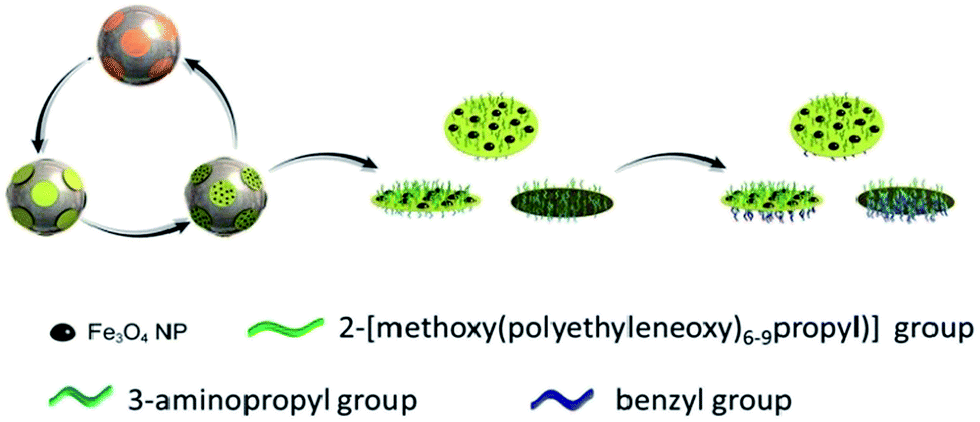 | ||
| Scheme 1 Synthesis of ultrathin Janus nanodiscs. The patchy magnetic Fe3O4@SiO2 core/shell microspheres as template can be recycled and the Janus nanodiscs are modified with Fe3O4 NPs and hydrophilic PEO group on the one side and hydrophobic benzyl group on the other side. | ||
Experimental
Materials
Iron(III) chloride hexahydrate (FeCl3·6H2O), iron chloride tetrahydrate (FeCl2·4H2O), ethylene glycol (EG), sodium acetate trihydrate (NaAC), sodium citrate, hydrochloride acid (HCl), ammonium hydroxide (25 wt%), tetraethyl orthosilicate (TEOS), sliver nitrate (AgNO3), nitric acid (HNO3), dimethyl sulphoxide (DMSO), potassium iodide (KI), sodium bicarbonate (NaHCO3), benzaldehyde, paraffin (52–54 °C), tetrahydrofuran (THF), N-heptane, sodium borohydride (NaBH4) and cyclohexane were purchased from Sinopharm Chemical Reagent. N-Butylamine was purchased from ACROS. 4-(chloromethyl)phenyltrimethoxysilane (PhCH2Cl–TMS) were purchased from Alfa Aesar. N-Octyltrimethoxysilane (C8–TMS), 3-aminopropyltrimethoxysilane (APTMS), fluorescein isothiocyanate isomer1 (FITC) and coumarine-6 were purchased from J&K. 2-[Methoxy(polyethyleneoxy)6–9propyl]trimethoxysilane (PEO-TMS) was purchase from Fluorochem.Characterization
Morphology of the samples were characterized by scanning electron microscopy (Hitachi S-4800 at 15 kV), which with an energy dispersive X-ray (EDX) analyser and transmission electron microscopy (JEOL1011 at 100 kV). The samples for SEM observation were ambient dried and vacuum sputtered with Pt. The samples for TEM observation were prepared by spreading very dilute emulsions in ethanol onto carbon-coated copper grids. FTIR spectroscopy was performed on the sample/KBR pressed pellets after scanning samples for 32 times using a BRUKER EQUINOX55 spectrometer. Thickness and shape of the samples was measured by AFM of Bruker Multimode 8. Emulsion was characterized by inverted fluorescence microscopy (Olympus IX83) and Olympus optical microscope. Size contribution was measured by Zeta-sizer (Nano Series, Malvern Instruments) at 25 °C.Results and discussion
The Fe3O4@SiO2 microsphere is prepared by solvothermal method following a SiO2 layer coating via sol–gel process.10 The representative Fe3O4 microsphere (200 nm in diameter) is selected as a paramagnetic core for easy separation (Fig. S1†). Onto the Fe3O4 microsphere surface, SiO2 is detected by EDX (Fig. S2a and b†). The size contribution of Fe3O4 and Fe3O4@SiO2 microspheres is measured by DLS, which is consistent with TEM results (Fig. S2c†). The thickness of SiO2 layer is about 30–40 nm (Fig. S3a†). Onto the Fe3O4@SiO2 microsphere, Ag NPs are achieved by reducing AgNO3 with n-butylamine at 50 °C (Fig. S3b†).11 The diameter of Ag NPs can be adjusted with reaction time. The exposed bare SiO2 surface is treated to be hydrophobic with n-octyltrimethoxysilane (C8–TMS). Then, a C8–(Fe3O4@SiO2)–Ag composite microsphere is achieved (Fig. 1a). Upon removal of Ag NPs by dissolution, surface of the microsphere becomes smooth, and fresh SiO2 domains are exposed (Fig. S3c†). The fresh SiO2 domains are further modified with 4-(chloromethyl)phenyltrimethoxysilane (PhCH2Cl–TMS) to graft benzyl chloride groups (Fig. S3d†). Benzaldehyde groups are derived from the benzyl chloride groups by Kornblum oxidation.12 A patchy C8–(Fe3O4@SiO2)–PhCHO microsphere is achieved (Fig. 1b). The surface remains smooth. 3-Aminopropyltrimethoxysilane (APTMS) can form a monolayer onto the patchy domains since the aldehyde group can form a dynamic imine bond with the functional amine-group of APTMS. The amine-groups are prevented thereby. The pedant silane groups can form a molecular thick silica layer via sol–gel process. The sphere surface remains smooth (Fig. S3e and f†). After adding acid, the imine bond is broken. Then, the silica nanodiscs are released from the microsphere. Using a magnet, the patchy template microspheres are recycled, while the nanodiscs are purified.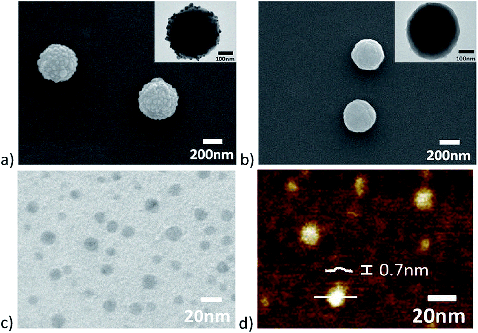 | ||
| Fig. 1 Morphologies of paramagnetic microspheres and as-formed Janus nanodiscs: SEM and inset TEM images of (a) patchy C8–(Fe3O4@SiO2)–Ag microspheres; (b) patchy C8–(Fe3O4@SiO2)–PhCHO microspheres; TEM (c) and AFM (d) images of the as-formed Janus nanodiscs. | ||
The compositions of different microspheres are measured by FTIR spectrum (Fig. S4†). Characteristic peaks at 2840 cm−1 and 2920 cm−1 are respectively assigned to –CH3 group and –CH2– group, which prove that the patchy C8–(Fe3O4@SiO2) microspheres are prepared. Characteristic peaks at 1600–1400 cm−1 are assigned to phenyl group, which proves that the patchy C8–(Fe3O4@SiO2)–PhCH2Cl microspheres had been prepared. Meanwhile, the characteristic peak at 1750 cm−1 is assigned to –CHO group, it is shown that the patchy C8–(Fe3O4@SiO2)–PhCHO microspheres are achieved. The peaks at 3404 cm−1 and 3520 cm−1 are assigned to –NH2 group of APTMS which is grafted onto the –PhCHO group region of patchy C8–(Fe3O4@SiO2)–PhCHO microspheres.
When pH is tuned to about 4.5, aminopropyl/hydroxyl composited Janus nanodiscs are released from the templated microspheres for the dynamic imine bond is cleaved. The average size of Janus nanodiscs is about 15 nm in diameter (Fig. 1c). The thickness of Janus nanodiscs is about 0.7 nm (Fig. 1d). Shape and size of the aminopropyl/hydroxyl composited Janus nanodiscs are uniform. Because of the as-formed Janus nanodiscs are too thin to observe in SEM, they can only be characterized by TEM and AFM in morphology. The compositions of the as-formed Janus nanodiscs are measured by FTIR spectrum (Fig. S5†). The peaks at 3448 cm−1 and 3360 cm−1 are assigned to –NH2 group, the peaks 3155 cm−1 are assigned to –OH group, and the peak at 1090 cm−1 are assigned to –Si–O–Si– bond.
The single molecular scale thickness Janus nanodiscs are prepared from the domains where the Ag NPs are. So the size of Janus nanodiscs can be adjusted by controlling the size of Ag NPs. It is found that the Ag NPs grow bigger with the increase of reaction time. When the reaction time is 40 min, the diameters of Ag NPs are about 5 nm (Fig. S6a†). Then the as-formed Janus nanodiscs are about 5 nm in diameter (Fig. 2a). When the reaction time is increased to 60 min, the average diameters of both Ag NPs and as-formed Janus nanodiscs are about 25 nm (Fig. S6b,† and 2b). Thus, the size of the single molecular thickness Janus nanodiscs can be precisely controlled. However, the thickness of the Janus nanodiscs with different sizes remains 0.7 nm (Fig. 2c and d).
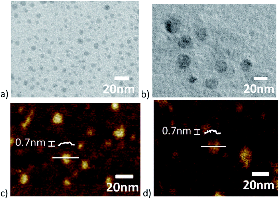 | ||
| Fig. 2 Size control of the as-formed Janus nanodiscs. TEM and AFM images of the aminopropyl/hydroxyl composited Janus nanodiscs are achieved at different reaction time: (a) and (c) are at 40 min; (b) and (d) are at 60 min. | ||
Two sides of the as-formed Janus nanodiscs are easily to be selectively modified with different compositions, respectively. Firstly, Fe3O4 NPs are grafted onto the hydroxyl group side by coprecipitation method.13 Fe3O4 NPs are barely recognized on the surface of patchy C8–(Fe3O4@SiO2)–SiO2 microsphere (Fig. 3a). When pH is tuned to about 4.5, aminopropyl/Fe3O4 NPs composited Janus nanodiscs are released from the templated microspheres (Fig. 3b). It is easy to find the Fe3O4 NPs exist on the surface of the Janus composite nanodiscs. In order to enhancing hydrophilic property of the Janus composite nanodiscs, 2-[methoxy(polyethyleneoxy)6–9propyl]trimethoxysilane (PEO-TMS) are grafted on the Fe3O4 NPs side via sol–gel reaction. The patchy C8–(Fe3O4@SiO2)–SiO2–Fe3O4 NPs-PEO microspheres are achieved (Fig. S7†). Turn pH to about 4.5, the Fe3O4 NPs-PEO modified Janus composite nanodiscs are achieved from the templated microspheres. Then, benzaldehyde is grafted to the aminopropyl group via Schiff base reaction, and further imine bonds are reduce with NaBH4. So the benzyl/Fe3O4 NPs-PEO Janus nanodiscs are fabricated (Fig. 3c). Hydrophilic PEO group and hydrophobic benzyl group are respectively grafted onto the two sides of the as-formed Janus nanodiscs. For the existence of Fe3O4 NPs, PEO and benzyl groups, thickness of the Janus nanodiscs increase to about 2.5 nm (Fig. 3d). But the size of Janus nanodiscs does not change obviously. The compositions of different Janus nanodiscs are proved by FTIR spectrum (Fig. S8†). The character peak at 506 cm−1 indicates Fe–O bond, and peak at 1090 cm−1 indicates –Si–O–Si– bond. Meanwhile, the peaks at 1390 cm−1 and 1690 cm−1 respectively indicate C–N and C![[double bond, length as m-dash]](https://www.rsc.org/images/entities/char_e001.gif) N bonds.
N bonds.
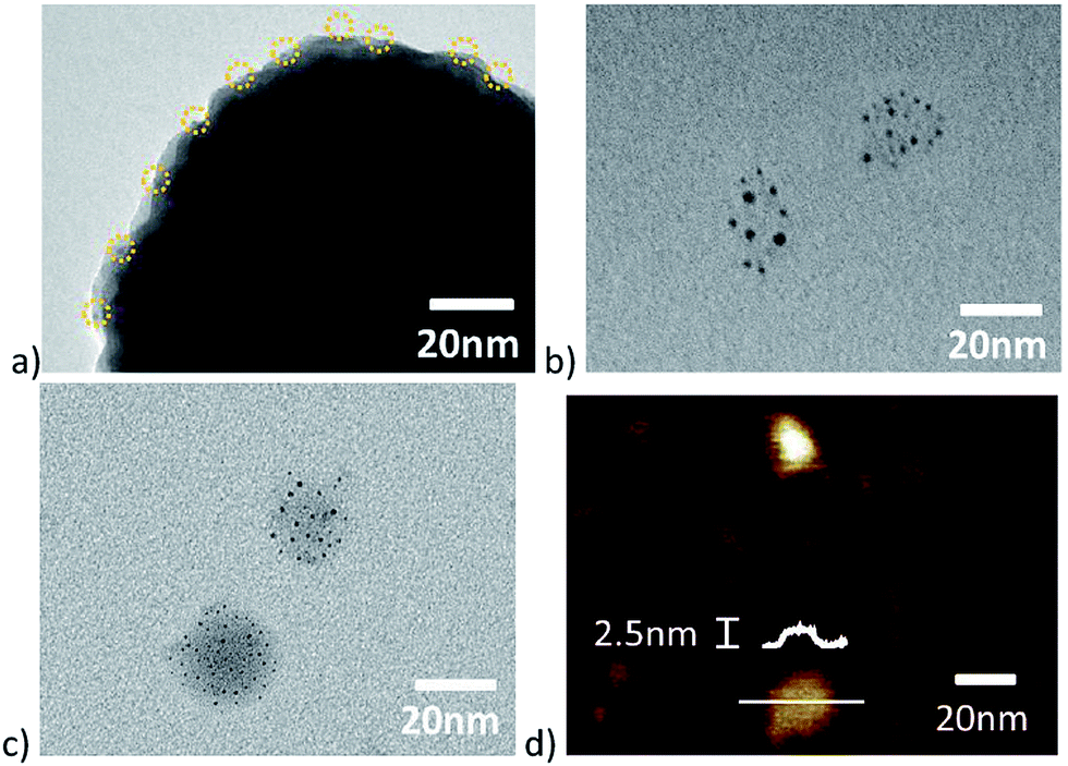 | ||
| Fig. 3 Morphologies of different Janus nanodiscs: (a) TEM image of patchy C8–(Fe3O4@SiO2)–SiO2–Fe3O4 NPs microsphere; (b) TEM image of Fe3O4 NPs modified Janus nanodiscs; TEM (c) and AFM (d) images of benzyl/Fe3O4 NPs-PEO Janus composite nanodiscs. | ||
In order to prove their amphiphilic property which originated from their Janus structures, aggregation structures of Janus nanodiscs in different solvents are studied. When the Janus nanodiscs are dispersed in selective solvent such as water, the hydrophobic benzyl side is stacked together while the hydrophilic PEO side exposes to the aqueous phase to form back-to-back stacked aggregates for their amphiphilic property originated from their Janus structure (Fig. S9a†). Similarly in cyclohexane, the hydrophilic PEO side is stacked together while the hydrophobic benzyl side exposes to the cyclohexane phase to form large size aggregates (Fig. S9b†). When the Janus nanodiscs are dispersed in co-solvent THF, Janus nanodiscs are mono-dispersed as single particles (Fig. S9c†). Meanwhile, DLS data shown that Janus nanodiscs aggregate together to form large size aggregated particles in selective solvents and are dispersed as single particles in co-solvent, respectively (Fig. S9d†).
For the magnetic responsive property of Fe3O4 NPs, the as-prepared Janus nanodiscs can be easily manipulated with external magnetic field (Fig. S9†). The as-prepared Janus nanodiscs can be employed as solid surfactants to stabilize emulsion. Taking water and n-heptane as immiscible solvents, they are emulsified to oil-in-water emulsion (Fig. 4a). For their magnetic property, Janus composite nanodiscs can be collected with magnet and reused. In order to observe the as-stabilized emulsion droplets more clearly, melt paraffin (Tm = 52–54 °C) is used as oil phase. Then a paraffin-in-water emulsion forms upon 70 °C and stirring at 2000 rpm for 10 min. The paraffin-in-water emulsion could be stable for several months and the diameter of paraffin droplets is 2–5 μm (Fig. 4b). After the emulsion is cooled down to room temperature, the paraffin is solidified (Fig. 4c). FITC is employed to observe the positions of as-used Janus nanodiscs in emulsion system. It is shown that all the Janus nanodiscs aggregate at the interface of emulsion (Fig. 4d). So the as-prepared Janus nanodiscs are a kind of efficient solid emulsion stabilizer.
 | ||
| Fig. 4 Performance demonstration of the as-prepared Janus nanodiscs: (a) magnetic performance: the as-prepared Janus nanodiscs are served as solid surfactant in the n-heptane-in-water emulsion, the n-heptane is dyed by coumarine-6 (left), and the as-used Janus nanodiscs are easily collected by magnet (right); (b) polarizing optical image of the paraffin-in-water emulsion; (c) SEM image of the paraffin droplets; (d) inverted fluorescence microscope image of the paraffin-in-water emulsion. | ||
Conclusions
In summary, single molecular scale thickness Janus nanodiscs are successfully achieved from patchy C8–(Fe3O4@SiO2)–PhCHO microspheres as template. The sizes of Janus nanodiscs can be controlled by tuning the patchy domains. Meanwhile, paramagnetic Fe3O4 NPs are introduced onto the hydrophilic side of the Janus nanodiscs thus achieving magnetic response. Further, benzyl groups and PEO groups are grafted respectively onto the hydrophobic and hydrophilic sides of the as-formed Janus nanodiscs to enhance their amphiphilic performance. The as-prepared Janus composite nanodiscs can be used as recyclable solid surfactants to stabilize emulsion. Most importantly, the patchy C8–(Fe3O4@SiO2)–PhCHO templated microspheres can be easily recovered and reused to fabricate more Janus nanodiscs.Acknowledgements
This work was supported by the National Natural Science Foundation of China (51233007, 51622308).Notes and references
- S. Jiang and S. Granick, Janus particle synthesis, self-assembly and applications, RSC Press, London, England, 2012 CrossRef CAS PubMed; A. Walther and A. H. E. Müller, Chem. Rev., 2013, 113, 5194 CrossRef CAS PubMed; F. X. Liang, C. L. Zhang and Z. Z. Yang, Adv. Mater., 2014, 26, 6944 CrossRef PubMed.
- A. Walther, K. Matussek and A. H. E. Müller, ACS Nano, 2008, 2, 1167 CrossRef CAS PubMed; A. Walther, M. Drechsler and A. H. E. Müller, Soft Matter, 2009, 5, 385 RSC; A. Walther, M. Hoffmann and A. H. E. Müller, Angew. Chem., Int. Ed., 2008, 120, 711 CrossRef PubMed.
- A. C. Trindade, R. Craveiro, A. P. C. Almeida, J. P. Canejo, A. Paiva, S. Barreiros and M. H. Godinho, J. Supercrit. Fluids, 2017, 120, 125 CrossRef CAS.
- B. Zhao, H. Zhou, C. Y. Liu, Y. Long, G. Q. Yang, C. H. Tung and K. Song, New J. Chem., 2016, 40, 6541 RSC.
- F. X. Liang, K. Shen, X. Z. Qu, C. L. Zhang, Q. Wang, J. L. Li, J. G. Liu and Z. Z. Yang, Angew. Chem., Int. Ed., 2011, 50, 2379 CrossRef CAS PubMed.
- F. X. Liang, J. G. Liu, C. L. Zhang, X. Z. Qu, J. L. Li and Z. Z. Yang, Chem. Commun., 2011, 47, 1231 RSC.
- Y. Nonomura, S. Komura and K. Tsujii, Langmuir, 2004, 20, 11821 CrossRef CAS PubMed.
- A. Walther, X. Andre, M. Drechsler, V. Abetz and A. H. E. Müller, J. Am. Chem. Soc., 2007, 129, 6187 CrossRef CAS PubMed.
- Y. J. Liu, F. X. Liang, Q. Wang, X. Z. Qu and Z. Z. Yang, Chem. Commun., 2015, 51, 3562 RSC; Y. J. Liu, Q. Wang, X. Z. Qu, F. X. Liang and Z. Z. Yang, Sci. China Mater., 2015, 58, 126 CrossRef CAS.
- Y. H. Deng, D. W. Qi, C. H. Deng, X. M. Zhang and D. Y. Zhao, J. Am. Chem. Soc., 2008, 130, 28 CrossRef CAS PubMed.
- K. Kim, H. S. Kim and H. K. Park, Langmuir, 2006, 22, 8083 CrossRef CAS PubMed.
- W. W. Epstein and F. W. Sweat, Chem. Rev., 1967, 67, 247 CrossRef CAS.
- R. Massart, IEEE Trans. Magn., 1981, 17, 1247 CrossRef.
Footnote |
| † Electronic supplementary information (ESI) available: Fig. 1–9. See DOI: 10.1039/c7ra03116e |
| This journal is © The Royal Society of Chemistry 2017 |