DOI:
10.1039/C6RA24551J
(Paper)
RSC Adv., 2016,
6, 104969-104978
Efficient drug delivery system for bone repair by tuning the surface of hydroxyapatite particles
Received
2nd October 2016
, Accepted 14th October 2016
First published on 25th October 2016
Abstract
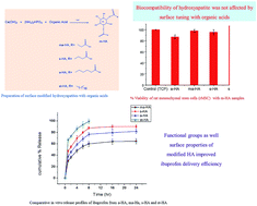
A limited blood flow to skeletal tissues results in minimal therapeutic effect of drugs being administered to a patient using conventional ways. To obtain sufficient amount of drug at an effected site, implanted drug delivery systems based on biomaterials can be used. In this study, surface modified hydroxyapatites (m-HA) were prepared and evaluated as drug delivery systems. The effect of modifiers on surface properties of HA and their in vitro drug delivery efficiency were investigated. For synthesis of m-HA, a simple in situ co-precipitation method was used. Hydroxyapatite was subjected to surface modification by various carboxylic acids such as adipic acid, malonic acid, succinic acid and stearic acid. This surface modification affected its surface properties such as surface area, pore size, pore volume, particle size and crystallinity. The m-HA were characterized by Fourier transform infrared spectroscopy (FT-IR), X-ray diffraction (XRD) and thermogravimetric analysis (TGA). Brunauer–Emmett–Teller (BET) technique was used to compute surface properties of m-HA. The highest BET surface area of 143 m2 g−1 has been found for HA modified with malonic acid and the lowest surface area of 37 m2 g−1 was calculated for stearic acid modified HA. The BET adsorption average pore size (17–20 nm) of m-HA confirmed its mesoporous nature. The biocompatible nature of the prepared m-HA was assessed by 3-(4,5)-dimethylthiahiazo(-z-yl)-3,5-di-phenytetrazoliumromide (MTT) assay. To evaluate the influence of functional groups and surface properties of m-HA on drug delivery efficiency, ibuprofen was used as a model drug. In vitro drug delivery experimental results indicated that drug loading and release efficiency relied on functional groups, surface area, and porosity of m-HA. The percentage loading of ibuprofen was good for samples containing free –COOH groups and high surface area. A drug loading of 22 mg g−1 has been found for malonic acid modified HA (ma-HA) having high surface area, pore volume, whereas a poor loading of 2.03 mg g−1 has been observed for stearic acid modified HA (st-HA) sample having low surface area and pore volume. A sustained drug release profile showed that 61% drug had been released from malonic acid modified HA (ma-HA) in 24 hours. A 100% drug release was observed for st-HA in 8 hours. Succinic acid modified HA and adipic acid modified HA exhibited intermediate drug release profiles. The drug release behavior of m-HA followed Fick's laws of diffusion.
1 Introduction
Drug therapy is required after an orthopedic treatment to avoid infection, pain and swelling. In oral drug administration, a high drug dose and periodic intake are usually required to maintain sufficient concentration of drug at less blood irrigated bone tissues. Fabrication of bioactive drug eluted bone grafts is the most efficient therapeutic approach in hard tissue engineering. This technique helps to improve the functions of bone implants as well as bone healing process.1–4 An ideal drug eluted bone graft should help in bone healing and sustained drug release, while maintaining its biocompatibility.
Porous materials, particularly mesoporous nanomaterials, are excellent drug delivery systems. The surface area, pore volume and pore size facilitate to host the sufficient amount of drugs and their controlled release.5 Commonly exploited drug delivery materials are mesoporous silica,6–9 bioactive glasses,10 hydroxyapatites3,11 and titania.12,13 Hydroxyapatite (HA) [Ca10(PO4)6(OH)2] is regarded as one of the preferred material for hard tissue regeneration due to its excellent biocompatibility, osteoconductivity, osseointegrativity and possibility of tailoring bioactivity by ionic substitution.14,15
Frequently exploited strategies to prepare mesoporous materials focus on using structure directing agents during synthesis, such as, surfactants with organic chelants, and finally the elimination of templates by dissolution with appropriate solvents or pyrolysis to create porosity.16–18 Although the templating agents provide extraordinary results, yet an additional step is required to remove templates causing a threat to environment. Non-templating approaches used for the synthesis of porous materials, particularly for porous HA, include surface modification of apatites with organic chelants19–21 and synthesis of metal doped apatites.22,23 The surface modification carried out with organic chelants and polymers19–21 can tailor surface area, pore size, pore volume and also introduce surface charge and surface interfaces such as hydrophobicity or hydrophilicity. These factors play a key role in drug delivery efficiency of a material.19,24,25 In addition, surface modification introduces functional groups on the surface of material used that facilitate better drug loading, controlled drug release, and improved physicochemical interaction with proteins participating in bone formation.26,27
Chemicals used for surface tuning of bioceramics should be physiologically nontoxic and biocompatible. Furthermore, these modifiers should not decrease bioactivity of bioceramics. The most widely exploited organic chelants used for HA surface tuning are carboxylates,28 amino acids,29 and silane coupling agents27 etc. Silane coupling agents have some biotoxic effects30 whereas the nontoxic carboxylic acids are the most promising modifiers used for surface tuning of HA.26,28 There are a number of studies reported in the literature highlighting the potential of functionalized hydroxyapatite as drug delivery system by taking ibuprofen as a benchmark anti-inflammatory drug.31–33 After combing through the literature survey, it is concluded that organic carboxylic acid modified mesoporous hydroxyapatite particles have not been evaluated as ibuprofen drug carriers.
This study is featured with the development of hydroxyapatite based drug delivery system with improved drug carrying ability and sustained release. A simple single step in situ co-precipitation method is used to introduce carboxylic acids of different carbon chain lengths at the surface of hydroxyapatite. Ibuprofen is used as a model drug to evaluate drug delivery capacity of m-HA. Implanted bone repair materials usually show inflammatory response. Therefore, the biomaterials inherited with in situ release of anti-inflammatory drug constitute an effective alternative compared to systemic drug dose.34 Ibuprofen (IBU) is a common anti-inflammatory and analgesic drug being used to treat bone diseases and postoperative orthopedic pain.35–37 It is widely used as model drug to evaluate new sustained release delivery systems because of its good pharmacological activity.35
2 Materials and methods
2.1. Chemicals and reagents
Adipic, malonic, stearic and succinic acids of Analar grade were purchased from Merck. Calcium hydroxide, diammonium hydrogen phosphate and ethanol of Analar grade were purchased from Across, Sigma Aldrich and BDH respectively. All reagents and chemicals were used without further purification. Phosphate-buffered saline (PBS) was purchased from Sigma Aldrich.
2.2. Experimental procedure for in situ synthesis of modified hydroxyapatite
An in situ co-precipitation method was used to synthesize m-HA. 1.67 M Ca(OH)2 and 1 M (NH4)2HPO4 solutions were separately prepared in deionized water at room temperature. A stoichiometric molar ratio of Ca/P 1.67 was used for the synthesis of m-HA.
After adding carboxylic acid (0.25 mol equivalent) to Ca(OH)2 solution, the mixture was stirred for 1 hour followed by dropwise addition of (NH4)2HPO4 solution (10 drops per min). The reaction mixture was stirred for 1 hour at room temperature and left for 48 hours for aging. Finally, the precipitates were filtered and washed thoroughly with distilled water till neutral pH of the filtrate was achieved.28 The m-HA precipitates were dried at 80 °C for 24 hours. The carboxylic acid modified hydroxyapatites (m-HA) were named as a-HA (adipic acid modified hydroxyapatite), ma-HA (malonic acid modified hydroxyapatite), s-HA (succinic acid modified hydroxyapatite) and st-HA (stearic acid modified hydroxyapatite).
2.3. Biocompatibility test
2.3.1. In vitro culture of rat mesenchymal stem cells (rMSC). Using direct adherence method. The femur was isolated under sterile conditions. A disposable aseptic syringe was used to draw antibiotic supplemented L-DMEM medium repeatedly in and out of bone marrow cavity. The cell fraction was collected in a sterile Petri dish. The obtained cell suspension was centrifuged at 250 × g for 5 minutes. The cell pellet was re-suspended in DMEM (Gibco) containing 10% FBS (Gibco), 0.1% penicillin and streptomycin (Gibco) and transferred to T25 tissue culture flask. The flasks were incubated at 37 °C in a 5% CO2 incubator. The first medium was changed after 4 days, followed by the medium change on alternative days until the cells became 70–80% confluent. MSC were sub-cultured at 70–80% confluency. The cells were trypsinized, counted (dead cells excluded by trypan blue assay) and passaged in T-75 flasks. Second- or third-passage MSC were used for cytotoxicity and SEM analysis. In this study, all cell culture experiments were performed in compliance with the Biosafety, Ethical Rules and Regulations administered by the Ethical Committee of Animal Handling for Experimentation, University of Veterinary and Animal Sciences, Lahore. All the experiments were approved by the Ethical Review Committee for the Use of Laboratory Animals (ERCULA), University of Veterinary and Animal Sciences (UVAS), Pakistan. The present study was restricted to rats and no human trials or experiments were carried out.
2.3.2. Cytotoxicity assay. Cellular toxicity was determined by 3-(4,5-dimethylthiazol-2-yl)-2,5-diphenyltetrasodium bromide (MTT) assay. Prior to cell culture, approximately 10 mg of a m-HA was sterilized by 1 mL of 70% ethanol. Before cell seeding the scaffolds were washed and pre-conditioned in DMEM medium. MSC were seeded in 24-well cell culture plate with 5 × 10⁴ cells per well with or without m-HA. Cells seeded in 24-plate wells without HA were used as positive control (tissue culture plate, TCP). Post day 7, the medium was discarded and cells were washed with 1 mL PBS. MTT solution, 1 mL (0.5 mg mL−1) was added to each well and the plate was incubated at 37 °C for 3 hours. The MTT solution was discarded and the cells were washed once with 1 mL of PBS. To solubilize the formazan crystals, 0.3 mL of dimethyl sulfoxide (DMSO) (Sigma Aldrich) was added to each well and the plate was kept on a shaker for 15–20 minutes. The optical density (OD) of the dissolved crystals was measured by using microplate reader at 590 nm. The assay was set up in duplicates with MSC derived from 2 different rats for each sample. The % viability is represented as mean ± SD of 2 independent experiments. The % viability was calculated by using the following eqn (1): |
 | (1) |
2.4. Ibuprofen loading and release studies
The drug adsorption was performed using 0.3 g of dry powder of m-HA. The m-HA sample was dipped in 1000 ppm ibuprofen ethanol solution at room temperature. The sample was oscillated for 16 hours at 37 °C at a speed of 120 rpm by using K-201BS oscillator. Ibuprofen adsorption capacity was evaluated by UV-vis absorption spectroscopy at 263 nm. The loading of ibuprofen (1 mg of drug per gram of sample) was calculated as: Q = (Co − C)V/W, where Co and C were concentrations of drug before and after drug loading respectively, V volume of drug solution used for loading experiments, Q amount of drug loaded on m-HA mg g−1 and W represented weight of HA sample taken. To evaluate drug release profile, drug loaded samples were immersed in 10 mL of PBS at 37 °C. At different time intervals of 2, 4, 6, 8 16 and 24 hours specific amount of the drug eluted solution was withdrawn and replaced with same amount of fresh PBS solution. The drug release quantity was checked by UV-vis absorption spectroscopy at 263 nm.
2.5. Characterization of m-HA
The m-HA was characterized by Fourier transform infrared spectroscopy (FT-IR), X-ray diffraction (XRD), Thermo Gravimetric Analysis (TGA) and Brunauer, Emmett and Teller (BET) and Scanning Electron Microscopy (SEM) techniques. FT-IR spectra were recorded at 8 cm−1 resolution on Thermo Nicolet 6700™ FT-IR spectrometer (Thermoscientific) in conjunction with a photoacoustic sampling cell, which omits the need of sample preparation and neat samples can be analysed within the spectral range of (4000–400 cm−1). XRD analysis was carried out by using MPD XP'ERT PRO™ diffractometer of PANALYTICAL limited with monochromatic Cu-Kα (λ = 0.15418 nm). The scans were taken with a step size 0.02° per second at 2θ range of 20–80°. The crystallite size of m-HA samples was determined by using Scherrer equation (D = kλ/β
cos
θ), where D is crystalline size (Å), k is shape factor, λ is diffraction wavelength (0.015404 nm), θ is diffraction angle (radian) and β is full width at half maximum (FWHM) value in radians. The crystallinity index of m-HA samples was determined with the formula: Xc = (KA/β002)3, where KA is a constant (0.24) and β002 is the (FWHM) of (002) peak in degree.38 Thermogravimetric investigations were carried out with dried samples using ramp rate of 10 °C min−1 up to 1000 °C. Approximately 3–10 mg of m-HA sample was placed in alumina pans. The specific surface area and particle size of samples were determined by BET Tristar II of Micromeritics using the data between 0.001–0.99 with degassing conditions of 1.5 hours duration, 10 °C ramp rate at 100 °C.
3 Results and discussion
3.1. Carboxylic acid modification of hydroxyapatite
The ability of carboxylic groups to interact with calcium ions by making a complex is a useful tool to tune HA surface properties.26,28 Surface modified HA particles were synthesized by a co-precipitation method. In this method, a carboxylic acid modifier solution was added to 1 M calcium hydroxide solution followed by dropwise addition of 0.6 M phosphate solution to prepare m-HA (Fig. 1). This is a simple and economical method compared to hydrothermal synthesis as this does not require high temperature and specific reactor.
 |
| Fig. 1 Preparation of surface modified hydroxyapatites with organic acids (m-HA) via in situ co-precipitation method. | |
3.2. Characterization
FTIR spectroscopy was used for chemical structural characterization of m-HA (Fig. 2). Characteristics peaks for PO43− group of HA such as 1090, 1025, 960, 605, 555 and 575 cm−1 were present in all FTIR spectra of m-HA.39,40 A broad band in the range of 2800–3500 cm−1 corresponded to a characteristic stretching vibration of O–H of HA,41 organic acids and adsorbed water. An asymmetric vibration band of COO− of a-HA appeared at 1550 cm−1 and a symmetric vibration band of COO− at 1427 cm−1 overlapped with carbonate band.28,42 In s-HA asymmetric vibration band of COO− appeared at 1560 cm−1 and its symmetric band and –CH signals at 1438 cm−1.28,43 In ma-HA asymmetric vibration band of COO− appeared at 1595 cm−1 and its symmetric band and carbonate signals at 1421 cm−1. In st-HA, peaks at 1556 and 1427 cm−1 were vibration bands for COO− and the spectral peaks present at 1427, 1546, 2923 and 2854 were attributed to CH vibrations.44,45 A small peak at 2316 cm−1 represented an asymmetric vibration of carbon dioxide present in air. Stearic acid is a monocarboxylic acid and its –COOH functional group interacts with calcium ions. In other dicarboxylic acid modifiers (such as adipic acid, succinic acid and malonic acid), only one carboxylic group interacts with calcium ion and other is free (Fig. 1). This is evidenced by FTIR spectra where carbonyl group of free carboxylic acid appears around 1640 cm−1 for s-HA, a-HA and ma-HA and there is no such peak for st-HA in this region (Fig. 2a and b).
 |
| Fig. 2 (a) Fourier transform infrared spectra of surface modified hydroxyapatites with organic acids (m-HA). (b) Enlarged FTIR region in wave number range 1800–1200 cm−1. | |
The FT-IR spectrum of ibuprofen loaded a-HA showed strong peaks in the region of 2800–3000 cm−1, these were characteristic symmetric and asymmetric stretching vibrations of alkyl chain of ibuprofen. In addition, characteristic vibrations near 1407 cm−1 were corresponding to aromatic ring of ibuprofen. The FT-IR spectrum of ibuprofen loaded a-HA showed that drug had been successfully loaded on it46 (Fig. 3).
 |
| Fig. 3 FT-IR spectrum of ibuprofen loaded adipic acid modified HA showing loading of ibuprofen. | |
XRD was employed to analyze the effect of modifier on crystallinity and particle size of HA. The XRD patterns of as prepared m-HA samples after drying at 80 °C were shown in Fig. 4. XRD patterns of m-HA samples showed characteristic 2θ values at 26, 31, 33, 40, 46 and 54.47 These values confirmed that the intrinsic properties of m-HA had been conserved. Diffraction data gave a good match to pattern number ICDD 09432 corresponding to HA. The grafting of HA with organic ligands decreased the growth of HA particles by limiting the further addition of hydroxyl, calcium and phosphate on HA particle surface resulting in reduced particle size and crystallinity.28,48 Peak broadening in XRD patterns of m-HA samples also indicated decrease in particle size and crystallinity. The crystallite size of m-HA was determined by using the Debye–Scherrer equation. The well separated reflection (002) was used to calculate crystallinity index (Xc).38 The crystal size of prepared m-HA was found in nanocrystalline range of 173–193 Å. The numbers only represent the general trend and the values are approximate. It was anticipated that carboxylic acid modifiers had influenced the crystallinity index of m-HA. The lowest Xc value of 0.124 was observed for st-HA and the highest value of 0.168 for s-HA (Table 1).
 |
| Fig. 4 XRD patterns of m-HA describing peaks of hydroxyapatites. | |
Table 1 Crystallinity and crystalline size of m-HAa
Sample |
FWHM (002) |
Crystallinity (Xc) |
Crystalline size (Å) |
Values (mean ± standard deviation of three replicates). |
st-HA |
0.485 ± 0.02 |
0.124 ± 0.03 |
173 ± 2.08 |
s-HA |
0.435 ± 0.01 |
0.168 ± 0.02 |
193 ± 2.12 |
ma-HA |
0.437 ± 0.01 |
0.165 ± 0.02 |
191 ± 1.41 |
a-HA |
0.463 ± 0.02 |
0.139 ± 0.01 |
180 ± 2.12 |
The peak broadening at reflection (002) was the highest in st-HA sample (Table 1, entry 1) representing the lowest crystallinity of st-HA. The lowest crystallinity could be attributed to the long chain of stearic acid. The side long alkyl chain of stearic acid rotates freely and causes steric hindrance to the crystal packing during crystallization of HA. This resulted in disrupting the arrangement of HA crystals.49 A similar trend of lower crystallinity was reported in literature by Lee et al. as they described that crystallinity of HA was decreased when modified with long chain amino acids.48 Among the other three dicarboxylic acids, it is envisaged that adipic acid, having the highest pKa value and low ionization, hinders the addition of calcium and phosphate on the growing particle surface resulting in low crystallinity and particle size.
Thermogravimetric analysis on modified HA powder has been carried out within the range of room temperature to 1000 °C. TGA curves of m-HA were shown in Fig. 5. All m-HA samples showed similar weight loss patterns except st-HA. There was a continuous weight loss till 1000 °C for all m-HA samples, corresponding to gradual dehydroxylation from HA.50 The weakly bounded and absorbed water losses occurred between 90 to 200 °C. The % water loss from m-HA was in the range of 1.8–2.5 wt%. A weight loss between 200–500 °C corresponded to decomposition of organic/carbon contents grafted in the apatite layers.28 A sharp dip in this range for st-HA sample corresponded to more carbon loss arising from the long chain of stearic acid. The percentages of carbon content and total weight loss were shown in Table 2. The highest carbon contents and weight loss were found for st-HA due to long carbon chain. A total weight loss of 37.64% was recorded for st-HA. DSC results were presented in Fig. 6. DSC curves were flat till 500 °C after that there was a rapid fall indicating the beginning of crystallization of m-HA and this fall remained up to 900 °C in all m-HA except s-HA. In s-HA the fall in the DSC curve continued till 1000 °C.51 Extended decrease of DSC curve implies that crystallization continues till 1000 °C for HA modified with succinic acid. The presence of succinic acid on HA surface has changed the temperature of crystallization resulting in extension of DSC curve till 1000 °C.
 |
| Fig. 5 TGA thermograms of m-HA describing the weight loss behavior of a-HA, ma-HA, s-HA and st-HA. | |
Table 2 Percentage of total weight loss, organics and adsorbed water losses from m-HA
Sample |
Total% weight loss |
% carbon lossa |
% adsorbed water |
Weight loss in temperature region 200–500 °C. |
a-HA |
11.25 |
2.38 |
2.47 |
ma-HA |
9.53 |
1.91 |
2.35 |
s-HA |
9.75 |
1.59 |
1.83 |
st-HA |
37.64 |
25.59 |
2.06 |
 |
| Fig. 6 DSC analysis of a-HA, ma-HA, s-HA and st-HA from rt to 1000 °C. | |
Scanning Electron Microscopy (SEM) was used to investigate the morphology of the synthesized m-HA (Fig. 7). The SEM images of m-HA indicated their existence in the form of aggregated granules of featureless nanopowder.28
 |
| Fig. 7 Scanning Electron Microscopy (SEM) image of m-HA showing aggregate granules nanopowder. | |
Specific surface area of m-HA was investigated by BET analysis. The BET and BJH results indicated conclusively that surface properties of HA had been tailored by its surface modification with carboxylic acids (Table 3). The BET specific surface area of ma-HA was 143.59 m2 g−1, being the highest among the prepared m-HA samples. The lowest surface area of 37.13 m2 g−1 was found for st-HA sample (Table 3). Adsorption average pore widths 4 V A−1 for ma-HA were 20.21 nm, and approximately 17 nm for s-HA, a-HA and st-HA. These pore width values of the prepared m-HA confirmed their mesoporous nature. BJH adsorption cumulative volume of pores was approximately 0.47 cm3 g−1 for a-HA and s-HA, 0.71 cm3 g−1 for ma-HA and 0.15 for st-HA sample. The lowest pore volume for st-HA might be due to blockage of pores owing to longer alkyl chain of stearic acid (Table 3). This porosity of material was crucial and provided space for drug holding and delivery.8,9
Table 3 BET surface area, pore size, pore volume and nanoparticle size of m-HAa
Sample |
BET surface area (m2 g−1) |
BET adsorption average pore width (nm) |
BJH adsorption cumulative volume of pores (cm3 g−1) |
Nanoparticle size from BET (nm) |
Values (mean ± standard deviation of three replicates). |
a-HA |
114.81 ± 1.60 |
16.90 ± 0.50 |
0.475 ± 0.015 |
53.02 ± 1.52 |
ma-HA |
143.59 ± 2.67 |
20.33 ± 1.31 |
0.716 ± 0.02 |
42.19 ± 2.55 |
s-HA |
106.44 ± 1.91 |
17.71 ± 1.24 |
0.454 ± 0.011 |
59.75 ± 3.17 |
st-HA |
37.13 ± 2.55 |
17.31 ± 1.14 |
0.152 ± 0.017 |
160.16 ± 2.56 |
3.3. Cell viability
MTT assay has been employed to inspect the viability of the rat mesenchymal stem cells (rMSC) grown with m-HA. MSC have the potential to differentiate into multiple cell lineages such as bone, cartilage, tendon, muscle, and adipose. Our results demonstrated no significant difference in % viability of cells grown with the m-HA compared with a control (TCP) (Fig. 8). The tool used to define the significance of the cellular data is prism graph pad. The software provides a robust combination of biostatistics, curve fitting (linear regression) and scientific graphing. For the statistical analysis of cellular data in the current study we employed one-way ANOVA and (Tukey's multiple comparison test). The data showed non-significant difference (p values are greater than 0.05) in % viability of cell grown with m-HA compared with a (tissue culture plate) TCP control, as shown in Fig. 8. These results further suggested that none of the m-HA included in study altered the proliferation capacity and viability of rMSC. These findings confirmed the biocompatible nature of our prepared m-HA.
 |
| Fig. 8 % viability of rat mesenchymal stem cells (rMSC) seeded in control (Tissue Culture Plate-TCP), with m-HA samples, determined by MTT assay after 7 days of culturing. Bars represent mean cell viability normalized to control cells and error bars depict the standard deviation of three independent experiments. | |
3.4. Drug loading
Ibuprofen loading onto m-HA samples was carried out in ethanol at 37 °C. Loading efficiency depends upon drug material interaction via hydrogen bonding, surface area and material porosity. A highest loading capacity of ibuprofen was 21.9 mg g−1, recorded for ma-HA having higher surface area and porosity. It was found that the samples having free –COOH groups exhibited better loading capacity due to stronger interaction between drug and substrate.25 The st-HA has no free –COOH groups at side chain and therefore was loaded with the least amount of drug comparatively. Furthermore, st-HA showed low surface area and pore volume among the prepared samples and thus did not provide sufficient space for drug holding (Fig. 9).
 |
| Fig. 9 Loading of ibuprofen (mg g−1) on a-HA, ma-HA, s-HA and st-HA for 16 hours at 37 °C in ethanol. | |
3.5. Drug release
Simulated body fluid (SBF, pH = 7.4) was used to investigate drug release profiles.52 Fig. 10 showed cumulative percentage release of ibuprofen in PBS from m-HA samples against different soaking intervals for 24 hours. In vitro drug release from m-HA was observed in a biphasic fashion. The physically adsorbed drug on m-HA surface resulted in an initial burst release and then there was a sustained release.35 The delivery rate was fast for st-HA sample, 100% of ibuprofen has been released in 8 hours. A fast release in this case indicated that drug was physically adsorbed on it. For st-HA, no free carboxylic group was available to interact with the drug. A sustained drug release was observed for ma-HA, 28.11% drug release in 4 hours and 64.5% after 24 hours. The relatively higher pore volume (0.716 cm3 g−1) and pore diameter (20.33 nm) of ma-HA provided more surface and interacting sites for the drug with carboxylic groups and helping in sustained drug release. An intermediate drug release profile was shown by s-HA and a-HA samples having comparable pore volume and pore size. The drug release was 90.12% and 81.82% after 24 hours from s-HA and a-HA respectively.
 |
| Fig. 10 Comparative in vitro release profiles of ibuprofen from a-HA, ma-HA, s-HA and st-HA up to 24 hours at 37 °C. | |
3.6. Release mechanism
Mathematical models are used to predict the drug release mechanism to develop a rational formulation and to minimize the need of extensive bio-studies. In general, drug diffusion and material degradation are main driving forces for drug release from a matrix. The obtained in vitro release data was analyzed by applying different kinetics models such as zero-order, first-order, Higuchi, Hixson–Crowell and Peppas kinetics. These models are represented by following eqn (2)–(6):
Zero order model:
|
 | (2) |
First order model:
|
 | (3) |
Higuchi model:
|
 | (4) |
Hixson–Crowell model:
|
 | (5) |
Korsmeyer–Peppas model:
|
 | (6) |
where
Mi/
Mt are the fractions of drug eluted from matrix at time
t,
k is a constant and
n is an indication of such mechanism. Higuchi model was previously used to explain drug release behaviour of planar systems but now it is extended to different geometrics as well as porous systems.
53 Hixson–Crowell model is used to explain drug release profile of particles or tablets with different surface area and diameter.
54 The Korsmeyer–Peppas model explains drug release by considering diffusion process. This model is frequently used in literature to explain drug release from mesoporous silica based systems.
55,56 The result obtained from the linear fit of these models were listed in
Table 4. The low
R2 values obtained with
eqn (2)–(6) rejected the linear fit of these models to the release data. To further investigate the release mechanism the non linear fit of Korsmeyer–Peppas model was applied to the release data as given in
eqn (7):
Korsmeyer–Peppas model:
|
 | (7) |
where,
Mt is amount of drug released at interval
t and
M∞ represents the amount of drug released at infinite interval, and
n an exponent that represents the diffusion mechanism. If the value of
n ≤ 0.45 the drug release patterns follows a Fickian diffusion.
56 The value of
n 0.45 <
n < 0.89 represents a non Fickian diffusions and a value >0.89 represents a typical zero-order release.
Table 4 Summary of the kinetics of ibuprofen released in SBF solution (SBF at pH 7.2) from the m-HA, correlation coefficient (R2) and exponent (n) for 24 h at 310 K ± 1
Sr. No. |
Linear fit |
Nonlinear fit |
Zero order |
First order |
Higuchi |
Hixson–Crowell |
Peppas |
Korsmeyer–Peppas |
R2 |
R2 |
R2 |
R2 |
R2 |
R2 |
k |
n |
ma-HA |
0.42 |
0.53 |
0.74 |
0.49 |
0.87 |
0.96 |
0.34 ± 0.031 |
0.18 ± 0.037 |
a-HA |
0.53 |
0.74 |
0.83 |
0.67 |
0.93 |
0.97 |
0.39 ± 0.034 |
0.23 ± 0.034 |
st-HA |
0.44 |
0.76 |
0.76 |
0.68 |
0.87 |
0.95 |
0.56 ± 0.062 |
0.20 ± 0.044 |
S-HA |
0.36 |
0.58 |
0.69 |
0.51 |
0.80 |
0.95 |
0.55 ± 0.061 |
0.17 ± 0.044 |
High correlation coefficient (R2) values of 0.95 to 0.97 obtained from the non-liner fit of Korsmeyer–Peppas model to the release profile showed that the release mechanism was diffusion based. The diffusion exponent (n) value ranging from 0.17 to 0.23 also suggested Fickian diffusional release of ibuprofen from HA materials. These results also ruled out the possibility of solubilisation or erosion of the prepared m-HA into the release medium.
4 Conclusions
Surface modified hydroxyapatite (m-HA) particles were synthesized via an economical in situ co-precipitation method. The surface properties of m-HA were successfully tailored by surface functionalization using different organic acids. To find the impact of functional groups and surface properties on drug delivery, ibuprofen was used as a model drug. Drug loading and release efficiency of m-HA showed dependence on functional groups, pore volume and surface area. A better loading efficiency was exhibited by the samples having free –COOH groups, high surface area and pore volume. A maximum drug loading and its sustained release was recorded for ma-HA sample having free –COOH groups, high surface area and pore volume. On the contrary, st-HA sample showed minimum loading and the fastest drug release rate due to absence of free carboxylic groups and low porosity. All the tested m-HA followed Fickian diffusion laws and thus ruling out their possibility of solubilization or erosion. These results are encouraging to spur further exploration of organic–inorganic mesoporous materials as efficient drug delivery systems.
Acknowledgements
We acknowledge COMSATS Institute of Information Technology, Pakistan (grant number 16-65/CRGP/CIIT/LHR/12/958), Higher Education Commission Pakistan (HEC PC-1) and Ministry of Science and Technology Pakistan (MoST PC-1) for their financial support.
References
- M. Vallet-Regí, Chem.–Eur. J, 2006, 12, 5934–5943 CrossRef PubMed.
- D. Loca, J. Locs, A. Dubnika, V. Zalite and L. Berzina-Cimdina, in Hydroxyapatite (Hap) for Biomedical Applications, ed. M. Mucalo, Woodhead Publishing, 2015, pp. 189–209 Search PubMed.
- P. Yang, Z. Quan, C. Li, X. Kang, H. Lian and J. Lin, Biomaterials, 2008, 29, 4341–4347 CrossRef CAS PubMed.
- M. Mir, S. A. Siddiqi, T. Hussain, A. A. Chaudhry, I. U. Rehman, A. S. Khan and G. Abbas, Ceram. Int., 2014, 40, 10719–10725 CrossRef CAS.
- M. Manzano and M. Vallet-Regí, Prog. Solid State Chem., 2012, 40, 17–30 CrossRef CAS.
- M. Vallet-Regí, F. Balas and D. Arcos, Angew. Chem., Int. Ed., 2007, 46, 7548–7558 CrossRef PubMed.
- J. L. Vivero-Escoto, I. I. Slowing, B. G. Trewyn and V. S. Y. Lin, Small, 2010, 6, 1952–1967 CrossRef CAS PubMed.
- M. Vallet-Regí, Chem.–Eur. J, 2006, 12, 5934–5943 CrossRef PubMed.
- M. Vallet-Regí, F. Balas and D. Arcos, Angew. Chem., Int. Ed., 2007, 46, 7548–7558 CrossRef PubMed.
- A. López-Noriega, D. Arcos and M. Vallet-Regí, Chem.–Eur. J, 2010, 16, 10879–10886 CrossRef PubMed.
- F. Ye, H. Guo, H. Zhang and X. He, Acta Biomater., 2010, 6, 2212–2218 CrossRef CAS PubMed.
- A. Peterson, T. Lopez, E. O. Islas and R. D. Gonzalez, Appl. Surf. Sci., 2007, 253, 5767–5771 CrossRef CAS.
- S. Simovic, D. Losic and K. Vasilev, Chem. Commun., 2010, 46, 1317–1319 RSC.
- R. Hussain, S. Tabassum, M. A. Gilani, E. Ahmed, A. Sharif, F. Manzoor, A. T. Shah, A. Asif, F. Sharif and F. Iqbal, Appl. Surf. Sci., 2016, 364, 117–123 CrossRef CAS.
- R. Nazir, A. S. Khan, A. Ahmed, A. Ur-Rehman, A. A. Chaudhry, I. U. Rehman and F. S. Wong, Ceram. Int., 2013, 39, 4339–4347 CrossRef CAS.
- X. Wu, X. Song, D. Li, J. Liu, P. Zhang and X. Chen, J. Bionic Eng., 2012, 9, 224–233 CrossRef.
- G. F. Andrade, V. S. Gomide, A. C. da Silva Júnior, A. M. Goes and E. M. B. de Sousa, J. Mater. Sci.: Mater. Med., 2014, 25, 2527–2540 CrossRef CAS PubMed.
- F. Tang, L. Li and D. Chen, Adv. Mater., 2012, 24, 1504–1534 CrossRef CAS PubMed.
- L. Chen, J. M. Mccrate, J. C. Lee and H. Li, Nanotechnology, 2011, 22, 105708 CrossRef PubMed.
- W.-H. Lee, C.-Y. Loo, K. L. Van, A. V. Zavgorodniy and R. Rohanizadeh, J. R. Soc., Interface, 2012, 9, 918–927 CrossRef CAS PubMed.
- D. K. Khajuria, R. Razdan and D. R. Mahapatra, Eur. J. Pharm. Sci., 2015, 66, 173–183 CrossRef CAS PubMed.
- K. Lin, Y. Zhou, Y. Zhou, H. Qu, F. Chen, Y. Zhu and J. Chang, J. Mater. Chem., 2011, 21, 16558–16565 RSC.
- K. Lin, P. Liu, L. Wei, Z. Zou, W. Zhang, Y. Qian, Y. Shen and J. Chang, Chem. Eng. J., 2013, 222, 49–59 CrossRef CAS.
- K. Kandori, M. Mukai, A. Fujiwara, A. Yasukawa and T. Ishikawa, J. Colloid Interface Sci., 1999, 212, 600–603 CrossRef CAS PubMed.
- W.-H. Lee, C.-Y. Loo and R. Rohanizadeh, Colloids Surf., B, 2014, 122, 823–834 CrossRef CAS PubMed.
- W.-H. Lee, C.-Y. Loo, A. V. Zavgorodniy, M. Ghadiri and R. Rohanizadeh, RSC Adv., 2013, 3, 4040–4051 RSC.
- T. Cao, W. Tang, J. Zhao, L. Qin and C. Lan, J. Bionic Eng., 2014, 11, 125–133 CrossRef.
- K. Achelhi, S. Masse, G. Laurent, A. Saoiabi, A. Laghzizil and T. Coradin, Dalton Trans., 2010, 39, 10644–10651 RSC.
- R. Gonzalez-McQuire, J.-Y. Chane-Ching, E. Vignaud, A. Lebugle and S. Mann, J. Mater. Chem., 2004, 14, 2277–2281 RSC.
- A. Dupraz, S. vd Meer, J. De Wijn and J. Goedemoed, J. Mater. Sci.: Mater. Med., 1996, 7, 731–738 CrossRef CAS.
- D. Li, Z. Liang, J. Chen, J. Yu and R. Xu, Dalton Trans., 2013, 42, 9877–9883 RSC.
- L. G. Bach, M. R. Islam and K. T. Lim, Mater. Lett., 2013, 93, 64–67 CrossRef CAS.
- D. Li, Y. Zhu and Z. Liang, Mater. Res. Bull., 2013, 48, 2201–2204 CrossRef CAS.
- S. Ladrón de Guevara-Fernández, C. V. Ragel and R. Vallet, Biomaterials, 2003, 24, 4037–4043 CrossRef.
- M. Öner, E. Yetiz, E. Ay and U. Uysal, Ceram. Int., 2011, 37, 2117–2125 CrossRef.
- S. L. de Guevara-Fernandez, C. Ragel and M. Vallet-Regı, Biomaterials, 2003, 24, 4037–4043 CrossRef.
- V. Slavic-Svircev, G. Heidrich, R. F. Kaiko and B. F. Rusy, Am. J. Med., 1984, 77, 84–86 CrossRef CAS PubMed.
- V. M. Rusu, C.-H. Ng, M. Wilke, B. Tiersch, P. Fratzl and M. G. Peter, Biomaterials, 2005, 26, 5414–5426 CrossRef CAS PubMed.
- R. Nazir, A. Samad Khan, A. Ahmed, A. Ur-Rehman, A. Anwar Chaudhry, I. Ur Rehman and F. S. L. Wong, Ceram. Int., 2013, 39, 4339–4347 CrossRef CAS.
- S. B. Qasim, R. M. Delaine-Smith, T. Fey, A. Rawlinson and I. U. Rehman, Acta Biomater., 2015, 23, 317–328 CrossRef CAS PubMed.
- M. Qidwai, M. A. Sheraz, S. Ahmed, A. A. Alkhuraif and I. U. Rehman, Dent. Mater., 2014, 30, e253–e263 CrossRef CAS PubMed.
- S. Saoiabi, K. Achelhi, S. Masse, A. Saoiabi, A. Laghzizil and T. Coradin, Colloids Surf., A, 2013, 419, 180–185 CrossRef CAS.
- I. Dolamic and T. Bürgi, J. Catal., 2007, 248, 268–276 CrossRef CAS.
- L. Borum-Nicholas and O. Wilson Jr, Biomaterials, 2003, 24, 3671–3679 CrossRef CAS PubMed.
- Y. Li and W. Weng, J. Mater. Sci.: Mater. Med., 2008, 19, 19–25 CrossRef CAS PubMed.
- F. Shao, L. Liu, K. Fan, Y. Cai and J. Yao, J. Mater. Sci., 2012, 47, 1054–1058 CrossRef CAS.
- A. A. Chaudhry, H. Yan, K. Gong, F. Inam, G. Viola, M. J. Reece, J. B. M. Goodall, I. ur Rehman, F. K. McNeil-Watson, J. C. W. Corbett, J. C. Knowles and J. A. Darr, Acta Biomater., 2011, 7, 791–799 CrossRef CAS PubMed.
- W. H. Lee, C. Y. Loo, A. V. Zavgorodniy and R. Rohanizadeh, J. Biomed. Mater. Res., Part A, 2013, 101, 873–883 CrossRef PubMed.
- S. Koutsopoulos and E. Dalas, J. Cryst. Growth, 2000, 217, 410–415 CrossRef CAS.
- I. Mobasherpour, M. S. Heshajin, A. Kazemzadeh and M. Zakeri, J. Alloys Compd., 2007, 430, 330–333 CrossRef CAS.
- C. Zhou, C. Deng, X. Chen, X. Zhao, Y. Chen, Y. Fan and X. Zhang, J. Mech. Behav. Biomed. Mater., 2015, 48, 1–11 CrossRef PubMed.
- M. Öner and U. Uysal, Mater. Sci. Eng., C, 2013, 33, 482–489 CrossRef PubMed.
- M. Grassi and G. Grassi, Curr. Drug Delivery, 2005, 2, 97–116 CrossRef CAS.
- P. Costa and J. M. S. Lobo, Eur. J. Pharm. Sci., 2001, 13, 123–133 CrossRef CAS PubMed.
- P. L. Ritger and N. A. Peppas, J. Controlled Release, 1987, 5, 23–36 CrossRef CAS.
- N. A. Peppas, Pharm. Acta Helv., 1985, 60, 110–111 CAS.
|
This journal is © The Royal Society of Chemistry 2016 |
Click here to see how this site uses Cookies. View our privacy policy here.