Understanding nucleation of the electroactive β-phase of poly(vinylidene fluoride) by nanostructures

M. S. Sebastian a, A. Larreab, R. Gonçalvescd, T. Alejob, J. L. Vilase, V. Sebastian*bf, P. Martins*c and S. Lanceros-Mendezag
aBCMaterials, Parque Científico y Tecnológico de Bizkaia, 48160-Derio, Spain
bDepartment of Chemical Engineering. Aragon INA, University of Zaragoza, Campus Río Ebro-Edificio I+D, C/ Poeta Mariano Esquillor S/N, 50018-Zaragoza, Spain
cCentro/Departamento de Física, Universidade do Minho, 4710-057, Braga, Portugal. E-mail: pmartins@fisica.uminho.pt
dCentro/Departamento de Química, Universidade do Minho, 4710-057, Braga, Portugal
eDepartamento de Química Física, Facultad de Ciencia Y Tecnología, Universidad del País Vasco/EHU, Apdo. 644, Bilbao, Spain
fCIBER de Bioingeniería, Biomateriales y Nanomedicina (CIBER-BBN), C/ Monforte de Lemos 3-5, Pabellón 11, 28029 Madrid, Spain
gIKERBASQUE, Basque Foundation for Science, Bilbao, Spain

Received 30th September 2016 , Accepted 21st November 2016

First published on 22nd November 2016


Abstract

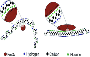
β-Poly(vinylidene fluoride) (PVDF) is of large technological relevance due to its piezoelectric, pyroelectric and/ferroelectric properties. In this way, a variety of methods have been developed to obtain such electroactive β-phase, being the addition of fillers one of the most popular, upscalable and innovative methods. The electrostatic interaction between negative charged fillers with the CH2 groups having a positive charge density has been the most widely accepted mechanism for the direct formation of polar β-phase on nanocomposites. Nevertheless some controversy remains in this matter as the dominating crystallization into the β-phase within PVDF is sometimes attributed to the interaction between the positively charged surfaces of the fillers and the CF2 dipoles in PVDF. In order to clarify such a controversial issue, this work uses two types of nanostructures, Fe3O4 nanorods and Fe3O4 nanoparticles, with distinct sizes and surface charges to study, isolate and evaluate the effects of the different ion–dipole interactions and shapes on the crystalline structures of PVDF. As a result it is shown that in the case of positive ion–CF2 dipole based β-phase nucleation, and beyond the effect of the intermolecular interactions, the rod-shape optimizes the crystallization in the electroactive conformation, thus promoting current development in PVDF-based electroactive devices.


1. Introduction

Electroactive polymers are among the most interesting classes of polymers used as smart materials in an increasing number of applications, such as sensors, actuators, filters, energy harvesters, membranes and as biomaterials in the biomedical field, among others.1–3 From the small class of polymers exhibiting piezo, pyro and/or ferroelectricity, such as Nylon-11, polylactic acid (PLLA) and poly(lactic-co-glycolic acid) (PLGA), poly(vinylidene fluoride) (PVDF) and its copolymers have the best all-around electroactive properties.4–6 Such properties, combined with its remarkable thermal stability, chemical resistance, high elasticity and transparency, biocompatibility, environmentally friendly and easy processing, make PVDF suitable for technological applications.7–10

This semi-crystalline polymer presents a complex structure and can reveal five different crystalline phases, each one related to different chain conformations designed as all trans (TTT) planar zigzag for the β-phase, TGTG (transgauchetransgauche) for the α and δ phases and T3GT3G for γ and ε phases.4,11–14

The β-phase is the one with the highest dipolar moment and spontaneous polarization per unit cell (8 × 10⁻³⁰ C m−2), the most electrically active phase and the one with highest piezoelectric coefficient (|d33| = 34 pC N−1),4,15 and for such reasons the technologically most interesting/desired one.16–19

With such remarkable properties, electroactive device applications are being used and developed based on the fine control of the processing conditions to achieve the β-phase of PVDF.4 Such β-phase of PVDF can be obtained by solvent-casting, mechanical stretching of the α-phase PVDF, and polarization under high electric fields,20 nevertheless all of the aforementioned methods usually induce undesired structural deformations or microstructural defects, or are complicated to implement for large scale industrial processing, which may hinder specific applications such as electro-optical sensors and non-volatile memories.4 In this way, alternative ways for obtaining the electroactive β-phase of PVDF have been established. One of the most innovative approaches was the addition of fillers such as BaTiO3, clays, hydrated ionic salts, PMMA, TiO2 and nanostructures (ferrite, palladium, gold and carbon nanotubes)4 into the polymer matrix during its melting processing,21 so that the material can be produced by conventional polymer processing technologies. Although the nucleation mechanism is not unambiguously stablished, most authors point to the fact that the essential factor for the nucleation of the β-phase on PVDF nanocomposites is the static electric interaction between the fillers with a negative surface potential and the CH2 groups having a positive charge density.22–27 The main evidence of such mechanism comes from of the inclusion of ferrite nanoparticles, that when were modified with positively charged molecules, lose the ability to induce the formation of the polar phase of PVDF.22

Nevertheless and surprisingly the induced formation of dominating β-phase in PVDF is sometimes attributed to the interaction between the positively charged surfaces of fillers and the CF2 dipoles in PVDF chains.20,24,28

Having this interesting discussion being centred on the surface charge of the nanostructures, a key factor was left out: the size and shape factor of the nanostructures, at the origin of specific electrical field geometries around the nanoparticle surface and to confinement effects.15,29 This issue is also particularly relevant when different nanoparticle shapes are increasingly being used for the development of novel sensors, such as anisotropic magnetoelectric magnetic field sensors.30

Considering also this parameter, our work proposes to create a unifying mechanism that could be used to explain the ambiguity and solve the existing controversy in the current literature about the nucleation of the β-phase of PVDF with nanostructures as well as to definitively set light on the significance of the key factors prompting the nucleation of the electroactive β-phase of this important electroactive polymer. In this way, this work will use two types of magnetic nanostructures, Fe3O4 nanorods and Fe3O4 nanoparticles, with distinct sizes and surface charges, to study, isolate and discuss the effects of the size, ion–dipole interactions and shape on the formation of the β-phase crystalline structure of PVDF.

2. Experimental methods

2.1 Materials

Potassium nitrate (KNO3, ≥99%) was purchased from Fluka (Buchs, Switzerland), ethanol absolute from Panreac (Barcelona, Spain), iron(III) acetylacetonate ([Fe(acac)3], ≥97%), ferrous sulfate heptahydrate (FeSO4·7H2O, ≥99%), FeCl2 4H2O (99%), sodium citrate tribasic dihydrate (99%), triethylene glycol (TREG, 99%), sodium hydroxide (NaOH, ≥98%), L-lysine crystallized (≥98%), hydrogen peroxide (H2O2, 30%), oleylamine (70%) branched polyethyleneimine (PEI, Mw 25[thin space (1/6-em)]000), sulfuric acid (H2SO4, 95–98%), hexane (99.5%), toluene (99.5%) and dimethylformamide (DMF, 99.8%) and ethyl acetate (HPLC grade) were purchased from Sigma-Aldrich (St. Louis, USA). All the chemicals were used as received without further purification.

2.2 Methods

2.2.1 Synthesis of 9 nm Fe3O4 magnetic nanoparticles (MNPs). Water-soluble superparamagnetic iron oxide nanoparticles were synthesized by a polyol-mediated method scaling up the synthesis procedure described previously.31,32 4.5 g of [Fe(acac)3] were vigorously mixed with 90 mL of TREG in a three-neck round-bottom flask equipped with a mechanical stirrer and degassed with Ar. The resulting mixture was heated at 15 °C min−1 to 180 °C and held at this temperature for 30 min in order to achieve the decomposition of the [Fe(acac)3] precursor. After that, the mixture was heated again at 5 °C min−1 to reach 280 °C and kept at this temperature for 30 min under reflux. The resulting dark solution was cooled to room temperature. After the reaction, the particles were washed 3 times with a mixture of ethyl acetate and ethanol (9[thin space (1/6-em)]:[thin space (1/6-em)]1) and separated using a magnet. The precipitated nanoparticles after washing cycle were redispersed in polar solvents such as water.
2.2.2 Synthesis of 30 nm and 50 nm Fe3O4 MNPs. Fe3O4 magnetic nanoparticles with an average particle size of 30 nm and 50 nm were synthesized by an oxidative hydrolysis method, following a synthesis pathway described in previous works.33 Briefly, 30 nm Fe3O4 MNPs were produced in a two-stage continuous flow PTFE-microreactor. The reagent solutions were prepared as follows. In a 500 mL vessel (solution 1), a 444 mL solution of 180 mM KNO3, 162 mM NaOH and 1.85 mM L-lysine was prepared using deionized water. In other 500 mL vessel (solution 2), a 444 mL solution of 13 mM ferrous sulfate heptahydrate and 3.38 mM sulfuric acid was prepared using deionized water. Argon gas was bubbled in each solution for 15 minutes. After deoxygenation, solution 1 and solution 2 were filled in a 60 mL plastic Becton Dickinson syringe. In the first stage, solutions were pumped at a flow rate of 1.87 mL min−1 with residence time of 1 min and were mixed in a PEEK polymer Y junction located in an ultrasound bath. Sonication was carried out by maintaining the temperature in between 25 and 30 °C, using a cooling bath. In a second stage a H2 gas stream was introduced in a PEEK Y junction to form a H2-liquid segmented flow and heated at 100 °C. The synthesized nanoparticles were centrifuged at 10[thin space (1/6-em)]000 rpm for 10 minutes, then washed twice with distilled water and finally re-suspended in distilled water.

50 nm Fe3O4 MNPs were produced in a mechanical mixed batch reactor. A 40 mL aqueous solution 0.1 mM KNO3, 90 mM NaOH and 1 mM of L-lysine were bubbled with Argon during 15 minutes. Subsequently, 4.4 mL of an aqueous solution containing 65 mM FeSO4·7H2O and of 17 mM H2SO4 was added dropwise under constant stirring. When the addition was completed, argon was allowed to pass for another 15 minutes and the suspension was heated at 90 °C for 1 hour in an oil bath. The synthesized nanoparticles were centrifuged at 10[thin space (1/6-em)]000 rpm for 10 minutes, then washed twice with distilled water and finally re-suspended in distilled water.

2.2.3 Synthesis of Fe3O4 nanorods. Magnetite (Fe3O4) nanorods (NRs) were obtained by reduction of as prepared feroxyhyte (β-FeOOH) NRs, according to the method reported by Aslam et al.34 In a typical synthesis, β-FeOOH NRs were prepared by mixing 5.6 g of FeCl3 6H2O and 0.2 mL of PEI in 100 mL of deionized water. The mixture was heated to 80 °C under stirring in a reaction flask during two hours. The resulting NRs were washed by centrifugation at 6000 rpm during 15 min.

β-FeOOH NRs were reduced to Fe3O4 NRs using oleylamine (9 mmol) mixed with 120 mg of β-FeOOH NRs in a three necked round bottom flask under argon atmosphere. The mixture was heated to 200 °C during 4 hours under vigorous stirring. The product was precipitated by magnetic separation in mixtures hexane[thin space (1/6-em)]:[thin space (1/6-em)]acetone (1[thin space (1/6-em)]:[thin space (1/6-em)]1) to remove the excess of oleylamine.


NRs surface modification with PEI. Fe3O4 NRs were treated to surface modification with polyethyleneimine (PEI) to make them stable. The ligand exchange was done by heating 100 mg of NRs dispersed in 10 mL of toluene and 2 mL of PEI in 10 mL of dimethylformamide (DMF). The solution was heated at 80 °C overnight under argon atmosphere. After the reaction solution was cooled to room temperature, NRs were washed by magnetic separation washing three times with ethanol to remove the excess of PEI, and finally dispersed in water.
2.2.4 Structural characterization of the nanoparticles. Transmission electron microscopy observations were performed with a T20-FEI microscope with a LaB6 electron source fitted with a “SuperTwin” objective lens allowing a point-to-point resolution of 2.4 Å. The phases of iron oxide nanoparticles were identified by powder X-ray diffraction (XRD). The X-ray patterns were collected between 20° and 80° (2θ) in a D-Max Rigaku diffractometer with Cu Kα radiation.
2.2.5 Fabrications of Fe3O4 nanostructures/PVDF composites. Aiming obtain a good nanostructure dispersion it was used and experimental procedure that ensures a good dispersion of nanofillers,30,35 briefly a mixture of magnetic nanostructures in DMF (Sigma-Aldrich) was first placed in an ultrasound bath for 8 h. After this period of time, PVDF powder (Solvay) was added to the solution. Complete dissolution of the polymer was achieved by using a Teflon mechanical stirrer incorporated in the ultrasound bath during 1 h. Flexible nanocomposite films with an average thickness of ≈50 μm and with filler content of 1 weight percent (wt%) were obtained by spreading the solution at room temperature on a clean glass substrate. Such wt% of magnetic nanofiller was chosen as previous studies show that such content is capable to effectively nucleate a significant amount of the polymer in the β-phase,4,21,22,36 suitable for the present investigation.

Four distinct magnetic/PVDF nanocomposites were obtained: composites with ≈9 nm magnetite (Fe3O4) nanoparticles (NP 9 nm); with ≈30 nm magnetite nanoparticles (NP 30 nm); with ≈50 nm magnetite nanoparticles (NP 50 nm); and with magnetite nanorods (NR).

Solvent evaporation was carried out, after sample melting, at a controlled temperature of 210 °C inside an oven (SELECTA, 2000).

2.2.6 Characterization of the iron oxide nanostructures/PVDF composites. The vibrational modes of the polymer, used to determine the polymer phase and phase content, were recorded by Fourier transformed infrared spectroscopy (FTIR) using a Thermo Nicolet Nexus 670 FTIR spectrophotometer from 650 to 4000 cm−1 with a resolution of 2 cm−1. 64 scans were performed for each sample. The thermal behaviour of the samples was determined by Thermo Gravimetric Analysis (TGA). Samples were transferred to open alumina pans with capacity of 60 μL and analysed using a TGA METTLER TOLEDO 822e, operating between room temperature and 975 °C. A heating rate of 10 °C min−1 and a nitrogen flow rate of 50 mL min−1 were used.

Zeta potential measurements were performed on a Malvern Zetasizer nano ZS, with an operating range between 0.3 nm to 10 microns, with a laser HeNe 633 nm, max 4 mW.

The morphology of composites was studied by scanning electron microscopy (SEM) using a Quanta 650 FEI electron microscope with acceleration voltage of 3 kV. Prior to SEM, the samples were coated with gold by magnetron sputtering.

3. Results/discussion

3.1 Nanoparticles

Fig. 1 shows representative TEM micrographs of the as-obtained magnetic nanomaterials. Fig. 1a, b, d and e show that the morphology of the nanoparticles produced by an oxidative hydrolysis method was octahedral with a rather uniform size. The particle size distribution was determined by statistical analysis of the dimensions of at least 100 nanoparticles measured on the TEM micrographs. Fitting to a log-normal distribution yielded an average particle size of 47.7 ± 9.6 nm and 29.5 ± 7.2 nm.
image file: c6ra24356h-f1.tif
Fig. 1 Morphology and magnetic characteristic of the obtained magnetic nanomaterials nanoparticles produced by oxidative hydrolysis: (a and b) TEM images of nanoparticles D = 50 nm, (c) magnetization curve at 300 K. (d and e) TEM images of nanoparticles D = 30 nm, (f) magnetization curve at 300 K. Nanoparticles produced by the polyol-mediated method. (g and h) TEM images of nanoparticles D = 9 nm. (i) magnetization curve at 300 K. Anisotropic growth of iron oxide nanostructures. (j and k) TEM images of anisotropic nanostructures , (l) magnetization curve at 300 K of anisotropic nanostructures.

Fig. 1g and h shows the nanoparticles produced by the polyol-mediated method, obtaining nanoparticles of roughly spherical morphology, rather uniform in size (average size 9.2 ± 2.3 nm), and well dispersed. The typical X-ray diffraction patterns of the 50 nm, 30 nm and 9 nm nanoparticles are shown in Fig. 2a. The XRD patterns of these samples were assigned to the phase of bulk magnetite (JCPDS card number 89-0691). The magnetization curves of the iron oxide nanoparticles, with average size of D = 50 nm and D = 30 nm show a ferromagnetic behaviour with a coercivity (Hc) of 110 and 65 Oe respectively (Fig. 1c and f). The saturation magnetization (Ms) at 40[thin space (1/6-em)]000 Oe is 84 emu g−1 for magnetite 50 nm and 82 emu g−1 for magnetite 30 nm. However, the magnetization curve of magnetite D = 9 nm shows a superparamagnetic behaviour with a small coercivity (Hc) of 9 Oe and a saturation magnetization (Ms) of 59.6 emu g−1 at 40[thin space (1/6-em)]000 Oe. These results are consistent with the literature37 and the fact that magnetite loses its permanent magnetism when its size is less than 20 nm, becoming superparamagnetic.38


image file: c6ra24356h-f2.tif
Fig. 2 XRD patterns of the prepared magnetic nanomaterials (the characteristic patters of magnetite Fe3O4 are included for comparison).

The anisotropic growth of magnetic nanomaterials has promising advantages over the spherical shape in areas such as lithium-ion batteries, gas sensors magnetic compasses,30,39 the use of NPs with an anisotropic configuration has not been properly demonstrated in the literature because their preparation is a challenging task as surface energy considerations favour the formation of spherical nanoparticles.34 The production of magnetic nanorods was conducted in this work by a novel and simple procedure, leading to FeOOH nanorods with an aspect ratio higher than 8 and a rod diameter of approx. 13 nm. After a thermal treatment, hematite nanorods were obtained, maintaining the morphological structure (Fig. 1j and k). The XRD pattern in Fig. 2b confirms the transformation of FeOOH to Fe3O4 (JCPDS card 89-0691).

The magnetization curves of Fe3O4 nanorods show a ferromagnetic behaviour with a coercivity (Hc) of 120 Oe and a saturation magnetization (Ms) of 0.8 emu g−1 at 40[thin space (1/6-em)]000 Oe. The low magnetization values of the NRs is been attributed to the existence of a surface spin disorder layer which decreases with the particle size.34

3.2 Fe3O4/PVDF nanocomposites

Fourier transform infrared spectroscopy (FTIR) has been proven to give quantitative information about PVDF structure allowing to distinguish and quantify the different crystalline forms. In particular, specific bands such as the ones at 766 and 840 cm−1 have been identified to correspond to the α and β-phase of PVDF, respectively.4 These specific bands have been used for identification and quantification of PVDF phases in the present work.

By assuming that the infrared absorption follows the Lambert–Beer law, for a system containing both α and β-phases, the relative β-phase content, F(β), can be determined using eqn (1):40

 
image file: c6ra24356h-t1.tif(1)
where F(β), represents the β-phase content; Aα and Aβ the absorbance at 766 and 840 cm−1; Kα and Kβ the absorption coefficients at the respective wavenumber (6.1 × 10⁴ and 7.7 × 104 cm2 mol−1), respectively.

Typical spectra and the variation of the relative fraction of the β-phase for the nanocomposites prepared with each different of filler are presented in Fig. 3.


image file: c6ra24356h-f3.tif
Fig. 3 (a) Infrared spectra of the magnetite/PVDF nanocomposites; (b) electroactive β-phase content of the magnetite/PVDF nanocomposites samples calculated from the infrared spectra by eqn (1). The inset reveals a typical SEM image of the magnetite/PVDF nanocomposites.

Fig. 3 shows that just Fe3O4 rod nanostructures (RD) successfully nucleate the β-phase of the polymer as indicated by the appearance of the β-phase peak at 840 cm−1 and the vanishing of the α-phase peak at 766 cm−1 (Fig. 1a). Additionally, the β-phase peak at 1279 cm−1 removes any possible confusion on the identification of the piezoelectric phase. The other piezoelectric δ-phase do not exhibit such peak “as a shoulder”.41

Fig. 3b shows that both nanocomposites with Fe3O4 nanoparticles with 50 nm and 30 nm (NP 50 nm and NP 30 nm) have the lowest percentage of β-phase (6%), and the nanocomposite with Fe3O4 nanoparticles with 9 nm reveals 10% of β-phase, being the remainder in the α-phase. FTIR results raise the possibility that can exist a shape factor in the mechanism of the β-phase formation, once nanocomposites with Fe3O4 nanorods (NR) reveal a much larger nucleation effect (F(β) ≈ 70%) than nanocomposites with Fe3O4 nanoparticles (NP 50 nm, NP 30 nm and NP 9 nm), independently of the size of the NP.

The absence of visible aggregates on the inset of Fig. 3b reflects the good distribution of nanostructures inside the polymer matrix.

Trying to investigate whether the amount of polymer that is electrostatically interacting the surface of the nanostructure (interface) has some influence on the β-phase nucleation ability, the percentage of polymer located at such interface was determined by TGA (Fig. 4).


image file: c6ra24356h-f4.tif
Fig. 4 (a) TG thermograms of magnetite/PVDF nanocomposites; (b) interface weight percentage for each of the magnetite/PVDF nanocomposites.

The mass fraction of the polymer located at the interface, mI, was determined following the procedure presented in,42 by using eqn (2):

 
image file: c6ra24356h-t2.tif(2)
where mI0 is the mass of the pristine polymer at the temperature at which the mass loss rate is maximum and m(x)I0 is the mass of the composite that has not degraded at the temperature at which the mass loss rate of the pristine polymer is maximum. The difference between mI0 and m(x)I0 values is related with the enhancement of the thermal stability of the polymer chains located at the interface. In this way, is this interface that is responsible for the overall increase of the thermal stability of nanocomposites.

For all Fe3O4/PVDF nanocomposites is observed the characteristic two step (i and ii) thermal degradation mechanism typical of PVDF.43 The first degradation step occurs between 400 and 500 °C, being the PVDF maximum degradation temperature dependent on the presence of the nanostructures within the polymer. In this first step, the degradation mechanism is chain-stripping where carbon-hydrogen and carbon fluorine scission occurs and the presence of both hydrogen and fluorine radicals leads to the formation of hydrogen fluoride,22 leading to the weight loss observed in the first degradation step.

In turn, the second degradation step occurs between 500 and 600 °C, and the detected differences in the plots when compared to the pure PVDF are attributed to the presence of the nanostructures, as the thermal degradation is independent of the polymer phase. This second step is a complex degradation procedure resulting in poly(aromatization). The polymeric sequence formed previously on the first degradation step is unstable and, therefore, the formed macromolecules undergo further reactions leading to scission followed by the formation of aromatic molecules.43 The residual weight that remains at high temperatures corresponds mainly to the nanostructures together with the residual char from the previous degradation steps.42

An extra degradation step (iii) at temperatures around 600 °C, when compared to the pure polymer was also identified in the nanocomposite samples. The emergence such new step of degradation in comparison to pure PVDF is related to the increase of an interphase in the interface volume between nanoparticles and polymer.

The onset temperature for the degradation of neat PVDF (452 °C) is lower than those of Fe3O4/PVDF nanocomposites (456 °C, 457 °C, 460 and 465 for the NP 30 nm, RD, NP 9 nm and NP 50 nm respectively), indicating that the thermal stability of the matrix is improved with the addition of Fe3O4 nanostructures. By using data from Fig. 4a and eqn (2), it is possible to determine that NP 9 nm shows the lower interface value (2%) and the other NP samples exhibited similar interface values (≈11%). The low interface value of the NP 9 nm sample can be related with the formation of clusters and aggregation of Fe3O4 nanoparticles. The RD composite sample revealed ≈50% of interface value, indicating the anisotropic structure of Fe3O4 rods potentiates the existence of more amount of polymer in the nanostructure/polymer interface. Such interface is related with the amount of polymer that is electrostatically interacting the surface of the nanostructure.

By comparing Fig. 3b and 4b, and contrary to other studies,42 there is no obvious relation between β-phase content and interface values.

Finally, zeta potential analysis was used to evaluate the electrostatic charge on the surface of the nanostructures in order to determine its influence on the β-phase nucleation mechanism, once previous studies indicated this as the main factor responsible for the β-phase nucleation.20,24,28,44

All nanostructures revealed positive surface charges, being 8 mV and 11 mV the values for NP 30 nm and NP 50 nm, respectively. Interestingly both NP 9 nm and RD nanocomposite revealed the same value of surface charge: 30 mV.

Previous works21,36 have shown that negative surfaces charges (−22 mV) on magnetic nanoparticles promoted F(β) ≈ 20% on PVDF composites with the same nanoparticles content (1 wt%). The low value of F(β) (≤10%) found for the present NP composites is thus related to the positive values of zeta-potential, once the electrostatic interaction between the positively charged magnetic spherical nanoparticles and the polymer is less intense. The low β-phase nucleation observed for NP nanocomposites confirms once again the previous experimental evidences detected CoFe2O4@cetrimonium bromide and CoFe2O4@citric acid magnetic PVDF nanocomposites: magnetic spherical nanoparticles with positive surface charge are not suitable for the nucleation of the β-phase of the polymer.22,36 Further, this fact is independent of the size of the spherical NP.

On the other hand, the positively surface charged Fe3O4 rods (30 mV) reveals a F(β) of ≈70%. This interesting fact indicates an underlying factor other than the electrostatic interaction between nanofillers and polymer for the β-phase formation on PVDF nanocomposites. The advantageous β-phase formation by Fe3O4 nanorods with respect to their spherical counterparts indicates directly at the relevance of the shape on the β-phase formation mechanism. Thus, it is possible to highlight that the crystallization of PVDF in its β-phase through positive ion–CF2 dipole interactions is assisted via nanostructures (in the form of substrates or templates) endowed with 1D (rod/tube) or 2D (planar) geometry through the organization of bonding sites, once β-phase conformation can extend beyond local ordering15 (Fig. 5b). Through arranged bonding sites, β-phase conformation can be induced and extended. In this way, can now be understood that radial intermolecular interaction surrounding positively charged Fe3O4 nanoparticles may not be able to retain the polar conformation in long range order with competing entropy.15 However, the Fe3O4 rod shape is capable to induce such β-phase conformation beyond the local ordering, thus leading to the formation of the polar β-phase along most of the polymer crystalline phase. Confinement effects on the crystallization of the PVDF with nanorod fillers may also allow a long range arrangement that optimizes the interaction between fillers and polymer chains, leading to the PVDF's β-form.15,29


image file: c6ra24356h-f5.tif
Fig. 5 (a) Zeta potential of the different magnetite nanostructures; (b) schematic representation of the β-phase nucleation mechanism through positive ion–CF2 dipole interactions.

Additionally the rod shape can induce a phase transition from an ambient coiled state to a stretched conformation (known as the coil-stretch transition) which is helpful for the conversion of the helix TGTG (α-phase) conformation to the zigzag TTT one (β-phase).45

In this way it is possible to conclude that:

(a) Negatively charged nanofillers promote the β-phase of PVDF nucleation thought negative ion–CH2 dipole interactions;

(b) Nanoparticles with positive surface charge fail to nucleate the β-phase of PVDF and;

(c) Rod or planar nanofillers with positive surface charge assist the nucleation of β-PVDF thought positive ion–CF2 dipole interactions, favoured by confinement effects.

Such conclusion is supported by the data of the present work and by other reported cases in the literature: (a) negatively charged ferrite nanoparticles nucleate the β-phase of the polymer and positively charged ferrite nanoparticles failed to induce the polar β-PVDF;21–23,36 (b) nearly spherical Ag and BaTiO3 cannot improve the β-phase formation;46,47 (c) multi-walled carbon nanotubes promote the β-phase growth even before post-processing;17 (d) graphene induces polar phase formation;48 (e) optimized β-phase formation by m-SiO2 nanorods when compared to their spherical counterparts15 and (f) flat surface of organoclays facilitates the β-phase formation on PVDF composites.28 Hence, both anisotropic shape and negatively charged surfaces benefit the β-phase conformation in PVDF nanocomposites.

4. Conclusions

In this work it was developed a simple and efficient method to synthesize β-PVDF films by incorporating Fe3O4 nanorods. The effects of surface charge and filler structure on the crystallization behaviour of PVDF were systematically studied by using two types of nanostructures, Fe3O4 nanorods and Fe3O4 spherical nanoparticles of different sizes, with distinct surface charges, in order to study, isolate and discuss the effects of the different ion–dipole interactions, size and shape on the nucleation of the β-phase of PVDF.

Having almost the same interface values (≈10%), different Fe3O4/PVDF composites exhibited distinct β-phase values. Fe3O4 nanoparticles with different sizes and distinct zeta potential values; 9 nm (30 mV), 30 nm (8 mV) and 50 nm (11 mV) failed to induce high fraction values of β-phase within the composites. In contrast Fe3O4 rods (30 mV) nucleated ≈70% of β-PVDF within the nanocomposites.

When compared to their spherical nanoparticle counterparts, the use of Fe3O4 nanorods has the advantages of anisotropic shape and organized crystallization that direct polar molecular conformation towards oriented growth of β-PVDF. Besides optimizing intermolecular interaction, nanorods promote a confinement effect essential to induce oriented ordering of molecular conformation towards long range order. The proposed β-phase nucleation mechanism could be used to explain unambiguously the existing controversy in current literature reports for the β-phase nucleation on PVDF nanocomposites and can be taken to advantage for the crystallization of β-PVDF form with a variety of intermolecular interactions, which largely benefits and dynamizes current development in PVDF based materials for electroactive devices applications.

Acknowledgements

The authors thank the FCT-Fundação para a Ciência e Tecnologia-for financial support in the framework of the Strategic Funding UID/FIS/04650/2013 and under project PTDC/EEI-SII/5582/2014. P. Martins and R. Gonçalves acknowledges also support from FCT (SFRH/BPD/96227/2013 and SFRH/BD/88397/2012 grants respectively). Financial support from the Basque Government Industry Department under the ELKARTEK Program is also acknowledged. VS thanks The People Program (CIG-Marie Curie Actions, REA grant agreement no. 321642) the Government of Aragon, and the European Social Fund.

References

  1. Y. Bar-Cohen and Q. Zhang, MRS Bull., 2008, 33, 173–181 CrossRef CAS.
  2. R. Kumaran, S. D. Kumar, N. Balasubramanian, M. Alagar, V. Subramanian and K. Dinakaran, J. Phys. Chem. C, 2016, 120, 13771–13778 CAS.
  3. T. W. Chuo, J. M. Yeh and Y. L. Liu, RSC Adv., 2016, 6, 55593–55598 RSC.
  4. P. Martins, A. C. C. Lopes and S. Lanceros-Mendez, Prog. Polym. Sci., 2014, 39, 683–706 CrossRef CAS.
  5. L. Dos Santos, O. Dubrunfaut, J. C. Badot and C. Laberty-Robert, J. Phys. Chem. C, 2016, 120, 6963–6970 CAS.
  6. X. Dai, J. Niu, Z. Ren, X. Sun and S. Yan, J. Phys. Chem. B, 2016, 120, 843–850 CrossRef CAS PubMed.
  7. C. Tsonos, C. Pandis, N. Soin, D. Sakellari, E. Myrovali, S. Kripotou, A. Kanapitsas and E. Siores, eXPRESS Polym. Lett., 2015, 9, 1104–1118 CrossRef CAS.
  8. J. Nunes-Pereira, P. Martins, V. F. Cardoso, C. M. Costa and S. Lanceros-Méndez, Mater. Des., 2016, 104, 183–189 CrossRef CAS.
  9. A. C. Lopes, C. M. Costa, C. J. Tavares, I. C. Neves and S. Lanceros-Mendez, J. Phys. Chem. C, 2011, 115, 18076–18082 CAS.
  10. M. Sharma, V. Srinivas, G. Madras and S. Bose, RSC Adv., 2016, 6, 6251–6258 RSC.
  11. M. Sharma, K. Sharma and S. Bose, J. Phys. Chem. B, 2013, 117, 8589–8602 CrossRef CAS PubMed.
  12. W. J. Kim, M. H. Han, Y. H. Shin, H. Kim and E. K. Lee, J. Phys. Chem. B, 2016, 120, 3240–3249 CrossRef CAS PubMed.
  13. Y. Hu, P. Xu, H. Gui, S. Yang and Y. Ding, RSC Adv., 2015, 5, 92418–92427 RSC.
  14. L. Li, M. Zhang, M. Rong and W. Ruan, RSC Adv., 2014, 4, 3938–3943 RSC.
  15. D. Yuan, Z. Li, W. Thitsartarn, X. Fan, J. Sun, H. Li and C. He, J. Mater. Chem. C, 2015, 3, 3708–3713 RSC.
  16. B. Fan, F. Bedoui, S. Weigand and J. Bai, J. Phys. Chem. C, 2016, 120, 9511–9519 CAS.
  17. Y. Ahn, J. Y. Lim, S. M. Hong, J. Lee, J. Ha, H. J. Choi and Y. Seo, J. Phys. Chem. C, 2013, 117, 11791–11799 CAS.
  18. A. Kuila, N. Maity, D. P. Chatterjee and A. K. Nandi, J. Phys. Chem. B, 2016, 120, 2557–2568 CrossRef CAS PubMed.
  19. P. Thakur, A. Kool, B. Bagchi, N. A. Hoque, S. Das and P. Nandy, RSC Adv., 2015, 5, 62819–62827 RSC.
  20. C. L. Liang, Z. H. Mai, Q. Xie, R. Y. Bao, W. Yang, B. H. Xie and M. B. Yang, J. Phys. Chem. B, 2014, 118, 9104–9111 CrossRef CAS PubMed.
  21. P. Martins, C. M. Costa and S. Lanceros-Mendez, Appl. Phys. A: Mater. Sci. Process., 2011, 103, 233–237 CrossRef CAS.
  22. P. Martins, C. M. Costa, M. Benelmekki, G. Botelho and S. Lanceros-Mendez, CrystEngComm, 2012, 14, 2807–2811 RSC.
  23. S. Roy, P. Thakur, N. A. Hoque, B. Bagchi and S. Das, RSC Adv., 2016, 6, 21881–21894 RSC.
  24. L. Yu and P. Cebe, Polymer, 2009, 50, 2133–2141 CrossRef CAS.
  25. Y. Li, J. Z. Xu, L. Zhu, G. J. Zhong and Z. M. Li, J. Phys. Chem. B, 2012, 116, 14951–14960 CrossRef CAS PubMed.
  26. C. Xing, L. Zhao, J. You, W. Dong, X. Cao and Y. Li, J. Phys. Chem. B, 2012, 116, 8312–8320 CrossRef CAS PubMed.
  27. Y. Wu, S. L. Hsu, C. Honeker, D. J. Bravet and D. S. Williams, J. Phys. Chem. B, 2012, 116, 7379–7388 CrossRef CAS PubMed.
  28. J. Wang, Q. Fu and Q. Zhang, Polymer, 2012, 53, 5455–5458 CrossRef CAS.
  29. M. C. García-Gutiérrez, A. Linares, I. Martín-Fabiani, J. J. Hernández, M. Soccio, D. R. Rueda, T. A. Ezquerra and M. Reynolds, Nanoscale, 2013, 5, 6006–6012 RSC.
  30. P. Martins, A. Larrea, R. Gonçalves, G. Botelho, E. V. Ramana, S. K. Mendiratta, V. Sebastian and S. Lanceros-Mendez, ACS Appl. Mater. Interfaces, 2015, 7, 11224–11229 CAS.
  31. W. Cai and J. Q. Wan, J. Colloid Interface Sci., 2007, 305, 366–370 CrossRef CAS PubMed.
  32. N. Miguel-Sancho, O. Bomati-Miguel, G. Colom, J. P. Salvador, M. P. Marco and J. Santamaria, Chem. Mater., 2011, 23, 2795–2802 CrossRef CAS.
  33. A. Larrea, V. Sebastian, A. Ibarra, M. Arruebo and J. Santamaria, Chem. Mater., 2015, 27, 4254–4260 CrossRef CAS PubMed.
  34. J. Mohapatra, A. Mitra, H. Tyagi, D. Bahadur and M. Aslam, Nanoscale, 2015, 7, 9174–9184 RSC.
  35. P. Martins, R. Gonçalves, S. Lanceros-Mendez, A. Lasheras, J. Gutiérrez and J. M. Barandiarán, Appl. Surf. Sci., 2014, 313, 215–219 CrossRef CAS.
  36. P. Martins, C. Caparros, R. Gonçalves, P. M. Martins, M. Benelmekki, G. Botelho and S. Lanceros-Mendez, J. Phys. Chem. C, 2012, 116, 15790–15794 CAS.
  37. A. Larrea, V. Sebastian, M. Arruebo and J. Santamaria, International Journal of Chemical, Molecular, Nuclear, Materials and Metallurgical Engineering, 2015, 103, 780–786 Search PubMed.
  38. R. Hao, R. J. Xing, Z. C. Xu, Y. L. Hou, S. Gao and S. H. Sun, Adv. Mater., 2010, 22, 2729–2742 CrossRef CAS PubMed.
  39. C. Wu, P. Yin, X. Zhu, C. OuYang and Y. Xie, J. Phys. Chem. B, 2006, 110, 17806–17812 CrossRef CAS PubMed.
  40. R. Gregorio Jr and M. Cestari, J. Polym. Sci., Part B: Polym. Phys., 1994, 32, 859–870 CrossRef.
  41. P. Martins, A. C. Lopes and S. Lanceros-Mendez, Prog. Polym. Sci., 2014, 39, 683–706 CrossRef CAS.
  42. P. Martins, C. M. Costa, M. Benelmekki, G. Botelho and S. Lanceros-Méndez, J. Mater. Sci., 2013, 48, 2681–2689 CrossRef CAS.
  43. G. Botelho, S. Lanceros-Mendez, A. M. Gonçalves, V. Sencadas and J. G. Rocha, J. Non-Cryst. Solids, 2008, 354, 72–78 CrossRef CAS.
  44. T. E. Quickel, L. T. Schelhas, R. A. Farrell, N. Petkov, V. H. Le and S. H. Tolbert, Nat. Commun., 2015, 6, 6562 CrossRef CAS PubMed.
  45. F. Bai, G. Chen, M. Nie and Q. Wang, RSC Adv., 2015, 5, 54171–54174 RSC.
  46. L. Gao, J. He, J. Hu and Y. Li, J. Phys. Chem. C, 2014, 118, 831–838 CAS.
  47. D. W. Chae and S. M. Hong, J. Polym. Sci., Part B: Polym. Phys., 2010, 48, 2379–2385 CrossRef CAS.
  48. L. Zhang, D. A. Zha, T. Du, S. Mei, Z. Shi and Z. Jin, Langmuir, 2011, 27, 8943–8949 CrossRef CAS PubMed.

Footnote

Equal contribution.

This journal is © The Royal Society of Chemistry 2016
Click here to see how this site uses Cookies. View our privacy policy here.