DOI:
10.1039/C6RA24282K
(Paper)
RSC Adv., 2016,
6, 113859-113870
Regeneration ability of valvular interstitial cells from diseased heart valve leaflets
Received
29th September 2016
, Accepted 16th November 2016
First published on 16th November 2016
Abstract
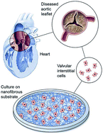
Regeneration of heart valves depends on the regeneration of cells residing in the heart valve leaflet, i.e., valvular interstitial cells (VICs). In this study, the regeneration capacity of VICs obtained from healthy and diseased valves of patients with various ages and genders were investigated. The cells obtained from the valves were cultured in vitro for 21 days on our developed standalone nanofibrous substrates. The substrates were pliable. Proliferation of cells, their morphologies, collagen deposition, and gene and protein expression were assayed to compare their regeneration capacity. VICs from healthy valves exhibited higher proliferation and cell spreading in comparison to VICs from diseased valves. However, collagen deposition by VICs from diseased valves was higher compared to the deposition by VICs from healthy valves, irrespective of age and gender of the patients. VICs on nanofibrous substrates showed high vimentin and collagen type I expression; expression of α-SMA showed the reverse trend demonstrating the importance of nanofibers in heart valve tissue engineering and regeneration. Although variations in proliferation, gene, and protein expression of VICs from healthy and diseased valves of patients with various ages and genders were observed, they were not statistically significant. Thus, VICs from a diseased valve maintain the potential to regenerate in a non-degenerative environment, providing an opportunity for tissue engineering of diseased valves.
Introduction
Heart valve disease is one of the cardiovascular diseases that causes substantial illness and death worldwide.1–3 The number of patients requiring heart valve replacement is expected to triple from approximately 290
000 in 2003, to over 850
000 by 2050.4 Degenerative pathology in developed nations and rheumatic fever in developing countries are the main causes of heart valve disease in adults.5,6 Congenital heart valve defects are found in 1 to 2% of the child population.7
Heart valve degeneration compromises valve integrity, resulting in stenotic or incompetent valves.8 This degeneration tends to occur primarily in the leaflet, consisting of circumferentially, randomly and radially arranged collagen fibrils in three successive layers with the primary cellular elements comprising of fibroblasts known as valvular interstitial cells (VICs).9 Heart valves cannot regenerate naturally.10 Currently, mechanical and bioprosthetic valves are the replacement options; however, they face some drawbacks.1–3,11 Mechanical valves have excellent durability but are limited by thrombogenicity and the patient's need for life-long anticoagulant treatment. Bioprosthetic valves, in contrast, are less durable and prone to degeneration, but are less thrombogenic. Both mechanical and bioprosthetic valves cannot grow or remodel, so they are not viable as a longterm solution for paediatric patients. Further, as bioprosthetic valves are less durable, adult patients need successive replacements of valves.
An alternative solution to the above problems could be artificial regeneration of the heart valve or a tissue engineering device.1–3,11 In heart valve regeneration, drug delivery techniques are applied.10 Drug delivery including administration of biologic and pharmacological agents is the standard procedure for treating the diseased valve.1,12 The success of this technique depends on the regeneration capability of the residing VICs, i.e. ability of VICs to proliferate, grow and deposit collagen fibrils adequately. To the best of our knowledge, no reports are available that explore their regeneration abilities through in vivo procedures. As an alternative, an in vitro mechanism using two-dimensional nanofibrous substrates with physical and mechanical properties similar to that of leaflets could be ideal to explore the regeneration capability of VICs. Nanofibers mimic the morphology of the extracellular matrix including collagen fibrils of the heart valve leaflets.9 For the same reason scaffold substrates made of nanofibers have potential in tissue engineering.13–15
Two-dimensional nanofibrous membrane-based substrates are often used to study the cellular behaviours such as phenotype, shape and size of the cells.16–18 However, the nanofibrous membrane cannot be used as a standalone membrane without any underlying supporting substrate such as glass coverslip. Higher mechanical properties of the underlying supporting substrate compared to a membrane could impact the cellular behaviours. In our previous work, we have shown the effect of glass coverslips used as an underlying supporting substrate of a circumferentially oriented nanofibrous membrane on cultured valvular interstitial cells.19 Therefore, we developed a novel method to produce standalone randomly oriented nanofibrous membrane-based substrates through an electrospinning technique and applied it to study the regeneration ability of VICs from diseased heart valve leaflets.
We hypothesize that regeneration ability of VICs differs with gender and age of the patient and with the nature of their heart valve diseases. Therefore, to verify this hypothesis, we collected VICs from diseased and healthy aortic heart valve leaflets and cultured them on the developed standalone randomly oriented nanofibrous substrate for 21 days. VICs from healthy heart valve leaflets were considered as a positive control. Proliferation, orientations, growth, and gene and protein expression of the VICs were investigated to assess their ability in heart valve regeneration.
Results
Standalone nanofibrous substrate and its properties
Nanofibrous membrane-based substrates were produced from polycaprolactone (PCL) polymer through an electrospinning setup described in detail in the method section. In brief, a metal ring with diametrically exiting holes (Fig. 1a) was fitted to a metal holder with two pins (Fig. 1b) to build a collector (Fig. 1c) which was used to develop a randomly oriented nanofibrous substrate (Fig. 1d) with this electrospinning technique. Randomly oriented nanofibers were deposited on the metal ring as well as on the inner area of the ring. In general, nanofibers were supposed to be deposited on the ring; however, comparatively longer nanofibers would come across the metal ring including its inner space and make a randomly oriented nanofibrous layer in the inner space area. Naturally, more nanofibers were deposited on the ring, than on the inner space area of the ring and thus, the nanofibrous layer on the metal ring was much thicker than the layer in the inner area of the ring. The thinner inner nanofibrous layer with an outer thicker supporting nanofibrous layer was detached from the collector to obtain a standalone randomly oriented nanofibrous substrate (Fig. 1e). The thicker layer worked as a supporting substrate for the thinner layer in the inner space of the ring. The substrates were standalone but pliable due to the presence of the thicker outer nanofibrous layer at the periphery of the thinner nanofibrous membrane layer (Fig. 1f). Only the thinner layer of the substrate from the inner space of the ring was characterized and cells were cultured on that layer. The SEM imaging of a nanofibrous membrane shows random orientation of the nanofibers in the membrane (Fig. 1g). The diameter of the nanofibers in the membranes was 457 ± 143 nm. Behavior of cells and other characteristics such as growth and phenotype depends strongly on mechanical properties of the membrane. Tensile stiffness and ultimate strength of the membranes were measured at 3.82 ± 1.03 MPa and 1.21 ± 0.39 MPa, respectively.
 |
| Fig. 1 Fabrication of a standalone randomly oriented nanofibrous substrate. (a) Photo image of a metal ring, scale bar: 4 mm. (b) Photo image of a two-pin metal holder, scale bar: 4 mm. (c) Photo image of a collector consisted of a metal ring with a two-pin metal holder, scale bar: 4 mm. (d) Photo image of a nanofibrous substrate made from deposited randomly oriented fibers on the inner area of the metal ring as well as on the ring, scale bar: 4 mm. The substrate was with the collector. (e) Photo image of a standalone nanofibrous substrate held by tweezers, scale bar: 4 mm. (f) Photo image of the standalone nanofibrous substrate showing its pliability, scale bar: 4 mm. (g) SEM image of the randomly oriented nanofibers in the nanofibrous substrate. The fiber diameter in the nanofibrous membranes was 457 ± 143 nm, scale bar: 4 μm. | |
Cell proliferation
VICs obtained from aortic valve leaflets of different male and female patients and various ages were cultured on standalone nanofibrous substrates for 21 days. Nomenclatures of the categories depending on gender, age and valve leaflet condition are listed in Table 1. Growth of cells is an important aspect in regeneration of diseased heart valve leaflets; thus, proliferation of the cultured VICs was investigated. Fig. 2 shows proliferation of the VICs on the randomly oriented nanofibrous substrates. While column graphs (left side) depict the proliferation of VICs from patients of different ages, genders and valve status on various substrates distinctly, line graphs (right side) show the proliferation trends. Although, proliferation of some VICs considering age, gender and valve status of the patients are significantly different (higher or lower) from the proliferation of other VICs (Fig. 2, left), there were no clear trends in their proliferation and proliferation of some VICs decreased at the 14 day time point (Fig. 2, right), therefore, proliferation data was not statistically analyzed. However, some inferences can be made from the figure. On the nanofibrous substrate, VICs from healthy valves of male patients showed higher proliferation compared to VICs from diseased (calcified) valves of male patients (Fig. 2a). Although, there was a nose dive trend in most VICs at the 14 day time point, their number increased at the 21 day time point. It is expected that they would proliferate continuously further. VICs from calcified valves of male patients showed little proliferation over 21 days. Similar observation was observed in the VICs from moderately or mildly calcified valves of female patients (Fig. 2b). VICs from non-calcified valves of female patients gradually proliferated on nanofibrous substrate.
Table 1 Nomenclature of the VICs depending on the gender and age of the patients and status of valves
Gender |
Age |
Leaflet condition |
Cultured on nanofiber |
Male |
81 |
Calcified |
M-81-C |
Male |
43 |
Calcified |
M-43-C |
Male |
79 |
Non-calcified |
M-79-nC |
Male |
65 |
Non-calcified |
M-65-nC |
Female |
80 |
Moderately calcified |
F-80-C |
Female |
54 |
Mild calcified |
F-54-mC |
Female |
83 |
Non-calcified |
F-83-nC |
Female |
59 |
Non-calcified |
F-59-nC |
 |
| Fig. 2 Proliferation of VICs on the nanofibrous substrates. (a) Proliferation (mean ± SD) of VICs from male patients; column graph (left) and line graph (right). (b) Proliferation (mean ± SD) of VICs from female patients; column graph (left) and line graph (right). | |
Cell morphology
After culturing for 21 days on a standalone nanofibrous substrate, VICs were imaged with SEM to investigate their morphology, shape, fusion and growth (Fig. 3). Cell morphologies on the nanofibrous substrates were diverse; some were flat and not distinct, some were oriented, spindle shaped and distinct. There were more gaps among the VICs from calcified valves of male patients than among VICs from non-calcified valves of male patients; consequently, the calcified cells were more distinct compared to the non-calcified cells (Fig. 3a). These observations from the SEM analysis confirm the higher proliferation of VICs from non-calcified valves of male patients compared to the VICs from calcified valves. Similarly, VICs from calcified valves of female patients showed elongation with orientation, whereas VICs from non-calcified valves of female patients were flat without any orientation (Fig. 3b). Cells proliferated and spread along the nanofibers randomly throughout the substrates. If the proliferation was high, over populated cells fused together leading to a flat morphology of the cells. Thus, cell morphology images shown in Fig. 3 were the follow up of the proliferation results shown in Fig. 2.
 |
| Fig. 3 SEM images of VICs on nanofibrous substrates after 21 days in culture. (a) VICs from male patients, scale bar: 2 μm. (b) VICs from female patients, scale bar: 2 μm. | |
Morphology of deposited collagen fibrils
Through SEM imaging, we also investigated the morphology of collagen fibril deposition from VICs on the nanofibrous substrate (Fig. 4). Irrespective of the sources, all VICs deposited collagen fibrils on the nanofibrous substrate in 21 days of culture. Scanning of samples through transmission electron microscope (TEM) confirmed the authenticity of collagen fibrils on the substrates. Diameter of the deposited collagen fibrils was 25.17 ± 3.43 nm irrespective of the sources of VICs. Collagen fibrils deposited by some VICs coalesced while others remained distinct. For the male patients, fibrils deposited by VICs from diseased valves coalesced (Fig. 4a). For female patients, coalesced fibrils were found to be deposited by VICs from elderly people (Fig. 4b). For coalescing of fibrils, they should be deposited with a sufficient amount so that they can be in very close proximity or touching each other. Therefore, it can be appraised that collagen deposition from VICs of calcified valves of male patients was more than the collagen deposition from non-calcified valves of male patients. A quite similar observation can be made for the VICs from female patients. In general, deposited collagen fibrils followed the guidance of nanofibers in the substrate and thus, oriented collagen fibrils were observed distinctly in some cases. It is interesting that VICs from diseased valves showed less proliferation compared to VICs from healthy valves; however, a reverse trend was observed in their collagen deposition.
 |
| Fig. 4 SEM images of collagen fibrils deposited by VICs on nanofibrous substrates during 21 days in culture. (a) SEM images of collagen fibrils deposited by VICs from male patients, scale bar: 200 nm. (b) SEM images of collagen fibrils deposited by VICs from female patients, scale bar: 200 nm. | |
Gene expression of cells
Phenotype of VICs is an important factor in heart valve tissue engineering and regeneration, so we evaluated mRNA expression levels of VICs on the nanofibrous substrate. When all the expression levels of vimentin, α-smooth muscle actin and collagen type I considering the age, gender and valve status of patients were scrutinized in detail, they were found to be diverse (Fig. 5).
 |
| Fig. 5 Gene expression of VICs on nanofibrous substrates after 21 days in culture. (a) Vimentin expression (mean ± SD) of VICs from male and female patients. (b) α-SMA expression (mean ± SD) of VICs from male and female patients. (c) Type I collagen expression (mean ± SD) of VICs from male and female patients. * denote statistical significances p < 0.05 by unpaired t-test. | |
VICs from male patients showed a slight variation in vimentin expression; among the male patients, vimentin expression of VICs on nanofibrous substrates was high in the older patients with calcified valves or in the younger patients with non-calcified valves; however, their differences had no statistical significance. For female patients, vimentin expression was almost similar irrespective of valve conditions.
Unlike vimentin expression, α-smooth muscle actin expression of VICs on nanofibrous substrate showed more variation. Interestingly, levels of smooth muscle actin and vimentin expression were quite opposite irrespective of age, gender and valve status of patients. Thus, α-smooth muscle actin expression of VICs from male patients on nanofibrous substrates was lower compared to that of VICs from female patients. Among the male patients, α-smooth muscle actin expression of VICs was low in the older patients with calcified valves or younger patients with non-calcified valves. Considering only female patients, α-smooth muscle actin expression of VICs was low in the younger patients with moderately calcified valves or with non-calcified valves.
Levels of collagen type I and vimentin expression was quite similar irrespective of age, gender and valve status of patients. Collagen expression of VICs was high in the male older patients with calcified valves or male younger patients with non-calcified valves. Similar observation can be made for female patients.
Overall, VICs from male patients showed higher vimentin and collagen type I expression compared to VICs from female patients. Conversely, VICs from male patients showed lower α-SMA expression compared to VICs from female patients. Despite these variations, the levels of expression were not significantly different.
Protein expression of cells
Qualitative protein expression of the VICs was investigated to confirm the above gene expression. VICs from both sources – female and male patients irrespective of calcified and non-calcified valve status, expressed vimentin (green color), which means the VICs showed fibroblast phenotype (Fig. 6). The figure also shows the morphology, elongation and spreading of the cells on the nanofibrous substrates. The VICs on most nanofibrous substrates were elongated. Most of the nanofibrous substrate samples were confluent with VICs from male patients whereas few substrate samples were confluent with VICs from female patients. This observation confirms the proliferations of VICs from different types of patients on nanofibrous substrates.
 |
| Fig. 6 Confocal images of vimentin stained VICs after 21 days in culture on nanofibrous substrates. (a) Vimentin stained VICs obtained from male patients, scale bar: 20 μm. (b) Vimentin stained VICs obtained from female patients, scale bar: 20 μm. | |
Expression of α-smooth muscle actin (α-SMA) protein (red color) by VICs on nanofibrous substrates is seen in Fig. 7. VICs showed lower α-SMA expression than expression of vimentin. On nanofibrous substrates, α-SMA expression was quite similar irrespective of age, gender and valve status of the patients. For female patients, non-calcified VICs showed a bit higher α-SMA expression than did the calcified VICs.
 |
| Fig. 7 Confocal images of α-SMA stained VICs after 21 days in culture on nanofibrous substrates. (a) α-SMA stained VICs obtained from male patients, scale bar: 20 μm. (b) α-SMA stained VICs obtained from female patients, scale bar: 20 μm. | |
Discussion
Heart valves cannot regenerate naturally.10 Surgical repair or replacement of the diseased valve by prosthetic substitutes is performed to avoid any catastrophic consequences. However, bioprosthetic heart valves are not perfect, and the main limitation is that they cannot grow or remodel in pediatric patients. Alternative procedures are tissue engineering or regeneration of heart valves.20,21 Development of autologous biologic heart valves through decellularization mechanisms, in vitro scaffold-based tissue engineering, or in-body tissue engineering has achieved importance.22,23 Clinical trials are investigating diseased valve regeneration through chemical, pharmacological and genetic administration and manipulation.10 However, no reports or investigations to date have considered the heart valve regeneration capability of the residing VICs. This study aimed to investigate VICs' regeneration capabilities including proliferation, growth and gene and protein expression on a randomly oriented nanofibrous substrate that mimics the morphology of extracellular matrix found in the human body, including heart valve leaflets.
Standalone randomly oriented nanofibrous (diameter: 457 ± 143 nm) membranes developed from polycaprolactone – a biocompatible and biodegradable polymer, were used as substrates. Previously, randomly oriented nanofibrous mesh has been used for heart valve tissue engineering.24–26 Compared to solid porous scaffolds and hydrogel scaffolds, nanofibrous mesh can easily be molded into steady valve-shaped scaffolds; thus, use of nanofibrous-based scaffolds in heart valve tissue engineering has been prevalent.2 Further, polycaprolactone has slow biodegradation characteristics, so the engineered tissue constructs would have long-term structural support from nanofibrous scaffolds.27 This type of structural support is useful for growing constructs during dynamic conditioning or for fully grown constructs after in vivo implantation. Thus, this study on regeneration of VICs on nanofibrous substrates could bring some positive results for the use of PCL nanofibrous substrates in heart valve tissue engineering. The microenvironment of a substrate, including its mechanical properties, structure and morphology, may influence cell behavior and this behavior is also cell type dependent.28,29 Phenotype of VICs depends on the mechanical properties of the substrates; VICs show undesirable pathogenic myofibroblast phenotype on substrates with high mechanical properties.30,31 VICs in the healthy leaflets (stiffness = 15.34 MPa in circumferential direction) do not show any pathogenic myofibroblast phenotype. Thus, VICs cultured on a substrate with leaflet structure and morphology are supposed to not show pathogenic myofibroblast phenotype if its stiffness is equal to or less than the stiffness of a healthy leaflet. It has been reported that VICs cultured on hydrogel, with mechanical properties greater than 8 kPa, showed myofibroblast phenotype.31 However, on our circumferentially oriented nanofibrous substrate, with tensile stiffness of 790 kPa, VICs did not show any myofibroblast phenotype after reaching a quiescent status (no proliferation), possibly because of structural and morphological similarities with the fibrosa layer of a leaflet.32 Standalone randomly oriented nanofibrous substrates developed in our lab were pliable as it did not require any supporting underlying substrates such as glass coverslips. The substrate's stiffness was 3.82 ± 1.03 MPa, which is lower than 15.34 MPa – the stiffness of the human aortic valve leaflet in the circumferential direction.33
Cell growth, proliferation and phenotype depend on the microenvironment constituted by the topography, mechanical properties and biological properties of the applying substrates as well as the status of the cells.14,16 Proliferation of VICs on nanofibrous substrates depended on status of the cells i.e. cells from diseased or healthy valves. VICs from calcific valves of male patients had less proliferation compared to their counterpart from non-calcific valve – possibly due to higher capability of non-diseased cells. Further, VICs from calcific valves could be more prone toward osteoblast-like phenotype, which unlike myofibroblast phenotype, could be less active.34 VICs from moderately and mildly calcified valves of female patients had higher proliferation and myofibroblastic expression than did the VICs from non-calcified valves of female patients. Mildly calcified VICs could be more active, leading to greater proliferation on the nanofibrous substrate.34 Nanofibrous substrates, prepared by electrospinning, may display variations in physical and structural properties. Even in the same substrate, pore size, diameter and density of nanofibers may vary. Therefore, for characterization purposes, measurements are taken in different regions on the same substrate as well as on different substrates to obtain average data. Such diversity in the characteristics of nanofibrous substrates, may affect the biology of the cells that are cultured on the substrates, leading to high standard deviations.
SEM images of the VICs from calcific valves or non-calcific valves of male and female patients endorsed the cell growth observed in the proliferation results. Stiffness and morphology of the substrates, phenotypes of the VICs and their activity levels seemed to be responsible for the outcome observed in the SEM images.9,30,31 These parameters were also responsible for varied amount of collagen deposition on the nanofibrous substrates. Collagen expression is high when the VICs are active.35,36 The VICs from diseased valves are active in order to fight against diseases; this leads to high collagen expression/deposition.35 Heart valve diseases occur in older patients due to various factors including age related changes and rheumatic fever. Conversely, in younger patient, heart valve diseases occur possibly due to environmental factors, genetic disorders etc. Thus, VICs from younger patients with calcified valves could be weaker i.e. less active compared to VICs from older patients with calcified valves. VICs from healthy valves of younger patients are active as they are healthy cells and proliferate immensely on nanofibrous substrates before reaching to maturity.36 During this period, they show high collagen expression/deposition for tissue formation and growth. However, when cultured on nanofibrous substrates, VICs from diseased leaflets are less active compared to the VICs from healthy younger patients and thus, they showed lower proliferation rate as well as reduced collagen deposition compared to VICs from healthy younger patients.
At the developing and remodelling stages of valve leaflets, VICs show active myofibroblast phenotype, characterized by high gene expression of vimentin (fibroblasts) and α-smooth muscle actin (smooth muscle cells).37 The active myofibroblast phenotype can be transformed to myofibroblast phenotype with contractile behavior in developing valve constructs or in restoring valve tissues if the biomechanical, biophysical and biochemical environments surrounding VICs are not appropriate.30,31 Higher mechanical properties also cause the myofibroblast phenotype with contractile behavior even if the construct is not in the developing or regenerating stage.9,38 After reaching maturity, VICs in the developed valve leaflets show quiescent fibroblast phenotype.39 Thus, for successful regeneration of heart valve leaflets, it is imperative to prohibit high α-smooth muscle actin expression of VICs owing to unfavorable environments such as high mechanical properties in the microenvironment. On nanofibrous substrates, VICs from all kinds of patients, irrespective of gender, age and valve status, showed high vimentin and collagen expression and low α-smooth muscle actin expression. From these results, it can be construed that nanofibrous substrates are good scaffold systems for tissue engineering or regeneration of heart valves.30,31
In the diseased valve, the resident VICs show myofibroblast and/or osteoblast-like phenotype whereas in the healthy valve, the resident cells show quiescent fibroblast phenotype. Thus, the obtained VICs had these phenotypes before they were cultured on nanofibrous substrates with ascorbic acid-suspended DMEM media (Fig. 8a and b). VICs from the diseased valve showed regeneration capacity on nanofibrous substrates in vitro; however, they cannot regenerate the heart valve in vivo so the valve continues to deteriorate with time. It is obvious that an in vivo degenerative microenvironment, which includes diffusible biochemical signals, physical matrix cues and cell–cell interactions, is not similar to an in vitro microenvironment (Fig. 8c).40 Valves have three layers: the fibrosa layer consisting of mainly collagen, the spongiosa layer of proteoglycan, glycosaminoglycans and collagen, and the ventricularis layer of elastin and collagen. Basal phenotypes of cells vary due to various compositions and mechanical properties of layers.41 The biochemical and biophysical properties of the collagen—the main component of leaflets—regulate the phenotypes of the resident cells and their behavior. Excessive deposition of collagen with disrupted alignment occurs at the diseased state and reduces the flexibility of the valvular layers.42 In valves with diseased state, proteoglycan and glycosaminoglycans may assist in development of atherosclerotic lesions through lipid retainment and enticement of inflammatory cells in the layers.43 Fragmentation and degradation of elastin occurs at this stage, and several biological phenomena occurring at this time induce calcification phenotype in VICs within the leaflets.44 At this stage, calcification in the layers increases mechanical properties that influence the phenotypes of the resident cells.2 The three layers in the leaflets hold various biochemical signals that come into play on demand and influence the resident cells.45
 |
| Fig. 8 Schematics of microenvironment. (a) In vitro tissue-culturing with nanofibrous substrate. (b) In vivo diseased heart valve leaflet. | |
The valvular cells—VICs and valvular endothelial cells (VECs)—maintain the homeostasis status of the valve by secreting required biochemical signals, matrix protein and matrix remodelling enzymes. At the early stage of valvular diseases such as sclerosis, regenerative micro-environments and the resident cells take steps for their repair; however, early-stage valvular diseases may worsen and move towards the formation of calcific aortic stenosis as the microenvironment degenerates.46–48 In both stages, proinflammatory cytokines, activated VECs, more soluble molecular signals, modified extracellular matrix and cell–cell interaction influence the microenvironment; and if it fails to regenerate, it degenerates.40 During degeneration, further calcification leads to more stiffening of the valve tissue and collapse of valve functions.2
If homeostasis of a heart valve is compromised, various cells take part in solving the problem. Immune cells infiltrate into the disturbed area and secret several proinflammatory cytokines including tumour necrosis factor (TNF) and interleukin-1β (IL-1β). Although IL-1β assists in production of matrix metalloproteinases (MMPs), TNF is responsible for promoting the calcification found in an in vitro study.49,50 When the diseased valve is in its advanced stage, several growth factors such as transforming growth factor-β1 (TGF-β1), bone morphogenetic protein 2 (BMP2) and valvular endothelial growth factor (VEGF) are secreted by valvular and inflammatory cells.51–53 These growth factors act on resident cells; in response, the resident cells change the composition and mechanics of valvular tissue by depositing fibrotic collagen fibrils.40
TGF-β1—a profibrotic cytokine in macrophages and resident mesenchymal stem cells—promotes fibrosis in the valve through collagen deposition and stress fiber expression.54,55 It also influences valvular and other progenitor cells to express profibrotic cytokines that accelerate the degenerative microenvironment in the diseased valve.56,57 Another growth factor, BMP2, is upregulated in the valve at advanced disease stages and influences the pro-osteogenic expression of resident cells such as RUNX2, causing their osteogenic differentiation.58 Investigation on the influence of other cytokines including VEGF, Wnt, lipids, fibroblast growth factor 2 and angiotensin II on valvular diseases is under way.51,59–62
In addition to the growth factors discussed above, endothelial damage also causes valve calcification and stenosis.63 Secretion of endothelial cells such as nitric oxide and C-type natriuretic peptide reduces the pathogenic phenotype of VICs.64 Similarly, VECs undergo endothelial-to-mesenchymal transition (EMT) to achieve fibroblast and myofibroblast phenotype to facilitate tissue regeneration.65 In vitro cell culture experiments have shown that co-culture of VICs and VECs stimulate each cell type, adopt quiescent phenotype and induce the reduction of any pathogenic environment. However, with sustained injuries to the heart valve, they express pathogenic phenotype and contribute to disease advancement.42 In presence of TGF-β1, co-existence of VICs produces cadherin-11, which promotes calcification.66
Hydrodynamic shear stress on the wall caused by blood flow provides biomechanical stimuli that are transduced into biological response by endothelial cells.67 Although laminar flow of blood does not injure the wall, turbulent flow makes atherosclerotic lesions on the wall. With valvular diseases, including the increased thickness and stiffness of the leaflets, the shape of the aortic valve orifice may alter, which may change the flow pattern from laminar to turbulent as flow pattern depends of leaflet geometry. Turbulent flow induces more lesions and damage to valvular tissue i.e. the resident cells are affected over time.
In comparison with an in vivo microenvironment, the in vitro microenvironment had only a 2D substrate and ascorbic acid-suspended cell culture media. Although interactions occurred among the cultured VICs on the 2D substrate, most of the biochemical signals, cytokines, or receptors responsible for advancement of diseases in in vivo microenvironments were absent in our studied in vitro microenvironment. We found no calcific nodule depositions in this study. Mechanical properties and morphology of the substrates contribute to the variation in the level of phenotype expression of VICs and their collagen deposition.31,68 The deposited collagen fibrils on the nanofibrous substrates were not fibrotic, which is generally observed in diseased valves. Perhaps calcific nodules and fibrotic collagen would appear over time. However, in another project that studied calcification of the same VICs for 21 days in the presence of calcific media and cytokines, the VICs showed the deposition of calcific nodules. Thus, it can be postulated that although VICs from diseased heart valves have the capability for heart valve regeneration, degenerative/non-conducive environments in the diseased leaflets alters them into degenerative cells, which produce growth factors and cytokines that hinder cell regeneration and cause calcification or other diseases.
Experimental
Ethics statement
This study was performed in accordance with authorization and guidelines of the Ethical Committee of Mayo Clinic, Rochester, MN, USA. Consent was given for the use of the patients' heart valve tissue. This study was performed conforming to the principles outlined in the declaration of Helsinki.
Fabrication of standalone nanofibrous substrate
From an aluminium plate of thickness 1 mm, a ring with inner diameter and outer diameter 27 and 30 mm, respectively was cut. Holes were made on the ring diametrically. A metal holder with two pins that fit to the holes diametrically was built. The metal ring attached to the metal holder was used as a collector in an electrospinning system to prepare standalone randomly oriented nanofibrous substrates.
A 9% (w/v) PCL solution in TFE was electrospun at a discharge rate of 0.3 ml h−1, voltage supply of 17 kV and with a 17 cm gap distance between a spinneret needle and the collector to deposit randomly oriented nanofibers. The spinneret needle was a blunt point, 18 gauges in diameter and 2 inches in length. Randomly oriented nanofibers were deposited on the inner area of the metal ring as well as on the ring and a randomly oriented nanofibrous substrate was prepared.
SEM imaging of substrate
Randomly oriented nanofibrous substrate samples were sputter coated with gold-palladium at 18 mA for 15 s and imaged with a scanning electron microscope (SEM, S-4700, Hitachi, Japan).
Tensile testing of substrate
A randomly oriented nanofibrous substrate sample (excluding the outer thick layer) was sandwiched between two paper window frames with dimension of 15 mm × 15 mm and with a window dimension of 11 mm × 10 mm. The size of the window was much less than the size of the nanofibrous substrate (diameter: ∼30 mm) and thus only nanofibrous membrane (not the ring-frame) was exposed inside the window frame for the test. A Microscale tensile tester (Bose Electroforce, USA) with a 150 g load cell was used for uniaxial tensile testing of the nanofibrous membrane. Test samples (n = 4) were loaded at the extension rate 0.1 mm s−1 until complete failure. For stress calculation, thickness of the samples were measured using the SEM imaging system.
Cell isolation and culture
Aortic heart valve leaflets were collected from different patients (n = 8) of various ages, gender, and valve conditions and were used for isolation of VICs.32 Gender and age of the patients and their valve conditions are listed in Table 1. Ex vivo qualitative evaluation of the leaflet conditions was performed by observation of the tissue: calcified areas of the leaflet are distinctly hard, brittle, thick and white colored compared to pink colored healthy leaflets. Diseased valves were collected from the patients who underwent heart valve surgeries. Healthy valves were collected from healthy donors i.e. the patients who suffered from other diseases and died of those diseases. All the valves were collected and processed according to the protocols approved by the Institutional Review Board, Mayo Clinic, USA. The leaflets were washed with phosphate buffer solution (PBS, Hyclone, USA) and then placed in trypsin (Invitrogen, USA) at 37 °C for 5 min. Both sides of each leaflet were then swabbed gently to remove the endothelial layer from their surfaces. They were then digested in 0.5% (w/v) type I collagenase (Worthington Biochemical, USA) in PBS at 37 °C for 5 h. Through centrifuging at 1000 rpm for 10 min, VICs were isolated from digestion.
Isolated cells were cultured in tissue culture (TC) media made of Dulbecco's modified Eagle's medium (DMEM, Corning, USA) supplemented with 10% fetal bovine serum (FBS, Atlas Biologicals, USA) and 1% penicillin-streptomycin (Life Technologies, USA). The nanofibrous substrates were disinfected by ethanol oxide. The disinfected nanofibrous substrates were incubated in 50% ethanol under an aseptic environment for 1 h at room temperature (25 °C) to make them wet. The ethanol was removed from the substrates by washing sterilely with PBS in an aseptic environment. 100
000 VICs in 50 μl TC media were seeded on each substrate. After 4 h of incubation for cell attachment, the substrates were placed in 3 ml of TC media with ascorbic acid (150 μg ml−1) (TC-A media). They were then cultured in TC-A media for three weeks with replenishing of the media every three days.
Cell proliferation study
Following protocol, AlamarBlue (AB, Invitrogen, USA) colorimetric assay was performed to study the proliferation of VICs on different substrates. Samples were rinsed gently in PBS and then incubated in 10% AB solution in TC-A media in an incubator for a determined time. 200 μl of the assay solution was then transferred to a 96-well plate in triplicate and measured at 560 nm (absorbance) in a spectrophotometer (SpectraMax Plus 384, Molecular Devices, USA). The measured absorbance data were transformed to cell numbers using a calibrated curve produced from the absorbance data of known cell numbers. At 7, 14, and 21 day time points, cell numbers were counted. After 21 days of culture, the samples were processed appropriately for further characterization.
SEM imaging of cell-cultured sample
For SEM imaging, samples of each category were then rinsed in PBS, fixed in 4% formalin, washed with PBS, dehydrated in a graded ethanol series and dried in a critical point drier. Dried samples were processed further for SEM imaging.
Gene expression
Gene expression of the cultured VICs was investigated following our previously described protocol.32 RNA from cultured VICs was extracted using a Pure Link RNA mini kit (Ambion, Life Technologies, USA). The extracted RNA was purified with DNase I (Life technologies, USA). Applying High-Capacity cDNA Reverse Transcription kit with RNase Inhibitor (Applied Biosystems, USA) on purified RNA, first-strand cDNA was synthesized. TaqMan assays for vimentin (VIM, Ss04330801_gH), α-smooth muscle actin (α-SMA, Ss04245588_m1), and type I collagen (COL1A1, Ss03373341_g1) were used to probe cDNA transcripts. A Lightcycler 480 (Roche Applied Science, USA) was used to perform pre-incubation (95 °C, 15 min) and 40 cycles of amplification (denaturation: 95 °C, 15 s; annealing: 60 °C, 1 min; and extension: 72 °C, 1 s) for thermocycling of the probes. The gene data was processed applying comparative cycle threshold (Ct) methods.
Immunostaining and imaging
For immunostaining, the cell-culture samples were fixed in 4% methanol-free formaldehyde overnight at 4 °C, incubated in 0.1% Triton X-100 for 2 min, and incubated in 10% goat serum for 30 min. They were then incubated in a mouse anti-vimentin IgM primary antibody (Novus Biologicals, USA) at a 1
:
500 dilution in PBS for 1 h, and incubated in AF 488 conjugated goat anti-mouse IgM secondary antibody (Abcam, USA) at 1
:
500 dilution in PBS for 45 min. All the incubations were performed at room temperature and washed between steps. The samples were mounted on glass slides using Prolong Gold Antifade reagent with DAPI (Invitrogen) mounting media. For smooth muscle actin staining, primary and secondary antibodies were mouse anti-human smooth muscle actin IgG (Dako, USA) and AF 594 conjugated goat anti-mouse IgG (Abcam, USA), respectively. The stained samples were imaged through a confocal microscopy (LSM 800, Zeiss, Germany).
Statistical analysis
Data are reported as mean ± standard deviation (SD) (n > 3). Unpaired t-test on data was applied for statistical analysis. Data were compared using one-way ANOVA followed by Duncan's posthoc tests for statistical significance through GraphPad Prism software. P values < 0.05 were considered as significance.
Conclusions
For heart valve regeneration, VICs in the leaflets should have the ability to regenerate, i.e. to proliferate and grow, to express appropriate genes and to deposit suitable proteins. To investigate their regenerative capacity, standalone randomly oriented nanofibrous substrates were developed and VICs from different patients irrespective of their age, gender and valve status (healthy and diseased), were cultured on the substrates for 21 days. Cells from most of the sources showed the capability to proliferate to a sufficient number required for regeneration. On the substrates cells elongated along the nanofibers, and with over population surface of cells changed to a flat shape. On nanofibrous substrates, levels of vimentin and collagen type I expression of VICs were high and α-smooth muscle actin expression was low. Their protein expression confirmed the gene expression results. These above results confirmed the efficacy of nanofibrous substrates for heart valve tissue engineering and regeneration. Although, these in vitro results convey the ability of VICs to regenerate from disease valves, the non-conducive environment in the diseased leaflets possibly prevents the VICs from being efficient in heart valve regeneration in vivo.
Acknowledgements
This work is supported by the HH Sheikh Hamed bin Zayed Al Nahyan Program in Biological Valve Engineering, and the Mayo Clinic Center for Regenerative Medicine.
Notes and references
- S. Jana, R. D. Simari, D. B. Spoon and A. Lerman, J. Controlled Release, 2014, 196, 307–323 CrossRef CAS PubMed.
- S. Jana, B. J. Tefft, D. B. Spoon and R. D. Simari, Acta Biomater., 2014, 10, 2877–2893 CrossRef CAS PubMed.
- S. Jana, R. T. Tranquillo and A. Lerman, J. Tissue Eng. Regener. Med., 2016, 10, 804–824 CrossRef CAS PubMed.
- M. H. Yacoub and J. J. Takkenberg, Nat. Clin. Pract. Cardiovasc. Med., 2005, 2, 60–61 CrossRef CAS PubMed.
- J. Lloyd, Circulation, 2011, 124, E425 CrossRef.
- E. Marijon, D. S. Celermajer, M. Tafflet, S. El-Haou, D. N. Jani, B. Ferreira, A. O. Mocumbi, C. Paquet, D. Sidi and X. Jouven, Circulation, 2009, 120, 663–668 CrossRef PubMed.
- J. Hoffman, Cardiovascular journal of Africa, 2013, 24, 141–145 CrossRef PubMed.
- A. G. Vouyouka, B. J. Pfeiffer, T. K. Liem, T. A. Taylor, J. Mudaliar and C. L. Phillips, J. Vasc. Surg., 2001, 33, 1263–1270 CrossRef CAS PubMed.
- S. Jana, A. Lerman and R. D. Simari, ACS Appl. Mater. Interfaces, 2015, 7, 20012–20020 CAS.
- C. Zaragoza, C. Gomez-Guerrero, J. L. Martin-Ventura, L. Blanco-Colio, B. Lavin, B. Mallavia, C. Tarin, S. Mas, A. Ortiz and J. Egido, J. Biomed. Biotechnol., 2011, 2011, 497841 Search PubMed.
- S. Jana and A. Lerman, Biotechnol. Adv., 2015, 33, 1503–1521 CrossRef PubMed.
- J. S. Borer and A. Sharma, Circulation, 2015, 132, 1038–1045 CrossRef PubMed.
- S. Jana, A. Cooper, F. Ohuchi and M. Q. Zhang, ACS Appl. Mater. Interfaces, 2012, 4, 4817–4824 CAS.
- S. Jana, M. Leung, J. Chang and M. Zhang, Biofabrication, 2014, 6, 035012 CrossRef PubMed.
- S. Jana and M. Zhang, J. Mater. Chem. B, 2013, 1, 2575–2581 RSC.
- A. Cooper, S. Jana, N. Bhattarai and M. Zhang, J. Mater. Chem., 2010, 20, 8904–8911 RSC.
- F. M. Kievit, A. Cooper, S. Jana, M. C. Leung, K. Wang, D. Edmondson, D. Wood, J. S. Lee, R. G. Ellenbogen and M. Zhang, Adv. Healthcare Mater., 2013, 2, 1651–1659 CrossRef CAS PubMed.
- M. Leung, S. Jana, C.-T. Tsao and M. Zhang, J. Mater. Chem. B, 2013, 1, 6516–6524 RSC.
- S. Jana and A. Lerman, Biomedical Physics & Engineering Express, 2016, 2, 045001 Search PubMed.
- S. P. Hoerstrup, A. Kadner, S. Melnitchouk, A. Trojan, K. Eid, J. Tracy, R. Sodian, J. F. Visjager, S. A. Kolb, J. Grunenfelder, G. Zund and M. I. Turina, Circulation, 2002, 106, I143–I150 Search PubMed.
- S. P. Hoerstrup, R. Sodian, S. Daebritz, J. Wang, E. A. Bacha, D. P. Martin, A. M. Moran, K. J. Guleserian, J. S. Sperling, S. Kaushal, J. P. Vacanti, F. J. Schoen and J. E. Mayer, Circulation, 2000, 102, 44–49 CrossRef.
- A. V. Ambardekar, K. S. Hunter, A. N. Babu, R. M. Tuder, R. B. Dodson and J. Lindenfeld, Circ.: Heart Failure, 2015, 8, 944–952 CrossRef PubMed.
- Y. Nakayama, Y. Takewa, H. Sumikura, M. Yamanami, Y. Matsui, T. Oie, Y. Kishimoto, M. Arakawa, K. Ohmuma, T. Tajikawa, K. Kanda and E. Tatsumi, J. Biomed. Mater. Res., Part B, 2015, 103, 1–11 CrossRef PubMed.
- V. L. Sales, B. A. Mettler, G. C. Engelmayr, E. Aikawa, J. Bischoff, D. P. Martin, A. Exarhopoulos, M. A. Moses, F. J. Schoen, M. S. Sacks and J. E. Mayer, Tissue Eng., Part A, 2010, 16, 257–267 CrossRef CAS PubMed.
- D. Schmidt, A. Mol, C. Breymann, J. Achermann, B. Odermatt, M. Gossi, S. Neuenschwander, R. Pretre, M. Genoni, G. Zund and S. P. Hoerstrup, Circulation, 2006, 114, I125–I131 CrossRef PubMed.
- B. Weber, M. Y. Emmert, L. Behr, R. Schoenauer, C. Brokopp, C. Drogemuller, P. Modregger, M. Stampanoni, D. Vats, M. Rudin, W. Burzle, M. Farine, E. Mazza, T. Frauenfelder, A. C. Zannettino, G. Zund, O. Kretschmar, V. Falk and S. P. Hoerstrup, Biomaterials, 2012, 33, 4031–4043 CrossRef CAS PubMed.
- C. W. Baird, P. O. Myers and P. J. del Nido, Seminars in thoracic and cardiovascular surgery. Pediatric cardiac surgery annual, 2012, vol. 15, pp. 9–19 Search PubMed.
- S. Jana, A. Cooper and M. Zhang, Adv. Healthcare Mater., 2013, 2, 557–561 CrossRef CAS PubMed.
- S. Jana, S. J. Florczyk, M. Leung and M. Q. Zhang, J. Mater. Chem., 2012, 22, 6291–6299 RSC.
- H. Wang, S. M. Haeger, A. M. Kloxin, L. A. Leinwand and K. S. Anseth, PLoS One, 2012, 7, e39969 CAS.
- H. Wang, M. W. Tibbitt, S. J. Langer, L. A. Leinwand and K. S. Anseth, Proc. Natl. Acad. Sci. U. S. A., 2013, 110, 19336–19341 CrossRef CAS PubMed.
- S. Jana, A. Lerman and R. D. Simari, ACS Appl. Mater. Interfaces, 2015, 7, 20012–20020 CAS.
- A. Hasan, K. Ragaert, W. Swieszkowski, S. Selimovic, A. Paul, G. Camci-Unal, M. R. Mofrad and A. Khademhosseini, J. Biomech., 2014, 47, 1949–1963 CrossRef PubMed.
- R. Sodian, J. S. Sperling, D. P. Martin, U. Stock, J. E. Mayer Jr and J. P. Vacanti, Tissue Eng., 1999, 5, 489–494 CrossRef CAS PubMed.
- S. McDougall, J. Dallon, J. Sherratt and P. Maini, Philos. Trans. R. Soc., A, 2006, 364, 1385–1405 CrossRef CAS PubMed.
- V. Renault, L. E. Thornell, P.O. Eriksson, G. Butler-Browne and V. Mouly, Aging Cell, 2002, 1, 132–139 CrossRef CAS PubMed.
- E. Rabkin-Aikawa, M. Farber, M. Aikawa and F. J. Schoen, J. Heart Valve Dis., 2004, 13, 841–847 Search PubMed.
- D. B. Lovett, N. Shekhar, J. A. Nickerson, K. J. Roux and T. P. Lele, Cell. Mol. Bioeng., 2013, 6, 230–238 CrossRef CAS PubMed.
- E. Aikawa, P. Whittaker, M. Farber, K. Mendelson, R. F. Padera, M. Aikawa and F. J. Schoen, Circulation, 2006, 113, 1344–1352 CrossRef PubMed.
- H. Wang, L. A. Leinwand and K. S. Anseth, Nat. Rev. Cardiol., 2014, 11, 715–727 CrossRef PubMed.
- C. Moraes, M. Likhitpanichkul, C. J. Lam, B. M. Beca, Y. Sun and C. A. Simmons, Integr. Biol., 2013, 5, 673–680 RSC.
- N. M. Rajamannan, F. J. Evans, E. Aikawa, K. J. Grande-Allen, L. L. Demer, D. D. Heistad, C. A. Simmons, K. S. Masters, P. Mathieu, K. D. O'Brien, F. J. Schoen, D. A. Towler, A. P. Yoganathan and C. M. Otto, Circulation, 2011, 124, 1783–1791 CrossRef PubMed.
- K. J. Grande-Allen, N. Osman, M. L. Ballinger, H. Dadlani, S. Marasco and P. J. Little, Cardiovasc. Res., 2007, 76, 19–28 CrossRef CAS PubMed.
- A. Simionescu, D. T. Simionescu and N. R. Vyavahare, Am. J. Pathol., 2007, 171, 116–123 CrossRef CAS PubMed.
- S. H. Kim, J. Turnbull and S. Guimond, J. Endocrinol., 2011, 209, 139–151 CrossRef CAS PubMed.
- J. D. Hutcheson, E. Aikawa and W. D. Merryman, Nat. Rev. Cardiol., 2014, 11, 218–231 CrossRef CAS PubMed.
- C. M. Otto, B. K. Lind, D. W. Kitzman, B. J. Gersh and D. S. Siscovick, N. Engl. J. Med., 1999, 341, 142–147 CrossRef CAS PubMed.
- S. M. Gharacholou, B. L. Karon, C. Shub and P. A. Pellikka, Am. J. Med., 2011, 124, 103–110 CrossRef PubMed.
- R. V. Freeman and C. M. Otto, Circulation, 2005, 111, 3316–3326 CrossRef PubMed.
- J. J. Kaden, R. Kilic, A. Sarikoc, S. Hagl, S. Lang, U. Hoffmann, M. Brueckmann and M. Borggrefe, Int. J. Mol. Med., 2005, 16, 869–872 CAS.
- N. M. Rajamannan, T. B. Nealis, M. Subramaniam, S. Pandya, S. R. Stock, C. I. Ignatiev, T. J. Sebo, T. K. Rosengart, W. D. Edwards, P. M. McCarthy, R. O. Bonow and T. C. Spelsberg, Circulation, 2005, 111, 3296–3301 CrossRef CAS PubMed.
- B. Jian, N. Narula, Q. Y. Li, E. R. Mohler 3rd and R. J. Levy, Ann. Thorac. Surg., 2003, 75, 457–465 CrossRef PubMed.
- E. R. Mohler 3rd, F. Gannon, C. Reynolds, R. Zimmerman, M. G. Keane and F. S. Kaplan, Circulation, 2001, 103, 1522–1528 CrossRef CAS.
- J. N. Clark-Greuel, J. M. Connolly, E. Sorichillo, N. R. Narula, H. S. Rapoport, E. R. Mohler 3rd, J. H. Gorman 3rd, R. C. Gorman and R. J. Levy, Ann. Thorac. Surg., 2007, 83, 946–953 CrossRef PubMed.
- G. A. Walker, K. S. Masters, D. N. Shah, K. S. Anseth and L. A. Leinwand, Circ. Res., 2004, 95, 253–260 CrossRef CAS PubMed.
- M. R. Dweck, N. A. Boon and D. E. Newby, J. Am. Coll. Cardiol., 2012, 60, 1854–1863 CrossRef PubMed.
- A. Leask and D. J. Abraham, FASEB J., 2004, 18, 816–827 CrossRef CAS PubMed.
- X. Yang, X. Meng, X. Su, D. C. Mauchley, L. Ao, J. C. Cleveland Jr and D. A. Fullerton, J. Thorac. Cardiovasc. Surg., 2009, 138, 1008–1015 CrossRef CAS PubMed.
- M. C. Cushing, P. D. Mariner, J. T. Liao, E. A. Sims and K. S. Anseth, FASEB J., 2008, 22, 1769–1777 CrossRef CAS PubMed.
- K. I. Bostrom, N. M. Rajamannan and D. A. Towler, Circ. Res., 2011, 109, 564–577 CrossRef PubMed.
- A. Leask, Circ. Res., 2010, 106, 1675–1680 CrossRef CAS PubMed.
- L. L. Demer and Y. Tintut, Arterioscler., Thromb., Vasc. Biol., 2014, 34, 715–723 CrossRef CAS PubMed.
- K. Pohle, R. Maffert, D. Ropers, W. Moshage, N. Stilianakis, W. G. Daniel and S. Achenbach, Circulation, 2001, 104, 1927–1932 CrossRef CAS PubMed.
- J. Richards, I. El-Hamamsy, S. Chen, Z. Sarang, P. Sarathchandra, M. H. Yacoub, A. H. Chester and J. T. Butcher, Am. J. Pathol., 2013, 182, 1922–1931 CrossRef CAS PubMed.
- J. Bischoff and E. Aikawa, Journal of Cardiovascular Translational Research, 2011, 4, 710–719 CrossRef PubMed.
- J. D. Hutcheson, J. Chen, M. K. Sewell-Loftin, L. M. Ryzhova, C. I. Fisher, Y. R. Su and W. D. Merryman, Arterioscler., Thromb., Vasc. Biol., 2013, 33, 114–120 CrossRef CAS PubMed.
- C. H. Hsu, B. S. Nguyen and H. H. Vu, Hemodynamics and mechanical behaviors of aortic heart valves: A numerical evaluation, 2011 4th International Conference on Biomedical Engineering and Informatics (BMEI), 2011, pp. 1101–1105 Search PubMed.
- J. A. Benton, H. B. Kern and K. S. Anseth, J. Heart Valve Dis., 2008, 17, 689–699 Search PubMed.
|
This journal is © The Royal Society of Chemistry 2016 |
Click here to see how this site uses Cookies. View our privacy policy here.