Tripeptide consisting of benzyl protected di-cysteine and phenylalanine forms spherical assembly and induces cytotoxicity in cancer cells via apoptosis

Biswadip Banerji*a, Moumita Chatterjeea, Chandraday Prodhanb and Keya Chaudhurib
aDepartment of Organic & Medicinal Chemistry, CSIR-Indian Institute of Chemical Biology, 4 Raja S. C. Mullick Road, Kolkata, India 700032. E-mail: biswadip.banerji@gmail.com
bMolecular & Human Genetics Division, CSIR-Indian Institute of Chemical Biology, 4 Raja S. C. Mullick Road, Kolkata, India 700032. Fax: +91 33 24735197; Fax: +91 33 24723967; Tel: +91 33 24995709

Received 26th September 2016 , Accepted 21st November 2016

First published on 22nd November 2016


Abstract

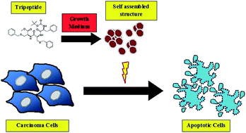
The study of peptide derived self-organized nanoarchitectures is an emerging scientific research area due to its potential application in advanced material science, biointerface engineering, therapeutics etc. In the present study, we report the synthesis and detailed biophysical properties of terminally protected tripeptide ‘N-Boc-L-Phe-S-benzyl-L-Cys-S-benzyl-L-Cys methyl ester’. The solid state FTIR spectrum reveals an intermolecular hydrogen bonded β-sheet like backbone in the solid state. Accordingly, secondary conformation of this tripeptide in the liquid state has been investigated by CD spectroscopy, which demonstrates that the tripeptide preferably exists as an unordered conformation in the liquid phase. Morphology of the self-assembled structure adapted by this tripeptide has been determined by atomic force microscopy (AFM), transmission electron microscopy (TEM) and field emission scanning electron microscopy (FESEM). AFM results revealed that the tripeptide self-assembly behaviour is concentration dependent but independent of the solvent system in which the self-assembled structure was formed. Concentrated methanol solution of the tripeptide produces oligomers 800–1050 nm in size, whereas diluted solutions of tripeptide in ethyl acetate solvent constructs oligomers 350–550 nm in size (diameter). Moreover, this tripeptide retains its self-assembled structure in biological environments i.e. DMEM and FBS also. Furthermore, the tripeptide shows potential cytotoxicity towards cancer cell lines. IC50 values were found to be 6.2 to 7.5 μM against breast (MCF-7, MDA-MB-231) and liver cancer (HepG2) cell lines, estimated by in vitro cell viability assay. Western blot analysis establishes that this tripeptide kills the cancer cells through apoptosis.


Introduction

Self-organization of biomolecules has inspired well-ordered templates at nano-dimensions for functional devices1,2 and is the most cultivated area in bio-nanotechnology. Proteins are naturally occurring biomolecules that can self assemble3–5 spontaneously to generate nano to microscopic materials. Among various organic molecules, small peptides are potential candidates for constructing well-ordered architectures via self-recognition process.6,7 This self-recognition event between the peptide molecules is mediated by weak interactions8 such as electrostatic, hydrogen bonding,9 hydrophobic, aromatic pi-stacking interactions. The various side chains of the amino acid residue regulate the nature of interactions involved in the self-arrangement process. Apart from that, peptide concentration and many physicochemical parameters such as pH,10 ionic strength,11,12 solvent,13 light,14 temperature,15 time16 also control the nature of the ultimate self-assembled structure. A wide range of three dimensional structures including hydro gels,17,18 tube, fibers, vesicles, spherical, rod and tape have been fashioned by altering the amino acid sequence and also the mentioned physicochemical parameters. These synthetic nano materials form the self assembled peptides, have wide range of applications19,20 in materials science21 and bio interface engineering22 including tissue engineering,23 drug delivery,24,25 biosensors,26 bioelectronics, imaging tools, bio mineralization,27 wound healing,28 surfactants29,30 etc. The wide applicability, biocompatibility, biodegradability31 and versatility in physical and chemical properties of the peptide molecule make them an attractive candidate in nanotechnology. It has been observed that the versatility in self assembled structure adopted by phenylalanine32–34 dipeptide (L-Phe-L-Phe) is due to the π–π interaction associated with phenyl group, which will exert the directionality and specific orientation required for molecular assembly of the peptide molecules. In an ongoing project we have recently shown that S-benzyl protected L-Cys-L-Cys dipeptide forms nanotube35 and S-benzyl protected L-Cys-L-Cys-L-Cys tripeptide forms nano sphere (unpublished work). We observed that side chain aromatic groups present in the amino acids hold a key π–π-stacking interaction to control the geometry of the newly formed self assembly structure. In the present study, we are interested to investigate the secondary conformation and the self assembly behaviour of the tripeptide containing L-phenylalanine and S-benzyl protected -L-cysteine dipeptide. This synthesized tripeptide is N-terminally protected by tert-butyloxycarbonyl group and C-terminally protected by methyl ester group. Phenylalanine moiety has been strategically incorporated into the tripeptide to examine the π–π interaction with S-benzyl group of the corresponding cysteine moiety and how it alter the ultimate self assembled structure compared to the dicysteine analogue.

Recently small peptides have gained attention as potential therapeutics for the treatment of diseases like cancer,36,37 diabetes38 etc. Tumour cell penetrating ability, biocompatibility, ease of synthesis and modification, morphological versatility make peptide as an attractive pharmacophore against these diseases. Accordingly, we are also interested to elucidate the cytotoxic effect of this tripeptide against cancer cell lines.

Materials and methods

Reagents

All chemicals were purchased from Sigma-Aldrich and used without further purification unless otherwise stated. Solvents were freshly distilled by the standard procedures prior to use. Column chromatography was performed on silica gel (Merck, 60–120 mesh) with the required eluent. Mass spectra were recorded on a Jeol MS station 700. All 1H and 13C-NMR spectra were recorded on Bruker 600 MHz spectrometer. All FT-IR are recorded on Bruker TENSOR27 spectrometer.

Synthesis of the tripeptide

To a well stirred solution of N-(tert-butoxycarbonyl)-S-benzyl-L-cysteine (1; 480 mg, 1.6 mmol) dissolved in N,N-dimethylformamide (8 mL), was added anhydrous hydroxybenzotriazole (HOBT; 300 mg, 1.92 mmol) slowly followed by 1-ethyl-3,3-(dimethylamino) propyl carbodiimide hydrochloride (EDC·HCl; 600 mg, 3.2 mmol) in cooled condition under nitrogen atmosphere. Then the stirring was continued for 10 minutes at ice-cooled condition and to this mixture triethylamine (TEA; 1 mL, 7.5 mmol) was added along with S-benzyl-L-cysteine methyl ester (2; 400 mg, 1.6 mmol), subsequently the reaction was further continued for 8 h at room temperature (monitoring via TLC). The reaction mixture was then concentrated to dryness and extracted with ethyl acetate (3 × 20 mL) from aqueous layer. Evaporation of solvent left the crude product, which was purified by column chromatography over silica gel (hexane/ethyl acetate 75[thin space (1/6-em)]:[thin space (1/6-em)]25) to afford the intermediate compound ‘3’ as white solid (yield = 70%). After that, deprotection of the N-(tert-butoxycarbonyl) group from the intermediate ‘3’ was mediated by 4 (M) HCl in 1,4 dioxane to afford the intermediate ‘4’. Evaporation of solvent and azeotrope with the toluene provide the crude product which was directly used to the next step reaction. To carry out the final reaction, anhydrous hydroxybenzotriazole (HOBT; 110 mg, 0.78 mmol) was added followed by the addition of 1-ethyl-3,3-(dimethylamino)propyl carbodiimide hydrochloride (EDC·HCl; 270 mg, 1.4 mmol) to a well stirred solution of N-(tert-butoxycarbonyl)-L-phenylalanine (5; 172 mg, 0.65 mmol) dissolved in N,N-dimethylformamide at cooled condition under nitrogen atmosphere. After stirring the solution for 10 minutes at ice-cooled condition triethylamine (TEA; 1 mL, 2.25 mmol) was added along with the addition of intermediate ‘4’ (2; 270 mg, 0.65 mmol), then the reaction was further continued for 8 h at room temperature (monitoring via thin-layer chromatography (TLC)). The reaction mixture was evaporated to dryness and extracted with ethyl acetate (3 × 20 mL) from aqueous layer. Evaporation of solvent left the crude product, which was further purified by column chromatography over silica gel (hexane/ethyl acetate 60[thin space (1/6-em)]:[thin space (1/6-em)]40) to afford the desired compound ‘6’ as white solid (yield = 50%) (Scheme 1).
image file: c6ra23911k-s1.tif
Scheme 1 Synthetic route to S-benzyl-protected cysteine and phenylalanine tripeptide. Reagents and condition (a) EDC·HCl, HOBt, TEA, 0 °C to r.t., 8 h, (b) 4 M HCl in 1,4-dioxane, 0 °C to r.t., 2 h, (c) EDC·HCl, HOBt, TEA, 0 °C to r.t., 8 h.

Circular dichroism (CD)

The CD spectrum of the tripeptide was recorded at JASCO-810 spectropolarimeter under constant nitrogen flow condition. Peptide dissolved in methanol placed at 1 mm path length quartz cuvette was used for all the CD spectra measurements. All the CD measurements were performed at 25 °C with an accuracy of ±0.1. The far-UV region was scanned between 190 to 250 nm using bandwidth of 1 nm. Each represented spectra was taken as the average of three individual scans.

FT-IR

The FT-IR spectra of the peptide were recorded on a Bruker TENSOR27 spectrometer by the KBr disc technique. Solid sample was mixed with KBr in a clean glass pestle and compressed to obtain a pellet. The spectra was scanned from 400–4000 cm−1 at 4 cm−1 resolution. Background spectra were obtained with a KBr pellet for each sample. Bruker software was used for data processing.

NMR method

1H and 13C NMR spectroscopic data were recorded with a Bruker DPX 300 MHz and 600 MHz spectrometer. Chemical shifts (δ) were reported in parts per million (ppm) and tetramethylsilane (δ = 0.00) used as the internal standard. All the 1H and 13C NMR spectra of the compound recorded in DMSO-D6 solvent having 1H NMR, δ = 2.50 ppm (s), 13C NMR, δ = 40 ppm (m) and all data were reported as follows: chemical shift, integration, multiplicity (s = singlet, d = doublet, t = triplet, q = quartet, m = multiplet) and coupling constant (s) in hertz (Hz). All the experiments were performed at room temperature (25 °C).

Atomic force microscope

AFM images were obtained on Vecco diCP II with a piezo scanner with the range of 100 μm. The images (256 × 256 pixels) were captured with a scan size of 5 μm to 25 μm at a scan speed rate of 0.5 to 0.6 Hz. Images were processed through flattening via Proscan 1.8 software. For this purpose, tripeptide solution incubated at room temperature for 24 h, and then the solution was applied to a mica foil, after drying the sample solution placed at mica foil, the specimen was observed through atomic force microscopy.

Transmission electron microscope

TEM image was acquired on Tecnai G2 Spirit Bio TWIN (Type: FP5018/40) at an acceleration voltage of 80 kV. For this purpose, 100 μM concentrated tripeptide in ethylacetate solution was aged at room temperature for 24 h and then placed on a 300-mesh carbon coated copper grid (Allied Scientific Product, USA) (5 μL, 10–15 min), gradually the excess samples were discarded carefully by tissue paper. Then it was finally dried and the images were captured.

Cell culture

Human breast carcinoma cell lines MCF-7, MDA-MB-231 and human hepatocellular carcinoma cell line Hep G2 were obtained from the National Centre for Cell Sciences (NCCS, Pune, India) and were maintained in Dulbecco's modified Eagle's medium DMEM supplemented with 10% fetal calf serum (Gibco, BRL) with antibiotics (penicillin-100 μg mL−1; streptomycin-50 μg mL−1) at 37 °C in 5% CO2 humidified incubator.

MTT assay

MCF-7, MDA-MB-231 and Hep G2 cells were seeded at a density of 105 per well in three separate 96-well plates. Cells were grown to 60–70% confluence, rinsed with phosphate buffered saline (PBS) and placed into serum-free medium overnight prior to treatments. After overnight incubation, all the cell line plates were treated with the tripeptide at the concentration of 0.5, 1, 2, 4, 6, 8, 10 μM. After 24 h, old medium was discarded and 50 μL of fresh medium was added along with 10 μL of MTT (5 mg mL−1). MTT solution was discarded after 4 h and the remaining purple crystals were dissolved in another 50 μL of DMSO. The absorbance of this purple solution was measured at test wavelength of 570 nm through Elisa Plate Reader (EMax Precision Microplate Reader, Molecular Devices). Cell viability was estimated by MTT (3-(4,5-dimethylthiazol-2-yl)-2,5-diphenyltetrazolium bromide), a yellow tetrazole assay where the viable cells were estimated by the reduction of the yellow MTT into purple formazan product by mitochondrial dehydrogenase present in metabolically active cells.

Western blotting

MCF7 cells were treated with increasing concentration (1, 3 and 6 μM) of the tripeptide and then incubated for 24 h. After the incubation with the tripeptide, MCF-7 cells were washed three times with ice-cold PBS and whole cellular protein was extracted using protein lysis buffer (62.5 mM Tris–HCl, pH 6.8, 2% SDS, 10% glycerol, 1× protease inhibitor cocktail). After incubation for 10 min on ice, the samples were centrifuged (13[thin space (1/6-em)]000 rpm at 4 °C for 20 min) and total proteins were measured with a Bio-Rad Bradford kit (Bio-Rad Laboratories, Hercules, CA, USA). About 20 μg of proteins were subjected to resolve in 12% SDS-PAGE and then electro transfer onto blotting nitrocellulose membranes. The membrane was washed twice with 1× TBS and with 0.1% Tween-20 (TBST, pH 7.4) and then preincubated with 5% bovine serum albumin in TBST blocking buffer at room temperature for 2 h. The blots were then incubated with rabbit polyclonal cleaved caspase 3, cleaved caspase 9 (Santa Cruz Biotechnology, Santa Cruz, CA) and mouse monoclonal β-actin (Sigma) primary antibodies at 1[thin space (1/6-em)]:[thin space (1/6-em)]1000 dilutions in TBST at 4 °C room overnight, followed by incubation with alkaline phosphatase-conjugated goat anti-rabbit along with goat anti-mouse IgG (1[thin space (1/6-em)]:[thin space (1/6-em)]10[thin space (1/6-em)]000) as the secondary antibody. Detection of the protein bands was performed using the chromogenic substrate 5-bromo-4-chloro-3′-indolylphosphate and nitro blue tetrazolium (BCIP/NBT). The intensities of the Western blots were quantified by using the image analysis software Image-J (NIH, MD, USA). The β-actin was used as internal control and relative intensity is expressed with respect to β-actin.

Results and discussion

Fourier transform infrared39 (FTIR), circular dichroism40 characterised the secondary conformation of this tripeptide in solid state and solution state respectively. From solid state FTIR spectrum, a wide number of vibrational bands were observed due to the presence of different functional groups such as CONH, COOCH3, aromatic ring, aliphatic groups (–CH2, –CH3) etc. The important FTIR band positions are marked in the Fig. 1 and the assignment of those band positions were given in Table 1. The characteristic vibration bands appeared at 3297, 3062, 3029, 2974, 2925, 1745, 1694, 1642, 1526, 1447, 1388, 1367, 1245, 1168, 701 cm−1 for the tripeptide. 3297 cm−1 band indicated the stretching motion of the N–H of the corresponding amide (CONH) groups, which was intermolecularly hydrogen bonded. Vibrational band at 3062 cm−1 signified to first overtone of N–H bending. The bands at 2974 cm−1 and 2925 cm−1 are appeared due to asymmetric and symmetric stretching vibrations of the aliphatic groups i.e. CH3 and CH2 groups. Vibrational band at 1694 and 1642 cm−1 originated due to amide C[double bond, length as m-dash]O stretching modes. These amide C[double bond, length as m-dash]O stretching vibration band at 1695 and 1641 cm−1 assigned as amide I band. Amide I band arises mainly due to the amide C[double bond, length as m-dash]O stretching vibration, but some minor contributions come from the out-of-phase C–N stretching motion, C–C–N deformation and the N–H in-plane bending motion also.
image file: c6ra23911k-f1.tif
Fig. 1 FT-IR spectra of the tripeptide at solid state.
Table 1 Assignment of the FT-IR bands of the tripeptide at solid state
IR frequency (cm−1) Modes of assignments IR frequency (cm−1) Modes of assignments
3297 Amide NH symmetric stretching 1526 Amide II (N–H bend in plane and C–N stretch)
3062 NH bending first overtone 1447 CH2 bending
3029 Aromatic C–H stretching 1388 CH3 symmetric bending
2974 CH3 antisymmetric stretching of tertiary butyl group 1367 CH3 of tertiary group symmetric bending
2925 CH2 antisymmetric stretching 1245 Amide III (N–H bend in plane and C–N stretch)
1745 C[double bond, length as m-dash]O stretching of the ester 1168 Ester C–O asymmetric stretch
1694 Amide C[double bond, length as m-dash]O stretching (amide I) 701 Out-of-plane NH bending
1642 Amide C[double bond, length as m-dash]O stretching (amide I)    


Accordingly, vibrational bands at 1526 cm−1 and 1245 cm−1 could be due to amide II and amide III respectively. It has been reported that peptides, having propensity toward β-sheet conformation, give two bands in amide I region, one is a strong transition between 1610–1643 cm−1 and the other one is a weak transition between 1680 and 1700 cm−1 in the FTIR spectra. In our case, the characteristic amide I bands appeared at 1642 cm−1 and 1694 cm−1 and the amide NH stretching band at around 3297 cm−1.

All these characteristic FTIR bands indicate that the tripeptide exists as intermolecularly hydrogen-bonded β-sheet like conformation in solid state. Moreover, amide II and amide III band at 1526 and 1245 cm−1 are also representative of β-sheet like structure. However, characteristic absorption band in the range 1610 to 1630 cm−1 for the aggregated amyloid-like peptides was not observed. Absence of this band in this region also signified that the tripeptide did not aggregate at solid state and rather exists as an ordered β-sheet like structure.

Circular dichroism spectrum revealed the most preferable secondary conformation of the tripeptide in solution state. For CD signal measurement, 200 μM concentrated tripeptide in methanol solution was used. This peptide solution showed a distinct sharp negative band at 198 nm along with some negative signals having low intensity at 201, 206 and 214 nm respectively as observed in Fig. 2a. Appearance of sharp CD signals at 198 nm suggested that the tripeptide prefers to exist as unordered conformation in solution state. Persistence of low signals at 201, 206 and 214 nm indicated that the tripeptide also have tendency to adopt some conformation where the geometric arrangement of the amide bond is different from the unordered pattern. This different type of amide bond arrangement as reflected from the CD spectrum synergistically indicated the existence of various type of molecular–molecular interaction present in various sizeed self assembled structure formed by the tripeptide molecules in solution state. Therefore, CD signal indicated the susceptibility towards self assembly of this tripeptide in solution phase.


image file: c6ra23911k-f2.tif
Fig. 2 (a) Circular dichroism spectra of the tripeptide solution (C = 0.2 mM) in methanol, (b) absorbance spectra of the tripeptide in methanol solution with concentration range of 20–60 μM.

Affinity towards self-assembly in solution phase for the tripeptide is further confirmed from absorption spectra measurement. For recording UV-vis spectra, tripeptide dissolved in methanol is used with various concentrations from 20 μM to 60 μM. In Fig. 2b, UV-vis spectra showed two characteristic peaks at 260 nm and 267 nm for the benzyl groups present in the tripeptide molecule. Scattering was also observed due to self-agglomeration of the tripeptide to generate well ordered oligomers. Moreover, scattering was enhanced with increasing tripeptide concentration because of increase in aggregation rate as the concentration increases.

The observed scattering in both the CD and UV spectrum suggested that the tripeptide has strong propensity for oligomerization in solution phase. Accordingly, morphology of the self assembled tripeptide was resolved through atomic force microscopy (AFM), transmission electron microscopy (TEM) and field emission scanning electron microscopy (FESEM).

A spherical architecture has been observed in solution phase under atomic force microscope for this tripeptide. Self assembly pattern generated by the tripeptide was validated in two organic solvent systems i.e. in ethyl acetate and methanol. Size of the spherical assemblies generated from tripeptide in ethyl acetate is in the range of 350–550 nm at 0.05 mM concentration. Similar spherical assembly with the diameter of 800–1050 nm were produced from higher concentrated tripeptide in methanol solution (0.5 mM). Fig. 3a and b depicted the concentration dependent self assembly of the tripeptide in ethyl acetate and methanol solution through AFM. From Fig. 3a, it has been observed that the diameter of spherical assemblies formed at lower concentration (0.05 mM) from ethyl acetate solution is less compared to higher concentrated tripeptide (0.5 mM) in methanol solution. i.e., self assembly of the tripeptide is concentration dependent in solution state. It seems that in higher concentrated tripeptide solution further aggregation of the discrete oligomers have taken place and formed large size spherical assembly compared to lower concentrated tripeptide solution. However the nature of the self assembled structure generated from this tripeptide in two solvents i.e. ethyl acetate and methanol is same. Therefore the nature of self assembly pattern of this tripeptide is solvent independent. TEM image (Fig. 3c) of this tripeptide in ethyl acetate solution after one day aging has shown that spherical assembly of 369 nm size has been formed by tripeptide molecules. Spherical assembly of different size has been shown under field emission scanning electron microscope (FESEM) in Fig. 3d, which also supported the results, got from the AFM scanning. In addition the histogram of the size distribution profile of the self assembled tripeptide in methanol and ethyl acetate obtained from AFM data has been shown in Fig. 4a and b. To get better insight into the size distribution pattern of the tripeptide assembly in different solvent system, DLS (dynamic light scattering) experiment has been performed (Fig. 5). The size distribution profile from the DLS scanning depicted the diameter of the assemblies generated in ethyl acetate solution of the tripeptide is in 295–531 nm range with maximum intensity at 396 nm in Fig. 5a. This various sized assembly indicate further aggregation of the unit assembly and is quite supportive of the AFM results.


image file: c6ra23911k-f3.tif
Fig. 3 (a) AFM image of the nanosphere of 350–550 nm size generated from tripeptide dissolved in ethylacetate solvent incubated for 24 h, (b) AFM image of the spherical assembly of 800–1050 nm size generated from tripeptide dissolved in methanol solvent incubated for 24 h, (c) and (d) TEM and FESEM image of the nanosphere generated from tripeptide dissolved in ethylacetate solvent incubated for 24 respectively.

image file: c6ra23911k-f4.tif
Fig. 4 Histogram of the size distribution of the assemble tripeptide in ethyl acetate and methanol solution. (a) Histogram of the size distribution pattern against concentration of the tripeptide in methanol solution (C = 0.5 mM), (b) histogram of the size distribution pattern against concentration of the tripeptide in ethyl acetate solution with (C = 0.05 mM).

image file: c6ra23911k-f5.tif
Fig. 5 DLS results of the assembled tripeptide in different solution. (a) Size distribution by intensity of the tripeptide assembly in ethyl acetate solution (C = 0.2 mM), (b) size distribution by intensity of the tripeptide assembly in methanol solution (C = 0.2 mM). (c) Size distribution by intensity of the tripeptide assembly in FBS (FBS[thin space (1/6-em)]:[thin space (1/6-em)]DMSO: 90[thin space (1/6-em)]:[thin space (1/6-em)]10) solution (C = 0.2 mM), (d) size distribution by intensity of the tripeptide assembly (C = 0.2 mM) in 10% FBS in DMEM (DMEM[thin space (1/6-em)]:[thin space (1/6-em)]DMSO: 90[thin space (1/6-em)]:[thin space (1/6-em)]10).

Similarly DLS scanning data of the tripeptide dissolved in methanol demonstrate the range of the assembled structure is 295–458 nm with maximum intensity at 342 nm (Fig. 5b). The discrepancy between AFM and DLS for the tripeptide solution in methanol possibly arises due to further aggregation of the unit oligomer in concentrated solution during AFM scanning compared to diluted solution in case of DLS scanning. These DLS results associated with the tripeptide suspended in different organic solvent also corroborate that the self assembly behaviour of this tripeptide is concentration dependent and solvent independent. In order to show biological activity, this self assembled tripeptide should circulate in the blood for a sufficiently long period of time, therefore assembled structure of this tripeptide should be intact throughout the circulation time.

For this purpose, self assembly propensity of this tripeptide has been investigated in biological medium including Dulbecco's Modified Eagles Media (DMEM) and Fetal Bovine Serum (FBS). The DLS scanning data revealed that the structure of the tripeptide assembly formed in DMEM and FBS medium is larger in size compared to the tripeptide assembly formed in organic solvent systems. From Fig. 5c, it has been observed that the tripeptide suspended in FBS (FBS[thin space (1/6-em)]:[thin space (1/6-em)]DMSO: 90[thin space (1/6-em)]:[thin space (1/6-em)]10) generated assemblies with wide distribution of size of 100–1400 nm along with maximum intensity at 200–500 nm. The tripeptide assembly of 200–500 nm size has been produced in the ethyl acetate and methanol medium also. Hence the nature of the assembly adopted by this tripeptide in presence of serum protein is same as in organic solvent systems. However the large size assemblies of 700–1400 nm size are the outcome of aggregation of the 200–500 nm sized assemblies. This further aggregation of the 200–500 nm size assemblies is promoted by the strong hydrophobic environment exerted by the serum protein (FBS). Similarly tripeptide suspended in 10% FBS in DMEM (DMEM[thin space (1/6-em)]:[thin space (1/6-em)]DMSO: 90[thin space (1/6-em)]:[thin space (1/6-em)]10) produced assembled structure of 90–300 nm size along with microsphere of 1–5 μm size (Fig. 5d). The size distribution pattern of the tripeptide assembly in biological mediums indicate that the serum protein present in FBS and DMEM exert strong hydrophobic environment and thus catalyse the rate of aggregation for the tripeptide. Therefore strong hydrophobic environment in the FBS and DMEM medium initially directed the tripeptides for self assembly to develop nano sized assembly and then close packing of these discrete assemblies leading to micro-assemblies. The morphology of the assembled tripeptide in DMEM has been resolved by the AFM scanning. From Fig. 6, it is clearly seen that the tripeptide formed nano-assembly of 60–110 nm size along with some large size particles of 220–250 nm size. This AFM data corroborates the DLS results of the tripeptide assembly in biological medium and the appearance of the large size assemblies as obtained from the DLS data is due to further aggregation of the unit assemblies. Therefore, this tripeptide retains its self assembled structure in biological medium also.


image file: c6ra23911k-f6.tif
Fig. 6 AFM image of the tripeptide suspended in 10% FBS in DMEM (DMEM[thin space (1/6-em)]:[thin space (1/6-em)]DMSO: 90[thin space (1/6-em)]:[thin space (1/6-em)]10) solution. (a) Topography of the tripeptide assembly of 220–250 nm size, (b) phase diagram of the mica film corresponding to 220–250 nm sized assembly, (c) topography of the tripeptide assembly of 60–110 nm size, (d) phase diagram of the mica film corresponding to 60–110 nm sized assembly.

This tripeptide showed significant cytotoxicity and cell death against different cancer cell lines. For this purpose, three different cancer cell lines i.e. MCF-7, HepG2, MDA-MB-231 were treated with this tripeptide and cell viability of these cancer cell lines after treatment with the tripeptide was evaluated by the MTT assay. Tripeptide dissolved in DMSO solvent was used for in vitro experiment and maximum 1% of DMSO concentration was maintained during in vitro experiment. Here, Fig. 7a shows the cell viability of different cancer cell lines after treatment with the tripeptide at the variable concentration (from 0.5 up to 10 μM) with respect to control. The corresponding IC50 value of this tripeptide against MCF-7, HepG2, MDA-MB-231 cell lines were found to be 6.26 μM, 6.5 μM and 7.44 μM respectively, obtained from the MTT assay.


image file: c6ra23911k-f7.tif
Fig. 7 (a) Cytotoxicity study of the tripeptide against MCF-7, HepG2, MDA-MB-231 cell lines, (b) expression level of cleaved caspase 3 and cleaved caspase 9 after treatment with increasing concentration of the tripeptide in MCF-7 cells compared to control or untreated MCF7 cells for 24 h by using western blot analysis. (c) Quantitative analysis and graphical representation (mean ± SD) of cleaved caspase 3 and cleaved caspase 9 level after treated with increasing concentration of the tripeptide in MCF7 cells compared to control or untreated MCF-7 cells for 24 h. Expression of β-actin was used as internal control.

In most of the cases, cell death is mediated by apoptotic pathways with caspase-3 as the commonly activated death protease, catalyzing the specific cleavage of many fundamental cellular proteins. Pathways for caspase-3 activation have been identified that are dependent/independent of mitochondrial cytochrome c release and caspase-9 function. To get an insight into the mechanism by which the tripeptide induced apoptosis against different cancer cell lines, we checked the expression of key molecules involved in cell apoptosis in MCF-7 cell line through western blotting (Fig. 7b and c). Significant enhancement in expression of apoptotic markers like cleaved caspase 3 and cleaved caspase 9 was observed in a dose-dependent manner. Moreover, it was also observed that the increase in effector cleaved caspase 3 level was better compared to proapoptotic cleaved caspase 9 level in MCF-7 cells after treatment with the tripeptide. Thus, the elevated expression of cleaved caspase 3 and caspase 9 levels suggested that the tripeptide can induce cancer cell death by apoptosis.

Conclusion

In summary, this newly synthesized tripeptide adopted intermolecularly hydrogen-bonded β-sheet like network in solid state and random coil (unordered pattern) in liquid phase. It produced well ordered spherical assembly at submicron region in different organic solvent in concentration depended manner. This tripeptide also forms self assemblies in presence of serum protein and biological medium. Moreover this tripeptide showed significant cytotoxicity toward different cancer cell lines and induced apoptosis to promote cancer cell death. Thus, this tripeptide may be used as potential candidate in the field of nano-drug delivery carrier, anticancer agent etc.

Acknowledgements

M. C. and C. P. thank University Grants Commission, India, for financial support. The authors would also like to thank the central instrumentation facilities of CSIR-Indian Institute of Chemical Biology for recording the spectra. The authors would like to thank CSIR-India for funding in this work in the form of a Network project, BSC206 (BenD).

References

  1. H. Liu, J. J. Schmidt, G. D. Bachand, S. S. Rizk, L. L. Looger, H. W. Hellinga and C. D. Montemagno, Control of a biomolecular motor-powered nanodevice with an engineered chemical switch, Nat. Mater., 2002, 1(3), 173–177 CrossRef CAS PubMed.
  2. B. Christian, E. Karl-Heinz, H. Henry and V. Viola, Lifetime of biomolecules in polymer-based hybrid nanodevices, Nanotechnology, 2004, 15(10), S540 CrossRef.
  3. J. J. McManus, P. Charbonneau, E. Zaccarelli and N. Asherie, The physics of protein self-assembly, Curr. Opin. Colloid Interface Sci., 2016, 22, 73–79 CrossRef CAS.
  4. Y. Bai, Q. Luo and J. Liu, Protein self-assembly via supramolecular strategies, Chem. Soc. Rev., 2016, 45(10), 2756–2767 RSC.
  5. J. A. Marsh and S. A. Teichmann, Structure, Dynamics, Assembly, and Evolution of Protein Complexes, Annu. Rev. Biochem., 2015, 84(1), 551–575 CrossRef CAS PubMed.
  6. X. Zhao, F. Pan and J. R. Lu, Recent development of peptide self-assembly, Prog. Nat. Sci., 2008, 18(6), 653–660 CrossRef CAS.
  7. D. Mandal, A. Nasrolahi Shirazi and K. Parang, Self-assembly of peptides to nanostructures, Org. Biomol. Chem., 2014, 12(22), 3544–3561 CAS.
  8. Y. Xie, X. Wang, R. Huang, W. Qi, Y. Wang, R. Su and Z. He, Electrostatic and Aromatic Interaction-Directed Supramolecular Self-Assembly of a Designed Fmoc-Tripeptide into Helical Nanoribbons, Langmuir, 2015, 31(9), 2885–2894 CrossRef CAS PubMed.
  9. S. E. Paramonov, H.-W. Jun and J. D. Hartgerink, Self-Assembly of Peptide–Amphiphile Nanofibers:[thin space (1/6-em)] The Roles of Hydrogen Bonding and Amphiphilic Packing, J. Am. Chem. Soc., 2006, 128(22), 7291–7298 CrossRef CAS PubMed.
  10. Y. Chen, H. X. Gan and Y. W. Tong, pH-Controlled Hierarchical Self-Assembly of Peptide Amphiphile, Macromolecules, 2015, 48(8), 2647–2653 CrossRef CAS.
  11. A. Abelein, J. Jarvet, A. Barth, A. Gräslund and J. Danielsson, Ionic Strength Modulation of the Free Energy Landscape of Aβ40 Peptide Fibril Formation, J. Am. Chem. Soc., 2016, 138(21), 6893–6902 CrossRef CAS PubMed.
  12. R. Li, C. C. Horgan, B. Long, A. L. Rodriguez, L. Mather, C. J. Barrow, D. R. Nisbet and R. J. Williams, Tuning the mechanical and morphological properties of self-assembled peptide hydrogels via control over the gelation mechanism through regulation of ionic strength and the rate of pH change, RSC Adv., 2015, 5(1), 301–307 RSC.
  13. R. Huang, W. Qi, R. Su, J. Zhao and Z. He, Solvent and surface controlled self-assembly of diphenylalanine peptide: from microtubes to nanofibers, Soft Matter, 2011, 7(14), 6418–6421 RSC.
  14. T. M. Doran, D. M. Ryan and B. L. Nilsson, Reversible photocontrol of self-assembled peptide hydrogel viscoelasticity, Polym. Chem., 2014, 5(1), 241–248 RSC.
  15. R. Huang, Y. Wang, W. Qi, R. Su and Z. He, Temperature-induced reversible self-assembly of diphenylalanine peptide and the structural transition from organogel to crystalline nanowires, Nanoscale Res. Lett., 2014, 9(1), 1–9 CrossRef CAS PubMed.
  16. J.-X. Wang, Q. Lei, G.-F. Luo, T.-T. Cai, J.-L. Li, S.-X. Cheng, R.-X. Zhuo and X.-Z. Zhang, Controlled Arrays of Self-Assembled Peptide Nanostructures in Solution and at Interface, Langmuir, 2013, 29(23), 6996–7004 CrossRef CAS PubMed.
  17. W. J. M. FrederixPim, G. G. Scott, Y. M. Abul-Haija, D. Kalafatovic, C. G. Pappas, N. Javid, N. T. Hunt, R. V. Ulijn and T. Tuttle, Exploring the sequence space for (tri-)peptide self-assembly to design and discover new hydrogels, Nat. Chem., 2015, 7(1), 30–37 CrossRef CAS PubMed.
  18. A. Lakshmanan and C. A. Hauser, Ultrasmall peptides self-assemble into diverse nanostructures: morphological evaluation and potential implications, Int. J. Mol. Sci., 2011, 12(9), 5736–5746 CrossRef CAS PubMed.
  19. S. Kim, J. H. Kim, J. S. Lee and C. B. Park, Beta-Sheet-Forming, Self-Assembled Peptide Nanomaterials towards Optical, Energy, and Healthcare Applications, Small, 2015, 11(30), 3623–3640 CrossRef CAS PubMed.
  20. P. P. Sharma, B. Rathi, J. Rodrigues and N. Y. Gorobets, Self-assembled peptide nanoarchitectures: applications and future aspects, Curr. Top. Med. Chem., 2015, 15(13), 1268–1289 CrossRef CAS PubMed.
  21. E. De Santis and M. G. Ryadnov, Peptide self-assembly for nanomaterials: the old new kid on the block, Chem. Soc. Rev., 2015, 44(22), 8288–8300 RSC.
  22. S. Lu and P. Chen, Constructing biomaterials using self-assembling peptide building blocks, Front. Mater. Sci. China, 2010, 4(2), 145–151 CrossRef.
  23. L.-L. Li, G.-B. Qi, F. Yu, S.-J. Liu and H. Wang, An Adaptive Biointerface from Self-Assembled Functional Peptides for Tissue Engineering, Adv. Mater., 2015, 27(20), 3181–3188 CrossRef CAS PubMed.
  24. N. Habibi, N. Kamaly, A. Memic and H. Shafiee, Self-assembled peptide-based nanostructures: smart nanomaterials toward targeted drug delivery, Nano Today, 2016, 11(1), 41–60 CrossRef CAS PubMed.
  25. Q. Wang, X. Zhang, J. Zheng and D. Liu, Self-assembled peptide nanotubes as potential nanocarriers for drug delivery, RSC Adv., 2014, 4(48), 25461–25469 RSC.
  26. M. Lian, X. Chen, Y. Lu and W. Yang, Self-assembled peptide hydrogel as a smart biointerface for enzyme-based electrochemical biosensing and cell monitoring, ACS Appl. Mater. Interfaces, 2016, 8(38), 25036–25042 CAS.
  27. M. Gungormus, M. Branco, H. Fong, J. P. Schneider, C. Tamerler and M. Sarikaya, Self assembled bi-functional peptide hydrogels with biomineralization-directing peptides, Biomaterials, 2010, 31(28), 7266–7274 CrossRef CAS PubMed.
  28. A. Schneider, J. A. Garlick and C. Egles, Self-Assembling Peptide Nanofiber Scaffolds Accelerate Wound Healing, PLoS One, 2008, 3(1), e1410 Search PubMed.
  29. X. Zhao, Design of self-assembling surfactant-like peptides and their applications, Curr. Opin. Colloid Interface Sci., 2009, 14(5), 340–348 CrossRef CAS.
  30. E. Melis Sardan, C. Goksu, K. Mohammad Aref and O. G. Mustafa, Self-assembled peptide nanostructures for functional materials, Nanotechnology, 2016, 27(40), 402002 CrossRef PubMed.
  31. S. Wang, J. Lin and P. Huang, Advances on the Use of Biodegradable Proteins/Peptides in Photothermal Theranostics, J. Nanomater., 2016, 2016, 10 Search PubMed.
  32. S. M. M. Reddy and G. Shanmugam, Role of Intramolecular Aromatic π–π Interactions in the Self-Assembly of Di-L-Phenylalanine Dipeptide Driven by Intermolecular Interactions: Effect of Alanine Substitution, ChemPhysChem, 2016, 17(18), 2897–2907 CrossRef CAS PubMed.
  33. P. Tamamis, L. Adler-Abramovich, M. Reches, K. Marshall, P. Sikorski, L. Serpell, E. Gazit and G. Archontis, Self-Assembly of Phenylalanine Oligopeptides: Insights from Experiments and Simulations, Biophys. J., 2009, 96(12), 5020–5029 CrossRef CAS PubMed.
  34. S. Marchesan, A. V. Vargiu and K. E. Styan, The Phe–Phe Motif for Peptide Self-Assembly in Nanomedicine, Molecules, 2015, 20(11), 19775–19788 CrossRef CAS PubMed.
  35. B. Banerji, S. K. Pramanik, U. Pal and N. C. Maiti, Dipeptide derived from benzylcystine forms unbranched nanotubes in aqueous solution, J. Nanostruct. Chem., 2013, 3(1), 1–6 Search PubMed.
  36. Y.-b. Huang, X.-f. Wang, H.-y. Wang, Y. Liu and Y. Chen, Studies on Mechanism of Action of Anticancer Peptides by Modulation of Hydrophobicity Within a Defined Structural Framework, Mol. Cancer Ther., 2011, 10(3), 416–426 CrossRef CAS PubMed.
  37. J. Thundimadathil, Cancer Treatment Using Peptides: Current Therapies and Future Prospects, J. Amino Acids, 2012, 2012, 13 Search PubMed.
  38. T. Billyard, P. McTernan and S. Kumar, Potential therapies based on antidiabetic peptides, Best Pract. Res., Clin. Endocrinol. Metab., 2007, 21(4), 641–655 CrossRef CAS PubMed.
  39. A. Adochitei and G. Drochioiu, Rapid characterization of peptide secondary structure by FT-IR spectroscopy, Rev. Roum. Chim., 2011, 56(8), 783–791 CAS.
  40. N. J. Greenfield, Using circular dichroism spectra to estimate protein secondary structure, Nat. Protoc., 2006, 1(6), 2876–2890 CrossRef CAS PubMed.

Footnote

Electronic supplementary information (ESI) available. See DOI: 10.1039/c6ra23911k

This journal is © The Royal Society of Chemistry 2016
Click here to see how this site uses Cookies. View our privacy policy here.