DOI:
10.1039/C6RA24502A
(Paper)
RSC Adv., 2016,
6, 112656-112666
Self assembly and hydrogelation of spermine functionalized aromatic peptidomimetics against planktonic and sessile methicillin resistant S. aureus†
Received
1st October 2016
, Accepted 23rd November 2016
First published on 23rd November 2016
Abstract
Peptide amphipathicity is a common characteristic of self assembling peptide nanomaterials and host defense cationic antimicrobial peptides. In the present work, we utilized amphipathicity of ultra short aromatic sequences with spermine as a cationic C-terminal tag for designing of antibacterial hydrogels against multidrug resistant bacteria. These designed peptidomimetics acquired single walled-nanofibres and nanotubes that formed hydrogels at higher concentrations between 0.5–1% w/v at physiological pH. In vitro antibacterial studies of the nanofibres exhibited potent activity (MIC: 14.2–227.2 μg mL−1) against Gram positive and Gram negative bacterial strains including clinically relevant pathogens such as methicillin resistant S. aureus (MRSA) and S. epidermidis. Bactericidal kinetic and scanning electron microscopy of peptidomimetics treated MRSA confirms the membrane active mode of action. These analogues also displayed a strong antibiofilm activity at the MIC level and demonstrated reduced viability of mature MRSA by >50% biofilms at 0.5–2% w/v hydrogels. Moreover, the nanofibres were neither cytotoxic nor hemolytic in nature. With selective modes of interaction of the designed nanostructure with bacteria these hydrogels could be potentially therapeutic against MRSA wound infections and also as implantable biomaterials.
Introduction
Self assembly is a ubiquitous process employed by nature which signifies various roles in the formation of a wide range of complex biological structures.1 More recently, self assembled peptide molecules and their supramolecular assemblies have attracted interest for use as biomaterials and drug delivery vehicles.2,3 Peptide building blocks have advantages over polymeric materials due to their biocompatibility, biodegradability and diversified structure.4 Peptide amphiphile based scaffolds are fascinating for hydrogelation because they can be self-assembled using various noncovalent interactions in water, including hydrogen bonding, van der Waals forces, electrostatic, or π–π interactions.5 Amphipathicity is a common characteristic between self assembled peptide building blocks and host defense cationic bactericidal peptides/peptidomimetics (HDCPs).6 HDCPs are promising candidates to combat the infections caused by multidrug resistant (MDR) pathogens.6 These agents primarily target the bacterial membranes via electrostatic/hydrophobic interactions resulting into disruption and rupture of membrane structure and killing of bacteria.7 Infection is one of the most common complications in chronic wounds which also delays healing further.8 Previously researchers had attempted on drying the wound site with absorptive gauzes dressing, however gauze dressing is heavily debated because of the neoepithelium (neoepithelial) pain and damage during removal.9,10 From the last decades, hydrogels have been extensively studied and used as alternative to the gauze dressing materials in wound dressing.11,12 Hydrogels have displayed advantages over conventional therapies to promote wound healing by creating a more favourable environment for tissue regeneration than uncovered wound.9 However, the high risk of infections in using hydrogels for wound care due to the moist environment, which could be further complicated when infection caused by MDR pathogens.9,13 Therefore utilization of common characteristics of antimicrobial peptides/peptidomimetics and peptide hydrogelators for making antimicrobial hydrogels can be promising solution in field of antimicrobial chemotherapy for the treatment of infected wounds caused by MDR pathogens. Numerous attempts have been made to develop hydrogels with potential antimicrobial properties, which simultaneously promoted the wound healing efficacy with eradication of infections.14–16 Ultra short peptide hydrogelators consist of two phenylalanine conjugated to a molecule of high aromaticity such as naphthalene (Nap) and 9-fluorenylmethoxy carbonyl (Fmoc), pyrene, benzyloxycarbonyl (Cbz).17 Diphenylalanine is the shortest structural recognition motif for the naturally occurring beta amyloid polypeptide, implicated in the formation of fibrous plaques in alzheimers.18 Researchers have harnessed the properties of aromatic groups for development of a range of diverse, short, nanomaterials structure in medical and technological research.17
Spermine is a low molecular weight biogenic polyamine which takes part in various processes in cells, including modulation of chromatin structure, DNA and RNA stabilization, gene transcription and translation, signal transduction and cell growth and proliferations in prokaryotes and eukaryotes.19 Spermine and their analogues exhibit diverse biological effects as antibacterials, antiparasites, antiendotoxin, and anticancer activities20–24 and it is well reported that exogenous polyamines (externally added to the medium) or conjugated polyamines enhances activity of synthetic antibacterials and clinically used antibiotics.25–27 By keeping in mind the above aspects we envisaged present study for designing of ultra short N-terminally modified aromatic peptidomimetics functionalized with spermine at C-terminal, which self assembled into nanofibres hydrogel at physiological pH and has the potential to be utilized as future antimicrobial biomaterials.
Materials and methods
Materials
Fmoc protected amino acids and 2-chlorotrityl chloride resin were purchased from Novabiochem (Germany). Spermine, N,N-diisopropyl carbodiimide (DIPCDI), boc anhydride [(Boc)2O], N-hydroxybenzotrizole (HOBt), di-isopropyl ethylamine (DIPEA), piperidine, trifluoroacetic acid (TFA), triisopropylsilane (TIS), 1,2-ethanedithiol (EDT), glucose, hydrazine, 4-(trifluoromethyl)phenyl acetic acid, decanoic acid, 2-naphthyl acetic acid, DMEM HAM F-12, fetal bovine serum (FBS) and TOX-7 kit (LDH release assay kit) were obtained from Sigma Chemical Co. (St Louis, MO, USA). 2-Acetyldimedone (Dde-OH), phenol and all the solvents were purchased from Merck. Tryptone soy broth (TSB), Mueller Hinton broth (MHB) and agar were purchased from DIFCO (Franklin Lakes, NJ, USA). Alamar blue reagent was purchased from Invitrogen (Molecular Probes, Eugene, OR, USA). Dichloromethane (DCM) and dimethylformamide (DMF) were freshly distilled prior to use.
Synthesis and purification of peptidomimetics
All peptidomimetics were synthesized using 2-chlorotrityl chloride resin as described previously with slight modifications.27,28 Peptide NF-1 was synthesized on 2-chlorotrityl chloride resin using routine procedure. For peptidomimetics NF-2 to NF-6 following strategy was employed. Briefly, 5 eq. of spermine (Sigma) were conjugated with 2-chlorotrityl linker of resin on pre swollen resin in DCM under N2 atmosphere for 4 h (Fig. 1).
 |
| | Fig. 1 Reagents and conditions: (1) 5 eq. spermine, DCM, 3 h; (2) MeOH for 30 min; (3) 2 eq. Dde-OH, DMF, overnight; (4) 6 eq. (Boc)2O, DCM : DMF (1 : 1), 3 h; (5) 2% hydrazine (DMF); (6) Fmoc-Glu(OtBu)-OH, HOBt, DIPCDI, DCM : DMF (1 : 1); (7) 20% piperidine (DMF); (8) Fmoc-Phe-OH, HOBt, DIPCDI, DCM : DMF (1 : 1), 1.5 h; (9) 3 eq. R-COOH, HOBt, DIPCDI, DCM : DMF (1 : 1), overnight; (10) 95% TFA, EDT, TIS, phenol; (11) 50% TFA/DCM. | |
Conjugation of spermine was confirmed by positive cold Kaiser test. Uncoupled spermine was removed by washing with DCM, DMF and methanol from resin. Capping of uncoupled resin was done using methanol for 45 min. Next, the orthogonal protection of primary amino group of conjugated spermine was done with 2 eq. of Dde-OH in DMF overnight. Remaining two secondary amino groups of spermine were protected with 6 eq. of (Boc)2O in presence of DIPEA for 4 h. Deprotection of Dde protection of primary amines was done using 2% w/v hydrazine in DMF. Further two couplings were done with Fmoc-Phe-OH in presence of HOBt and DIPCDI in DCM
:
DMF (1
:
1) for peptidomimetic NF-2. For synthesis of peptidomimetics NF-3 one coupling of Fmoc-Glu(tBu)-OH was performed using HOBt and DIPCDI and then two phenylalanine were coupled using same coupling reagents as above. After Fmoc deprotection using 20% piperidine in DMF, N-terminal taggings were done with 4 eq. of hydrophobic acids such as 2-naphthyl acetic acid, 4-(trifluoromethyl)phenyl acetic acid, and decanoic acid for synthesis of NF-4, NF-5, and NF-6, respectively (Fig. 1). Final deprotection of peptidomimetics from resin was performed using a cleavage cocktail (DCM
:
TFA
:
triisopropylsilane
:
phenol
:
water: in ratio 15
:
80
:
2
:
2
:
1). The crude products were filtered, precipitated and washed twice with cold ether and were desalted using LH-20 sephadex (Sigma) column. All the peptidomimetics were purified on RP-HPLC, using a semi preparative column (7.8 × 300 mm, 125 Å, 10 μm particle size) with gradient of 10–90% buffer 2, where, buffer 1 was water (0.1% TFA) and buffer 2 was acetonitrile (0.1% TFA) over 45 min. After purification, peptidomimetics were characterized by analytical HPLC (C18, 4.6 × 250 mm, 125 Å, 5 μm particle size) and ESI-MS (TripleTOF® 5600, AB Sciex). Analytical HPLC chromatograms and mass spectra of all peptidomimetics have been provided in ESI (Fig. S1–S12†).
Self assembly and hydrogelation of peptidomimetics
Lyophilized peptidomimetics (NF-2, NF-3, NF-4, NF-5 and NF-6) were dispersed in 1 mL sterile deionized water at three different concentrations i.e. 5 mg mL−1, 10 mg mL−1 and 20 mg mL−1. For NF-1 to dissolve the pH was raised using 0.1 N NaOH and after dissolution was brought down to 7.4 using 0.5 M HCl. The pH was verified using Whatman pH paper. Self assembly and the formation of a self-supporting gel were verified through the use of an inversion assay, as described previously.14 Further, the resulting materials were stored in 4 °C for characterization and biological activity determination.
High resolution-transmission electron microscopy (HR-TEM)
HR-TEM was performed using a FEI Morgagni 268 transmission electron microscope (FEI electronics, Burlington, Massachusetts, U.S.A.) using negative staining technique. Copper grids of 300 mesh coated with thick carbon film (∼35 nm) were used for this study. Samples (10 μL) were placed on the grid and allowed to absorb. Thereafter the grids were rinsed three times with double distilled water. The grid was stained by a 2% w/v aqueous solution of uranyl acetate. Excess of stained solution from the edge of the grid was removed using filter paper. Grids were allowed to dry overnight in desiccators before TEM examination.
Fourier transform infrared spectroscopy (FTIR)
Hydrogel samples (1% w/v) of each peptidomimetics were prepared for FTIR studies as described previously.29 Prepared hydrogels were lyophilized and dried powder was analyzed using FTIR (Bruker alpha series FTIR spectrometer), between the IR frequencies 4000–400 cm−1 with resolution 2 cm−1 under 128 scans on an average.
Circular Dichroism (CD)
CD was performed using a Jasco J815 Spectropolarimeter in quartz cuvette of 1 mm path length and a bandwidth of 1 nm over a range of wavelengths from 190 to 260 nm at 25 °C. Peptide samples were prepared 24 h before allowing them for self assembly and measurements were taken in water and buffer. Measurements were repeated three times and one representative graph after appropriate blank subtraction has been presented here.
Rheological studies of gels
The viscoelastic properties of the hydrogel NF-3 and NF-4 were determined using oscillatory rheology performed on aantonpaar MCR 302 rheometer at 25 °C. 1 mL of the prepared hydrogel were applied to the parallel plate using a pipetman. A frequency sweep experiment (from 0.1 rad s−1 to 100 rad s−1) was carried out on a parallel steel plate to calculate storage modulus (G′) and loss modulus (G′′) at a constant strain value of 1% applied from a gap of 0.2 mm. Measurements were repeated three times and mean ± standard deviation (SD) has been presented here.
Antibacterial activity under planktonic conditions
Antibacterial activity of designed peptidomimetics was evaluated by modifying serial broth dilution method, as reported previously.27,30 Bacterial strains used in this study were purchased from ATCC; Staphylococcus epidermidis (ATCC 12228), methicillin resistant Staphylococcus epidermidis (ATCC 51625), S. aureus (ATCC 29213), methicillin resistant S. aureus (ATCC 33591), S. aureus (ATCC BAA-44), E. faecalis (ATCC 7080), E. coli (ATCC 11775), P. aeruginosa (ATCC 25668) and A. baumannii (ATCC 19606). The initial inoculums for antibacterial assay were taken from mid-log phase bacterial cultures with ∼105 CFU mL−1 of bacterial suspension in MHB. The dilutions of peptidomimetics prepared in 0.01% v/v acetic acid and 0.2% bovine serum albumin (Sigma) over desired concentration range were added to the wells of 96 well micro titre plate. Thereafter mid log phase of bacterial suspension were added to the wells of the microplate. The plates were incubated overnight with agitation (180 rpm) at 37 °C. Absorbance was measured after 18 h at 630 nm. Cultures without test peptidomimetics were used as positive control. Un-inoculated MHB was used as negative control. Assays were performed in duplicate on three different days and minimum inhibitory concentration (MIC) is defined as the lowest concentration of peptidomimetics that completely inhibits growth. For comparison peptide antibiotic vancomycin (VAN) was also assayed under identical conditions.
Hemolytic activity
Hemolytic activity of the peptidomimetics was evaluated on human red blood cells (hRBC). Briefly, 100 μL of fresh hRBC suspension 4% v/v in PBS (35 mM phosphate buffer, 150 mM NaCl, pH 7.2) was placed in a 96 well plate containing serial dilution of peptidomimetics. After incubation of the test peptidomimetics (100 μL) in the hRBC suspension for 1 h at 37 °C, the plates were centrifuged and supernatant (100 μL) was transferred to fresh 96 well plate. Absorbance was read at 540 nm using ELISA plate reader (molecular devices). Percent hemolysis was calculated using the following formula:
| % hemolysis = 100[(A − A0)/(At − A0)] |
where, A represents absorbance of sample wells at 540 nm and A0 and At represent the zero percent and 100% hemolysis determined in PBS and 1% Triton X-100, respectively.
Cytotoxicity assay in human keratinocytes (HaCaT cells)
The cell viability was assessed by performing LDH assay using a TOX-7 kit (Sigma). HaCaT keratinocytes, 5000 cells per well, were seeded in 96-well plates in DMEM HAMS F12 media supplemented with 10% FBS to grow overnight. The next day media were aspirated and fresh incomplete media were added (50 μL per well). Serial two-fold dilutions of different test sequences (50 μL) were added to the wells and the plates were incubated at 37 °C with 5% CO2 for 18 h. After 18 h of incubation, the supernatant of each well were transferred to sterile 1.5 mL Eppendorf tubes, and assessed the release of LDH by using the kit, as described previously.31 The experiments were carried out in duplicate on three different days, and data is presented as a mean ± SD.
Bactericidal kinetics
The bactericidal effect of the peptidomimetics against MRSA (ATCC 33591) at 2 × MIC and 4 × MIC concentration was determined as described previously.32 Log-phase bacteria (1.2–3.0 × 10⁷ CFU mL−1) were incubated with peptidomimetics NF-3 and NF-4 at 2 × MIC and 4 × MIC in MHB. Aliquots were removed after 0.5, 1, 3 and 6 h, which were subsequently diluted with sterile normal saline before plating on the Mueller Hinton II agar and CFU were counted after 24 h incubation at 37 °C. The experiments were performed in duplicates repeated on two different days and curve was plotted between log10 CFU mL−1 versus time.
Scanning electron microscopy (SEM)
For SEM studies samples were prepared as described previously.27 Briefly, freshly inoculated MRSA (ATCC 33591) was grown on MHB up to OD600 ∼0.5 (corresponding to 108 CFU mL−1). Bacterial cells were then spun down at 4000 rpm for 15 min, washed thrice in PBS (10 mM phosphate buffer, 150 mM NaCl, pH 7.4) and re-suspended in equal volume of PBS. For SEM experiment, a higher bacterial inoculums (108 CFU mL−1) were used where the cells were incubated with test peptidomimetics NF-3 and NF-4 at respective 10 × MIC for 30 min. Controls were run in the absence of antibacterial agents. After 30 min, the cells were spun down and washed with PBS thrice. The washed bacterial pallet was re-suspended in 0.5 mL of 2.5% paraformaldehyde in PBS for cell fixation and was incubated at 4 °C overnight. After fixation, cells were spun down and washed with 0.1 M sodium cacodylate buffer twice and post fixed in 1% osmium tetraoxide in 0.1 M sodium cacodylate buffer at RT for 40 min in dark. Further the samples were dehydrated in series of graded ethanol solutions (30–100%), and finally dried in desiccators under reduced pressure. Upon dehydration the cells were air dried for 15 min in dark at RT after immersion in hexamethyldisilazane. An automatic sputter coater (Quorum-SC7640) was used for coating the specimens with thickness of 30 Å gold particles. Then samples were imaged via scanning electron microscope (Zeiss EVO LS15).
Biofilm susceptibility assay
For biofilm inhibition assay previously described protocol was adopted.33 In brief, freshly inoculated MRSA (ATCC 33591) was grown on biofilm growth media (TSB supplemented with 0.5% w/v NaCl and 0.25% w/v glucose) overnight. Next day, the culture was diluted in fresh biofilm growth media to 105 CFU mL−1. 200 μL of diluted culture was dispensed in wells of a 96 well polystyrene plate for biofilm formation. To evaluate the inhibition of biofilm formation, antibacterial agents at 2 × MIC and MICs were added initially with diluted culture following incubation at 37 °C without shaking. Another set of experiment was performed by addition of fresh medium containing antibacterial agents at 0.5%, 1% and 2% w/v gel concentrations after gently washing by sterile PBS buffer (35 mM phosphate buffer, 150 mM NaCl, pH 7.4) to 24 h preformed biofilm. The cultures of biofilm were re-incubated at 37 °C for 24 h. After removal of gel and medium, the biofilms were further washed twice with sterile PBS and assessed for metabolic activity (alamar blue assay) as follows:
The plates were sonicated in an ultrasonic bath (Elmasonic, Germany) for 5 min at 37 °C at 30 kHz to ensure detachment of bacteria from the biofilms before adding 10% (v/v) alamar blue reagent (according to the manufacturer's instructions). The plates were further incubated at 37 °C for 2 h. After 2 h, absorbance was measured at 570 nm and 600 nm to calculate percentage reduction of alamar blue (% cell viability) by using formula as per the manufacturers' instruction. The experiment was repeated thrice on three different days and results are given as mean ± SD.
Results and discussion
Design and synthesis of peptidomimetics
Previously, Fmoc protected diphenylalanine (Fmoc-FF-COOH, NF-1) as shortest hydrophobic hydrogelator has been explored for various biomedical and bioengineering applications.17,18,34 Based on the minimal pharmacophore model of antimicrobial peptidomimetics35 after conjugation of N-terminal modified dipeptide hydrogelators (hydrophobic residues) with spermine (positive charge moiety) we aimed to design peptide amphiphiles which could have common features of self assembly and antimicrobial properties. The resulting motif has minimum +2 to +3 charges and sufficient hydrophobicity (based on RP-HPLC retention time) to interact the negatively charged bacterial lipid membrane. According to the structure gelation relationship of short peptide hydrogelators C-terminal requires to be free carboxylic acid, which helps in self assembly with maintaining the polarity of the molecule.36 Hence we also incorporated one glutamic acid between diphenylalanine motif and spermine in (NF-3, NF-4, NF-5, and NF-6). The forces responsible for hydrogelation include aromatic stacking, H-bonding and hydrophobicity which promote secondary structure formation and self assembly in to nanofibres lead to hydrogelation. In aqueous solutions amphipathicity with a balance between polar and hydrophobic segments play the driving force although too hydrophobic sequences do not form hydrogels. To avoid precipitation it is desirable to have an ionisable functionality present in the peptidomimetics. For comparision we also synthesized one peptidomimetic NF-2, devoid of glutamic acid. Fmoc and Nap are well reported moieties for the nanostructure assembly which help for intermolecular π–π stacking in self assembling process.37,38 Decanoic acid and 4-(trifluoro methyl) phenyl acetic acid group were selected on the basis of our previous work for the designing of potent membrane active antimicrobial peptidomimetics.27 Aromatic residues with electron withdrawing substituent such as fluorine as in 4-(trifluoro methyl) phenyl acetic acid are reported to lead to better hydrogelation therefore we incorporated this tagging in the designed peptidomimetics.39
All the peptidomimetics were successfully synthesized by the solid phase peptide synthesis method (Fig. 1). After cleavage from solid phase of all the peptidomimetics were desalted by LH-20 column using methanol as eluting solvent. The purity and correct sequences were characterized by RP-HPLC and ESI-MS (Table 1 and Fig. S1–S12†). All peptidomimetics were lyophilized and stored in −20 °C till further use for gelation and biological activity.
Table 1 LC-MS characterization of synthesized peptidomimeticsa
| Peptidomimetics |
Sequence |
Molecular weight [M + H+] |
HPLC retention time (min) |
| Calculated |
Observed |
| Retention time obtained from run of reverse phase high performance liquid chromatography (RPHPLC), CF3 = 4-trifluorophenyl acetic acid, Dec = decanoic acid. |
| NF-1 |
Fmoc-FF-COOH |
535.2227 |
535.2261 |
36.2 |
| NF-2 |
Fmoc-FF-Spn |
719.4279 |
719.4316 |
31.2 |
| NF-3 |
Fmoc-FFE-Spn |
848.4705 |
848.4750 |
30.6 |
| NF-4 |
Nap-FFE-Spn |
794.4600 |
794.4629 |
28.3 |
| NF-5 |
CF3-FFE-Spn |
812.4317 |
812.4386 |
28.7 |
| NF-6 |
Dec-FFE-Spn |
780.5382 |
780.5450 |
30.3 |
Self assembly and hydrogelation of peptidomimetics
The synthesized peptides were evaluated for the tendency to gelation in distilled water at pH 7.4 (Table 2). NF-1 has been previously characterized to form a self supporting gel at concentrations as low as 0.5% w/v in water, due to extensive π–π stacking between aromatic groups present on the Fmoc and phenylalanine residues.38 Peptidomimetic NF-2 with C-terminal spermine did not formed hydrogels and remained soluble upto very high concentrations. Peptidomimetics NF-3 and NF-4 with a free carboxylic acid group formed stiffed hydrogel rapidly after dissolving lyophilized powder at 1% w/v concentrations. NF-1 which has previously been reported to form hydrogels was a water insoluble peptide at neutral pH or physiological conditions whereas our designed peptidomimetics with polyamine functionalization (NF-3 and NF-4) were water dispersible and readily formed hydrogels at concentrations above 1% w/v in water. The minimum gelation concentrations (MGC) of the individual peptidomimetics were dependent on the N-terminal tagging as the peptidomimetics which were tagged with aromatic moiety such as Fmoc (NF-3) and Nap (NF-4) showed minimum gelation concentration with 1% w/v in water, whereas NF-5 and NF-6 with 4-(trifluoro methyl) phenyl acetic acid and decanoic acid, respectively were a solution even at more than 2% w/v concentrations in water and buffer (Table 2).
Table 2 Gelation results of peptidomimetics in water
| Peptidomimetics |
State |
MGCa (% w/v) |
CACb (μg mL−1) |
| Minimum gelation concentrations. Critical aggregation concentrations. |
| NF-1 |
Opaque gel |
0.5 |
— |
| NF-2 |
Solution |
— |
63.09 |
| NF-3 |
Translucent gel |
1 |
15.84 |
| NF-4 |
Transparent gel |
1 |
79.42 |
| NF-5 |
Solution |
— |
89.12 |
| NF-6 |
Solution |
— |
79.40 |
We further determined critical aggregation concentration (CAC) of NF-2 to NF-6 in Mueller Hinton broth media to correlate the in vitro antibacterial activity and self assembling concentrations. To determine the CAC fluorescence based static light scattering was performed by measurement of scattering intensity at 402 nm upon excitation of samples at 400 nm. The graph between concentrations versus relative change in fluorescence intensity demonstrated CAC of all peptides was in the range of 15.8–89.1 μg mL−1 (Table 2 and Fig. S13–S17†). HR-TEM of dried sample of self assembled peptidomimetics showed different nanostructures under the microscopes. As previously reported, NF-1 formed nanofibres with highly cross linked nanofibres with various widths from 35–80 nm.38
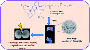
NF-3 and NF-4 assembled in uniform nanofibres with ∼5 nm width with clear and defined boundaries. Peptidomimetics NF-5 and NF-6 also formed nanofibres with relatively shorter length. We also observed some super coiling of fibres with different length for peptide NF-5 (Fig. 2d). The peptidomimetics without glutamic acid (NF-2) did not formed ordered aggregates in TEM under identical conditions (data not shown here).
 |
| | Fig. 2 HRTEM of hydrogels and optical images of their respective hydrogels have shown in inset, where (a) NF-1, (b) NF-3, (c) NF-4, (d) NF-5 and (e) NF-6. | |
As per the previous report on NF-1 supramolecular assembly that two main structural features are responsible for fiber formation: anti-parallel β-sheets and anti-parallel π-stacked fluorenyl groups.38
This model explains the formation of cylindrical structure formed by the interlocking through lateral π–π interactions of four twisted anti-parallel β-sheets. Our designed peptidomimetics were different from NF-1 due to presence of cationic spermine moiety which makes surface of each nanofibres to be highly cationic charged which in turn inhibits π–π interlocking of aromatic hydrophobic taggings resulting into single walled nanofibres. This was confirmed by measurement of surface zeta potential of the nanofibres solution in water. The measured zeta potential for NF-3 and NF-4 were found to be highly cationic with +83 and +73 mV (Fig. S22 and S23†).
The supramolecular arrangement and secondary structure of all peptidomimetics in solutions were confirmed by CD (Fig. 3) and FT-IR (Table 3 and Fig. S18–S21†) spectroscopy.
 |
| | Fig. 3 (a) CD spectroscopy of peptides in buffer, (b) frequency sweep analysis of hydrogels by rheometer. | |
Table 3 Characteristic peaks in FTIR spectra of xerogels
| Peptidomimetics |
C O stretching frequency (cm−1) |
NH bending frequency (cm−1) |
NH stretching frequency (cm−1) |
Assignments |
| NF-3 |
1669 |
1528 |
3299 |
β-Turn |
| NF-4 |
1643 |
1535 |
3275 |
β-Sheet |
| NF-5 |
1654 |
1540 |
3279 |
Random coil |
| NF-6 |
1647 |
1536 |
3278 |
Random coil |
The CD spectra were acquired at 0.2% w/v hydrogels in 10 mM PBS at pH 7.4. In buffer two negative maxima in the range of 203–207 nm and 210–215 nm and a positive peak below 195 nm for peptides NF-3 and NF-4 were observed. CD signals of α-helical peptides with β-turn shows two minima at 207 and 215 nm with the extended wavelength due to n–π* transition usually found for α-helices and β-sheets in the 215–230 nm wavelength range.40 Baldwin and coworkers has also reported that blue shift of negative maxima at 222 nm to 219, 217 and 215 nm as the sequence becomes shorter from 11 to 8 to 4 helical peptide units. Recently Gazit and coworkers also reported α-helical turn structure of ultra short peptide (SHR-FF) showing two minima at 205 nm and 218 nm and a positive peak at 195 nm.41 Our studies support the CD signals of NF-3 and NF-4 arises due to the helical conformation of the peptide in solution. As per deconvolution results for the rest of the designed sequences mixed structures with random coil or β-strand conformation were observed.
Frequency sweep rheological analysis of the hydrogel NF-3 and NF-4 conducted at 0.1–100 rad s−1 at a constant strain of 1% (w/v) at 25 °C showed a difference of high magnitude in the value of G′ (storage modulus) and G′′ (loss modulus) demonstrating the elastic nature of hydrogel (Fig. 3b). The naphthalene conjugated hydrogel (NF-4) demonstrated the rigid, self-supporting hydrogel with higher G′ value than G′′ by an order of magnitude.
Fourier transforms infrared spectroscopy (FTIR) of the xerogel (lyophilized hydrogel) of NF-3 and NF-4 revealed a hydrogen-bonded supramolecular network within the hydrogel. For NF-3 the peak at 1669 cm−1 is a signature of C
O stretching for amide I and a characteristics of β-turn structure (Fig. S18†). The observation vibrational frequency at 1643 and 1535 cm−1 for NF-4 showed characteristic peaks for mixture of β-sheet and random coil due to the C
O stretching and N–H bending of amide I and amide II (Table 3 and Fig. S19†). Other peptidomimetics NF-5 and NF-6 has shown vibrational frequency at 1647 and 1654 cm−1 for C
O stretching of amide I and 1538 and 1540 cm−1 for N–H bending for amide II, respectively (Table 3 and Fig. S20–S21†). These peaks are characteristics of the random coil peptide secondary structure reported previously.42
Antimicrobial activity
Antimicrobial activity of designed peptidomimetics were determined against Gram positive and Gram negative bacteria including multidrug resistant pathogen by broth micro dilution assay as per the standard method from CLSI.30 The antibacterial activity of each peptidomimetics was reported here in terms of minimum inhibitory concentrations (MIC) and MIC was defined as the concentrations at which complete inhibition in growth of bacteria occurred (Table 4). In MIC (μg mL−1), error was <10% of listed values in Table 4 for all measurements.
Table 4 Antibacterial activity of designed peptidomimetics
| Seq. |
MICa (μg mL−1) |
| S. aureus (29213b) |
MRSA (33591) |
S. epidermidis (11228) |
MRSE (51625) |
E. faecalis (7080) |
A. baumanni (19606) |
MRSA (BAA-44) |
E. coli 11775 |
P. aeruginosa (25668) |
| MIC (μg mL−1) the estimated error in all values was <10% for all measurement. Number given to each bacterial strains represent the ATCC number, ND = not determined, VAN = vancomycin, Spn = spermine. |
| NF-1 |
>454.5 |
>454.5 |
>454.5 |
>454.5 |
>454.5 |
>454.5 |
>454.5 |
>454.5 |
>454.5 |
| NF-2 |
7.1 |
7.1 |
3.5 |
7.1 |
14.1 |
ND |
14.1 |
28.4 |
56.8 |
| NF-3 |
14.1 |
28.1 |
14.1 |
28.4 |
28.4 |
56.8 |
28.4 |
113.7 |
227.2 |
| NF-4 |
56.8 |
113.7 |
56.8 |
113.7 |
113.7 |
113.7 |
113.7 |
227.2 |
>454.5 |
| NF-5 |
227.2 |
227.2 |
227.2 |
227.2 |
454.5 |
454.5 |
227.2 |
>454.5 |
>454.5 |
| NF-6 |
14.2 |
14.2 |
7.1 |
14.2 |
ND |
ND |
14.2 |
113.7 |
227.2 |
| VAN |
0.4 |
0.8 |
0.4 |
0.8 |
ND |
56.8 |
0.8 |
113.6 |
ND |
| Spn |
>454.5 |
>454.5 |
ND |
>454.5 |
ND |
ND |
ND |
ND |
ND |
NF-1 and spermine were inactive as antibacterial against all tested strains upto 454.54 μg mL−1 concentrations. Previously it was also reported that antimicrobial activity of spermine was found to be MIC at 4 mM (804 μg mL−1) against S. aureus ATCC 33556, S. aureus Mu50, and S. aureus N315.25 Upon conjugation of N-terminal tagged di-peptides with spermine (NF-2) showed good antimicrobial activity against all strains in the range of 3.5–56.8 μg mL−1 concentrations. The peptidomimetics NF-3 and NF-4 with one glutamic acid were also showing potent antimicrobial activity against all Gram positive bacteria including methicillin resistant (MIC; 14.2–113 μg mL−1), whereas these are moderate active against Gram negative bacteria and MIC were in the range of 227.2–454.5 μg mL−1. Peptide tagged with decanoic acid, NF-6 exhibited potent antibacterial activity against Gram positive bacteria (MIC; 7.1–14.2 μg mL−1), whereas against Gram negative bacteria its activity was moderate with the MIC range of 113.7–227.2 μg mL−1. NF-5 was moderate active in Gram positive bacteria whereas this peptidomimetic was not able to inhibit the growth of Gram negative bacteria. The antibacterial activity of peptidomimetics was also correlated with the self assembly in MHB media by determination of critical aggregation concentrations (CAC) (Table 2 and Fig. S3†). The result of the CAC determination exhibited that peptidomimetics NF-3 and NF-4 were showing MICs above their CAC against all tested bacterial strains, whereas other peptidomimetics exhibited MICs at lower than their respective CAC. The CAC explains that NF-3 and NF-4 exhibited antibacterial activity after assembling of molecules, whereas other peptidomimetics were monomer at the time of growth inhibition of bacteria. In previous studies supramolecular assembly of peptidomimetics have been reported as viable platform for design and synthesis of antimicrobial peptides with balanced features of stability, lower cytotoxicity and high antimicrobial activity.43 Similar to our present work, accumulation of cationic charge on surfaces of self assembly has previously been established to be a key factor for enhanced antibacterial potency for amphiphilic peptides and polymers.44
The cell selectivity of the designed analogues on enucleated hRBCs was determined (Fig. 4a). Hydrogelators peptides, including NF-3 and NF-4 were found to be non-hemolytic up to the hydrogelation concentration of 2% w/v. Peptides NF-2, NF-6 and NF-5 caused significant hemolysis, leading to 97.3 ± 4.4%, 74.8 ± 5.2%, and 55.5 ± 4.5% damage to hRBCs, respectively, at 2% w/v. The presence of cationic surface of nanofibres and well defined self assembling pattern of NF-3 and NF-4 selectively interacted with the negatively charged bacterial membranes whereas it did not interacts with cell membrane of hRBCs.
 |
| | Fig. 4 (a) In vitro hemolytic activity of peptidomimetics against human RBCs (b) cell viability of human keratinocytes (HaCaT) after 24 hours treatment with different concentrations of gel as calculated from the LDH release assay. | |
The LDH release assay with human keratinocytes of non haemolytic peptides (NF-3 and NF-4) were also performed upto their 25 μL and 50 μL of 1% (w/v) gel (Fig. 4b). NF-3 was found to cause 17.0% and 7.4% leakage on cell surfaces at 50 μL (500 μg mL−1) and 25 μL (250 μg mL−1), respectively. At 50 μL of 1% (w/v) gel of peptidomimetics NF-4 did not cause any effect on the membrane of human keratinocytes.
Killing kinetics and membrane disruptive mode of action
Bactericidal kinetic of NF-3 and NF-4, at 2 times and 4 times their respective planktonic MICs were performed with exponentially growing S. aureus ATCC 33591 (Fig. 5a).
 |
| | Fig. 5 (a) Bactericidal kinetic of peptidomimetics against exponentially growing MRSA, (b) scanning electron microscopic images of MRSA (ATCC 33591) treated with active nanofibres at 10 × MIC for 30 min. Pointed arrows indicates the effects of peptidomimetics on cell surface of MRSA. | |
At 2 × MIC, NF-3 peptidomimetics was not showing bactericidal effect even after 6 h of incubation and at 4 × MIC, bactericidal effects with ≥3
log10 CFU mL−1 reductions was observed up on 6 h of incubation. NF-4 was showing fast bactericidal kinetic at 2 × MIC with >3
log10 CFU mL−1 reduction within 6 h of incubation with MRSA. At 4 × MIC, NF-4 was showing bactericidal effect with complete killing of initially inoculated MRSA. The minimum detectable amount of colony counting of bacteria was 2
log10 CFU mL−1. To get insight the effect of hydrogelator peptidomimetics on the membrane of MRSA, SEM studies were performed at 10 × MIC with 108 CFU mL−1 bacterial suspensions. The vehicle control bacteria were examined to have intact membrane with smooth appearance. After 30 min of incubation of NF-3 and NF-4 significant changes in the surface morphology of MRSA with blebs formation and lysates appeared (Fig. 5b). The deformation and surface rupturing mechanism of the peptidomimetics thus demonstrated ability of designed hydrogels to mimic nature of antimicrobial peptides.
Activity against MRSA biofilm
We further evaluated the efficacy of self assembling peptidomimetics NF-3 and NF-4 to prevent the formation of biofilms and to eradicate preformed MRSA biofilms (24 h) by using alamar blue as a redox indicator for assessment of metabolic activity of biofilms (Fig. 6). A well-characterized biofilm-producing reference strain of MRSA (ATCC 33591) was used for biofilms formation. Prior to this experiment, the MICs of peptidomimetic NF-3 and NF-4, in biofilm growth medium (TSB with 0.5% w/v NaCl and 0.25% w/v glucose) were evaluated.
 |
| | Fig. 6 Activity of peptidomimetics against MRSA biofilms by alamar blue assay, here (a) biofilms formation inhibition assay of peptides, (b) effect of peptide hydrogels on 24 h preformed MRSA biofilms. | |
The results showed 2-fold increases in the MICs of peptidomimetics NF-3 and NF-4 in high-salt medium (supplemented with 0.5% NaCl) were observed. For the biofilm formation inhibition assay, initial inoculums were added with 1 × MIC and 2 × MIC concentrations of the tested agents (Fig. 6a). Hydrogel NF-3 and NF-4 were able to inhibit biofilm formation at 1 × MIC, causing reductions in metabolic up to 37.1 ± 7.1%, and 28.0 ± 5.2%, respectively. At 2 × MIC, both peptidomimetics were able to inhibit the adhesion of biofilm, causing ∼90% reductions in measured viability.
The effects of hydrogels on the viability of 24 h-preformed mature biofilms were also evaluated at minimum gelation concentration i.e. 1.0% and 2% w/v gel. At 2% w/v hydrogel, the designed peptidomimetics NF-3 and NF-4 showed 40.3% and 32.2% viable cells, respectively (Fig. 6b).
Overall, the present work gives an impetus towards design of small molecule hydrogels by using minimalist approach. The use of biodegradable monomers (aromatic amino acids) present facile self assembly with low cost synthesis which ultimately may leads to easier translation towards hard to treat infections especially wounds and chronic infections and as drug delivery vehicles. In our previous work we have established potent in vitro activity of linear amphiphiles with N-terminal tagged di-peptidomimetics with C-terminal spermine against MRSA biofilms.27 In previously reported models also it has been shown that the Fmoc-dipeptide sequence gets buried in the hydrogel core whereas the anionic COOH group remain on the surface. The stable positive zeta potential observed in the case of NF-3 and NF-4 showed that these peptidomimetics also arrange with a inner hydrophobic bulk and outer positive surface.
Presence of carbamate functionality in Fmoc group has been reported to hinder a β-sheet secondary structure formation,38 we also observed a deviation in secondary structure between NF-3 and NF-4. Here we explored to fine tune the linear amphiphiles towards hydrogelation to expand the scope of their applicability on biofilms and wound infections.
Conclusions
Herein, we have reported spermine functionalized cationic hydrogel. Systematic analysis with special emphasis on the structural aspects as well as the mechanism of the gelation processes reveals that a different architectural change at the molecular level influences the self-assembling mechanism. Several spectroscopic and microscopic studies confirm the helical structures with beta turn arrangement of the self assembled peptides in the gel phase make it a cationic charged single walled uniform nanofibre which selectively interacts with Gram-positive and Gram-negative bacteria membranes. Peptidomimetics NF-3 and NF-4 showed membrane disrupting bactericidal potential against MRSA. Moreover these hydrogels were also able to eradicate MRSA embedded in biofilms matrix. Hence, the development of such soft biomaterials from N-terminally modified polyamine (spermine) conjugated peptidomimetics exhibited antibacterial activity which will have immense scope in the material science.
Acknowledgements
This work was financially supported for work and fellowship to the author RPD by a Council of Scientific and Industrial Research (CSIR) network project BSC-0120. We are very thankful to Dr Souvik Maiti and Dr Kausik Chakraborty for useful instrumentation facility. Dr M. A. Q. is acknowledged for microbial facility. Mrs Hemlata Gautam, CSIR-IGIB gratefully acknowledged for helping us to acquire transmission electron microscopy and scanning electron microscopic images.
Notes and references
- G. M. Whitesides and M. Boncheva, Proc. Natl. Acad. Sci. U. S. A., 2002, 99, 4769–4774 CrossRef CAS PubMed.
- H. Cui, M. J. Webber and S. I. Stupp, Biopolymers, 2010, 94, 1–18 CrossRef CAS PubMed.
- E. Busseron, Y. Ruff, E. Moulin and N. Giuseppone, Nanoscale, 2013, 5, 7098–7140 RSC.
- M. Rad-Malekshahi, L. Lempsink, M. Amidi, W. E. Hennink and E. Mastrobattista, Bioconjugate Chem., 2016, 27, 3–18 CrossRef CAS PubMed.
- A. Dehsorkhi, V. Castelletto and I. W. Hamley, J. Pept. Sci., 2014, 20, 453–467 CrossRef CAS PubMed.
- A. P. McCloskey, B. F. Gilmore and G. Laverty, Pathogens, 2014, 3, 791–821 CrossRef CAS PubMed.
- M. Zasloff, Nature, 2002, 415, 389–395 CrossRef CAS PubMed.
- A. R. Siddiqui and J. M. Bernstein, Clin. Dermatol., 2010, 28, 519–526 CrossRef PubMed.
- A. Pascual, J. P. K. Tan, A. Yuen, J. M. W. Chan, D. J. Coady, D. Mecerreyes, J. L. Hedrick, Y. Y. Yang and H. Sardon, Biomacromolecules, 2015, 16, 1169–1178 CrossRef CAS PubMed.
- R. Lalani and L. Liu, Biomacromolecules, 2012, 13, 1853–1863 CrossRef CAS PubMed.
- C. Helary, M. Zarka and M. M. Giraud-Guille, J. Tissue Eng. Regener. Med., 2012, 6, 225–237 CrossRef CAS PubMed.
- E. A. Kamoun, X. Chen, M. S. Mohy Eldin and E.-R. S. Kenawy, Arabian J. Chem., 2015, 8, 1–14 CrossRef CAS.
- C. Nathan and O. Cars, N. Engl. J. Med., 2014, 371, 1761–1763 CrossRef PubMed.
- G. Laverty, A. P. McCloskey, B. F. Gilmore, D. S. Jones, J. Zhou and B. Xu, Biomacromolecules, 2014, 15, 3429–3439 CrossRef CAS PubMed.
- H. Du, G. Zha, L. Gao, H. Wang, X. Li, Z. Shen and W. Zhu, Polym. Chem., 2014, 5, 4002–4008 RSC.
- A. C. Engler, N. Wiradharma, Z. Y. Ong, D. J. Coady, J. L. Hedrick and Y.-Y. Yang, Nano Today, 2012, 7, 201–222 CrossRef CAS.
- G. Fichman and E. Gazit, Acta Biomater., 2014, 10, 1671–1682 CrossRef CAS PubMed.
- M. Reches and E. Gazit, Science, 2003, 300, 625–627 CrossRef CAS PubMed.
- K. Igarashi and K. Kashiwagi, Int. J. Biochem. Cell Biol., 2010, 42, 39–51 CrossRef CAS PubMed.
- S. L. Nowotarski, P. M. Woster and R. A. J. Casero, Expert Rev. Mol. Med., 2013, 15, e3 CrossRef PubMed.
- S. Razin and R. Rozansky, Arch. Biochem. Biophys., 1959, 81, 36–54 CrossRef CAS PubMed.
- M. R. Burns, S. J. Wood, K. A. Miller, T. Nguyen, J. R. Cromer and S. A. David, Bioorg. Med. Chem., 2005, 13, 2523–2536 CrossRef CAS PubMed.
- R. A. Casero and P. M. Woster, J. Med. Chem., 2009, 52, 4551–4573 CrossRef CAS PubMed.
- R. Mukhopadhyay and R. Madhubala, Pharmacol. Res., 1993, 28, 359–366 CrossRef CAS PubMed.
- D.-H. Kwon and C.-D. Lu, Antimicrob. Agents Chemother., 2007, 51, 2070–2077 CrossRef CAS PubMed.
- O. N. Kostopoulou, E. C. Kouvela, G. E. Magoulas, T. Garnelis, I. Panagoulias, M. Rodi, G. Papadopoulos, A. Mouzaki, G. P. Dinos, D. Papaioannou and D. L. Kalpaxis, Nucleic Acids Res., 2014, 42, 8621–8634 CrossRef CAS PubMed.
- R. P. Dewangan, S. Joshi, S. Kumari, H. Gautam, M. S. Yar and S. Pasha, Antimicrob. Agents Chemother., 2014, 58, 5435–5447 CrossRef CAS PubMed.
- S. R. Chhabra, A. N. Khan and B. W. Bycroft, Tetrahedron Lett., 2000, 41, 1095–1098 CrossRef CAS.
- A. Baral, S. Roy, A. Dehsorkhi, I. W. Hamley, S. Mohapatra, S. Ghosh and A. Banerjee, Langmuir, 2014, 30, 929–936 CrossRef CAS PubMed.
- F. R. Cockerill, M. A. Wikler, J. Alder, M. N. Dudley, G. M. Eliopoulos, M. J. Ferraro, D. J. Hardy, D. W. Hecht, J. A. Hindler, J. B. Patel, M. Powell, J. M. Swenson, R. B. Thomson, M. M. Traczewski, J. D. Turnidge, M. P. Weinstein and B. L. Zimmer, Methods for Dilution Antimicrobial Susceptibility Tests for Bacteria That Grow Aerobically; Approved Standard- Ninth Edition, Clinical and Laboratory Standards Institute, Wayne, PA, 2012 Search PubMed.
- T. Decker and M.-L. Lohmann-Matthes, J. Immunol. Methods, 1988, 115, 61–69 CrossRef CAS PubMed.
- R. D. Pearson, R. T. Steigbigel, H. T. Davis and S. W. Chapman, Antimicrob. Agents Chemother., 1980, 18, 699–708 CrossRef CAS PubMed.
- J. Bauer, W. Siala, P. M. Tulkens and F. Van Bambeke, Antimicrob. Agents Chemother., 2013, 57, 2726–2737 CrossRef CAS PubMed.
- M. Zhou, A. M. Smith, A. K. Das, N. W. Hodson, R. F. Collins, R. V. Ulijn and J. E. Gough, Biomaterials, 2009, 30, 2523–2530 CrossRef CAS PubMed.
- B. E. Haug, W. Stensen, M. Kalaaji, Ø. Rekdal and J. S. Svendsen, J. Med. Chem., 2008, 51, 4306–4314 CrossRef CAS PubMed.
- D. J. Adams, L. M. Mullen, M. Berta, L. Chen and W. J. Frith, Soft Matter, 2010, 6, 1971–1980 RSC.
- Y. Zhang, Y. Kuang, Y. Gao and B. Xu, Langmuir, 2011, 27, 529–537 CrossRef CAS PubMed.
- A. M. Smith, R. J. Williams, C. Tang, P. Coppo, R. F. Collins, M. L. Turner, A. Saiani and R. V. Ulijn, Adv. Mater., 2008, 20, 37–41 CrossRef CAS.
- D. M. Ryan, T. M. Doran, S. B. Anderson and B. L. Nilsson, Langmuir, 2011, 27, 4029–4039 CrossRef CAS PubMed.
- N. E. Shepherd, H. N. Hoang, G. Abbenante and D. P. Fairlie, J. Am. Chem. Soc., 2005, 127, 2974–2983 CrossRef CAS PubMed.
- S. Mondal, L. Adler-Abramovich, A. Lampel, Y. Bram, S. Lipstman and E. Gazit, Nat. Commun., 2015, 6, 8615 CrossRef CAS PubMed.
- J. Kong and S. Yu, Acta Biochim. Biophys. Sin., 2007, 39, 549–559 CrossRef CAS PubMed.
- D. Xu, L. Jiang, A. Singh, D. Dustin, M. Yang, L. Liu, R. Lund, T. J. Sellati and H. Dong, Chem. Commun., 2015, 51, 1289–1292 RSC.
- L. Liu, K. Xu, H. Wang, P. K. Jeremy Tan, W. Fan, S. S. Venkatraman, L. Li and Y.-Y. Yang, Nat. Nanotechnol., 2009, 4, 457–463 CrossRef CAS PubMed.
Footnote |
| † Electronic supplementary information (ESI) available: HPLC chromatograms, mass spectrums, methods and graphs of critical aggregation concentrations, FTIR, and surface zeta potential. See DOI: 10.1039/c6ra24502a |
|
| This journal is © The Royal Society of Chemistry 2016 |
Click here to see how this site uses Cookies. View our privacy policy here.