Yuuya Nagata*,
Ryohei Takeda and
Michinori Suginome*
Department of Synthetic Chemistry and Biological Chemistry, Graduate School of Engineering, Kyoto University, Katsura, Nishikyo-ku, Kyoto 615-8510, Japan. E-mail: nagata@sbchem.kyoto-u.ac.jp; suginome@sbchem.kyoto-u.ac.jp
First published on 11th November 2016
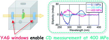
Circular dichroism (CD) spectroscopy at high pressure (≤400 MPa) was accomplished by using polycrystalline yttrium aluminum garnet (Y3Al5O12, YAG) as pressure-resistant optical windows. Conformational changes, including main-chain helix inversions of poly(quinoxaline-2,3-diyl)s (PQXs), in organic solvents at high hydrostatic pressure (>200 MPa) using a newly developed high-pressure CD cell with polycrystalline YAG windows were demonstrated.
The key requirements of a pressure-resistant optical window for CD spectroscopy are: (i) high transmittance for a wide wavelength range; (ii) high durability at high pressure, which can be estimated from the Young's modulus (YM) or apparent elastic limit of the window material; (iii) optical isotropy, so that artifact CD signals can be excluded. It should be noted here that (i) and (ii) are also common requirements for high-pressure optical windows for UV-vis-IR absorption and fluorescence measurements. So far, single-crystal alumina (α-Al2O3, sapphire) has been used as window material for high-pressure optical measurements, as it exhibits high transmittance for a broad wavelength range as well as a high YM (400 GPa).8 However, α-Al2O3 is not suitable as an optical window for CD spectroscopy due to its optical anisotropy arising from its crystal structure (trigonal crystal system, R3c).
In 1976, Harris and co-workers reported a pressure cell for CD measurements using fused silica (quartz glass, SiO2) windows,7a albeit that the pressure was limited to 17 MPa. In 2002, Hayashi and co-workers developed a universal high-pressure cell with exchangeable optical windows (Fig. 1a).7b In this system, the sample solution is enclosed in an inner quartz cell with a diaphragm tube, and the cell is pressurized by water from a high-pressure pump. The sample solution is pressurized through the deformation of the diaphragm tube (Fig. 1b).
![]() | ||
Fig. 1 (a) Schematic illustration of the universal high-pressure CD cell developed by Hayashi and co-workers. (b) Pressurization of a sample solution through deformation of the diaphragm tube. |
Such a high-pressure cell with windows made of diamond (cubic carbon; thickness, T = 1.0 mm), which exhibits an extremely high YM (1100 GPa)8 and optical isotropy (cubic crystal system, Fd3m), tolerated up to 400 MPa. However, as optical grade diamonds are very expensive, alternative materials are desirable. Hayashi's group also showed that a similar cell could tolerate pressures up to 200 MPa when using thick SiO2 windows (T = 8.0 mm, YM = 95 GPa).8
Yttrium aluminum garnet (Y3Al5O12, YAG) is widely used as a lasing medium for solid-state lasers upon doping with various lanthanides.9 Recently, transparent polycrystalline YAG has been developed as an efficient lasing medium,10 which is now commercially available at low cost. Undoped polycrystalline YAG shows good transmittance in a wide wavelength range and a high YM (308 GPa).11 Furthermore, because YAG exhibits optical isotropy (cubic crystal system, m3m), it should be a good prospective material for optical windows in high-pressure CD spectroscopy. In this paper, we report the optical properties of prospective materials for pressure-resistant optical windows including fused silica, sapphire, diamond, and polycrystalline YAG. In addition, we demonstrate high-pressure CD measurements using a pressure cell equipped with polycrystalline YAG windows.
Initially, we prepared disc-shaped windows of fused silica (1, SiO2 glass, T = 6.5 mm), sapphire (2, α-Al2O3, T = 7.0 mm), diamond (3, cubic C, T = 0.5 mm), and polycrystalline YAG (4, T = 5.0 mm) to measure UV-vis transmittance spectra (Fig. 2a). While 1 and 2 showed high transmittance in the entire UV-vis region (200–700 nm), 3 exhibited strong absorption despite its thinness (T = 0.5 mm). Although 4 also exhibited an absorption peak in the short-wavelength region (220 nm), the transmittance of 4 was higher than that of 3 (3: 1.2%, 4: 23.6%), even though the thickness of 4 was ten times higher than that of 3.
Subsequently, we carried out CD measurements on 1–4 (Fig. 2b). While 1 showed weak artifact signals arising from striae or interior strain of the fused silica, 2 exhibited strong artifact peaks due to its optical anisotropy. In addition, the sign of the artifact signal was dependent on the orientation of the disc-shaped sample (for details, see ESI†). Where its transmittance is sufficient (>10%), 3 afforded an almost flat CD spectrum (230–700 nm). It should also be noted that 4 furnished a CD spectrum without artifact peaks and high transmittance for a wider range of the UV-vis region (215–700 nm) than 3 (Fig. 2a), even though the thickness of 4 was ten times higher than that of 3.
Next, the mechanical strength of polycrystalline YAG as an optical window material was investigated (appropriate safety measures must be taken, see ESI†). Polycrystalline YAG discs with different thickness (T = 0.5 and 1.0 mm) were prepared and introduced in an unclamped-type flange with a circular hole (diameter, D = 5.0 mm). The hydrostatic pressure (P) was increased to reach the burst pressure of the disc (Fig. 3a). Average burst pressure values of 67 MPa and 125 MPa were found for T = 0.5 mm and T = 1.0 mm, respectively (Fig. 3b). The apparent elastic limit Fa can be expressed by the equation:
Fa = P × (D/T)2 × K/4 | (1) |
Finally, we performed high-pressure CD measurements on poly(quinoxaline-2,3-diyl)s (PQXs)13 dissolved in organic solvents using polycrystalline YAG windows (T = 5.0 mm). Recently, we reported solvent-14 and pressure-dependent helix inversion15 of the helical main chains in PQXs. For example, a PQX bearing (S)-2-butoxymethyl side chains (P1; Fig. 4) adopted P- or M-helical structures in a mixture of CHCl3/1,1,2-trichloroethane (CHCl3/1,1,2-TCE, v/v = 35/65) at ambient (0.1 MPa) or high pressure (200 MPa), respectively. However, its screw-sense induction behavior at pressures beyond 200 MPa could not be measured, given that we used SiO2 windows.
P1 was dissolved in CHCl3/1,1,2-TCE mixtures of varying proportion and subjected to CD measurements at 0.1, 200, and 400 MPa. The solvent without chiral solute showed almost flat CD spectra at these pressures, suggesting that the YAG windows were not distorted even at the high pressure (Fig. 4a). After the baseline correction, CD spectra of P1 at 0.1, 200, and 400 MPa were obtained (Fig. 4b, see ESI† for other samples). At ambient pressure (0.1 MPa), P1 showed a positive signal at 368.0 nm in a mixture consisting of 20–35% CHCl3, indicating that P1 adopted a P-helical conformation therein (Fig. 4c). In mixtures consisting of 40–100% CHCl3, P1 adopted an M-helical conformation at 0.1 MPa. At 200 MPa, the conformation of P1 was inverted to M-helical in a mixture containing 35% CHCl3. Finally, at 400 MPa, P1 adopted an M-helical conformation in mixtures consisting of 30–100% CHCl3.
We then carry out CD measurements on P1 in 1,2-dichloroethane (1,2-DCE) or 1-butylchloride (1-BuCl), and on P2 bearing (S)-2-pentyloxymethyl side chains in 1,2-DCE (Fig. 4d). P1 in 1,2-DCE exhibited a helix inversion around 100 MPa, and a negative CD signal was intensified at 300 MPa. Although the gabs value slightly increased up to 400 MPa, the screw-sense excess reached saturation at 300–400 MPa. P1 in 1-BuCl adopted an M-helical conformation at 0.1–200 MPa, but the main chain was inverted to a P-helical conformation at 400 MPa. The gabs value of P2 in 1,2-DCE gradually decreased with pressurization, albeit that helix inversion was not observed up to 400 MPa. These results suggest that the use of the high-pressure-resistant YAG windows affords more insight into the effect of pressure on the conformational change of helical PQXs.
Footnote |
† Electronic supplementary information (ESI) available: Experimental procedures, and UV-vis absorption and CD spectra. See DOI: 10.1039/c6ra23736c |
This journal is © The Royal Society of Chemistry 2016 |