Dual release of angiostatin and curcumin from biodegradable PLGA microspheres inhibit Lewis lung cancer in a mice model

Yue Caoa, Xiaoguang Yanga, Yannan Wuc, Jingwen Yia, Yin Wuc, Chunlei Yuc, Yanxin Huangb, Yongli Bao*c, Luguo Sun*b and Yuxin Li*a
aNational Engineering Laboratory for Druggable Gene and Protein Screening, Northeast Normal University, Jingyue Street 2555, Changchun 130117, P. R. China. E-mail: liyx486@nenu.edu.cn; Fax: +86-431-89165917; Tel: +86-431-89165917
bInstitute of Genetics and Cytology, Northeast Normal University, Renmin Street 5268, Changchun 130024, P. R. China. E-mail: sunlg388@nenu.edu.cn; Fax: +86-431-89165922; Tel: +86-431-89165922
cSchool of Life Sciences, Northeast Normal University, Renmin Street 5268, Changchun 130024, P. R. China. E-mail: baoyl800@nenu.edu.cn; Fax: +86-431-89165920; Tel: +86-431-89165920

Received 22nd September 2016 , Accepted 11th November 2016

First published on 16th November 2016


Abstract

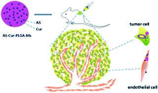
Lung cancer is an aggressive deadly disease worldwide. In this research, PLGA microspheres were used as a co-delivery system of angiostatin and curcumin for the synergistic treatment of lung cancer. The formulations were characterized by morphology, mean diameter, surface potential, and in vitro release kinetics. Furthermore, dual-drug loaded microspheres exhibited a higher antiproliferative activity in endothelial cells but not in HepG2 cells. More importantly, compared with single-drug loaded Ms (AS–PLGA-Ms and Cur–PLGA-Ms), dual-drug loaded Ms (As–Cur–PLGA-Ms) exhibited higher antitumor activity, which was further confirmed by the systemic histological and immunohistochemical analyses.


1 Introduction

Cancer is an aggressive and progressive deadly disease. As a leading cancer type, the global incidence of lung cancer is rising by 0.5% per year, and the mortality from lung cancer is expected to be over 50% by 2020.1 Thus, there are urgent needs to exploit new specific drugs or novel therapeutic methods for lung cancer treatment. It has been well established that tumor growth, including lung cancer, is critically dependent on angiogenesis for nutrients and oxygen supply. Therefore, antiangiogenesis has been regarded as a promising therapeutic strategy for clinical therapy of cancer.2–4

Angiostatin, a 38 KD protein, was originally purified from serum and urine of mice with primary Lewis lung carcinoma. It has been identified and characterized as a potent angiogenic inhibitor.5 In vitro, angiostatin inhibits the proliferation of capillary endothelial cells.6 Subsequently, angiostatin has been proven to efficiently suppress the growth of a broad spectrum of tumors in mice using either murine or human cell lines.7 By inhibiting the formation of new blood, angiostatin treatment generate increased apoptosis of the tumor cells. Just like other protein drugs, angiostatin have a lot of shortcoming to overcome, such as short half life, instability, expensive.8

Curcumin (Cur) is a low molecular weight natural pigment, a polyphenolic compound derived from the Indian spice turmeric (curcuma longa). Previous studies have reported that Cur showed inhibitory effect on a wide range of cancers, such as lung, pancreatic, prostate carcinoma and malignant glioma.9–11 The mechanisms of anticancer activity of curcumin have been studied for many years, including induction of tumor cell apoptosis, antiangiogenesis and inhibition of invasion and metastasis.12 However, the therapeutic efficacy of Cur is restricted due to its low solubility in aqueous solution and poor oral bioavailability.13,14

Recently, multi-agent therapies have been gaining more attention in cancer treatment. Combination with different mechanisms by utilize multiple drugs may treat cancer effectively.15,16 We recently reported that biodegradable polymersomes based on PEG–PCL diblock copolymers could efficiently co-encapsulate angiostatin and curcumin. Our data demonstrates a synergistic effect of angiostatin and curcumin in inhibiting angiogenesis, both in vitro and in vivo.17

The dual delivery of both lipophilic and hydrophilic agents in polymeric carriers to achieve synergistic effects is a new trend for anti-tumour therapy.18–21 Among them, many studies have been focused on the microspheres composed of poly(lactide-co-glycolide) (PLGA). PLGA is an efficient drug carrier owing its biodegradable and biocompatible and approved by FDA.22,23 PLGA microspheres have been used as a sustained delivery system of many proteins.24–27 Due to their size and spherical structure, PLGA microspheres can be easily injected to any site in vivo. Therefore, our strategy is to incorporated angiostatin and curcumin in PLGA microspheres in order to exert greater antiangiogenic and antitumorigenic effects through additive effects.

In this study, PLGA microspheres were prepared by a water-in-oil-in-water (W1/O/W2) double emulsion solvent evaporation technique, and then characterized for morphology analysis, size and zeta potential. We tested the cytotoxic effect of individual and combinational drugs in HMEC-1 cell line. The antitumor efficacy of microspheres was evaluated in a Lewis lung cancer cell bearing xenograft tumor model.

2 Materials and methods

2.1 Materials

Poly(D,L-lactide-co-glycolide) (PLGA; L/G = 75/25, MW 50[thin space (1/6-em)]000) was obtained from Changchun SinoBiomaterials Co., Ltd.; angiostatin kringles 1 to 3 (K1–3) were obtained from our laboratory (Northeast Normal University, China). Polyvinyl alcohol (PVA; MW 89[thin space (1/6-em)]000–98[thin space (1/6-em)]000), curcumin, fetal bovine serum (FBS) and 3-(4,5-dimethyl-2-thiazolyl)-2,5-diphenyl-2-H-tetrazolium bromide (MTT) were purchased from Sigma Aldrich (St. Louis, USA). HMEC-1 and HepG2 cell line was obtained from National Institute for the Control of Pharmaceutical and Biological Products. BCA™ Protein Assay Kit and 4′,6-diamidino-2-phenylindole (DAPI) were purchased from Beyotime (Shanghai, China). CD 31 antibodies (mouse anti-human) and ECL reagent were purchased from BD Company (New Jersey, USA). Other chemicals used were of analytical grade. Male C57B16/J mice (4–6 week) were purchased from the Animal Center of Jinlin University (Changchun, China).

2.2 Preparation of PLGA microspheres loaded with AS/Cur

Angiostatin and curcumin (AS–Cur) loaded PLGA microspheres (Ms) were prepared using a W1/O/W2 method.28–30 Briefly, 135 mg of PLGA, 13.5 mg curcumin and 24 mg Tween-80 were fully dissolved in 4.5 mL of dichloromethane (organic phase, O), mixed with 0.2 mL double-distilled water containing 13.5 mg angiostatin (inner aqueous phase, W1). Acoustic vibration in ice bath for 60 s using an ultrasonic oscillation instrument (KQ-600DE, Kun Shan Ultrasonic Instruments Co., Ltd, China) produced water-in-oil (W1/O) emulsion. Subsequently, the emulsion was added into 50 mL of 1% PVA solution (outer aqueous phase, W2) and homogenized in ice bath at 5000 rpm for 5 min with a high-speed homogenization dispersion machine (POLYTRON-2000, POLYTRON, Switzerland) to produce a double emulsion solution. Then, 100 mL double-distilled water was poured and gently stirred at room temperature for 8 h to evaporate the organic solvents. The PLGA nanoparticles were collected by centrifugation (Allegra 25R centrifuge, Backman coulter, American) at 12[thin space (1/6-em)]000 rpm for 5 min and rinsed three times. Finally, microspheres were freeze-dried and stored at 4 °C until use. The blank microspheres (without drugs) and microspheres loaded with angiostatin (AS–PLGA-Ms) or curcumin (Cur–PLGA-Ms) were prepared in a similar way.

2.3 Microsphere morphology and size

The shape and surface morphology of PLGA microspheres were studied by Philips FEI XL30 scanning electron microscopy. The dried microspheres were placed onto a silicon wafer using double-sided adhesive tap, sputter-coated with a gold–palladium film and then analyzed with SEM. The size and zeta potentials (ζ-potential) of the microspheres were measured using a 90Plus particle size analyzer.

2.4 Determination of loading efficiency

10 mg of the dual drug loaded microspheres (AS–Cur–PLGA-Ms) were dissolved in 1 mL DCM. The amount of loaded angiostatin was estimated using BCA protein assays according to the supplier's protocols. The amount of curcumin was determined by fluorescence measurements at 425 nm. Each sample was assayed three times. The drug loading content (DLC%) and the drug loading efficiency (DLE%) were calculated by the following eqn (1) and (2).
 
DLC (%) = (amount of drug in microsphere/amount of drug loaded microsphere) × 100% (1)
 
DLE (%) = (amount of drug in microsphere/amount of drug in feed) × 100% (2)

2.5 Release studies

About 100 mg of freeze-dried microspheres were incubated in 10 mL of PBS at different pH (pH 7.4 and pH 4.5) in a centrifuge tube. The tubes were kept in a water bath shaker in 120 rpm at 37 °C. At specific time intervals, the tubes were removed from water bathes, and centrifuged at 2000 rpm for 10 min. The supernatants were collected and replenished with the same amount of fresh buffer. The release amount of drug was analyzed by the same procedure of load test.

2.6 Inhibition of cell viability assay

HMEC-1 and HepG2 cells were cultured in the complete DMEM medium at 37 °C and 5% CO2. The cells were seeded into 96-well plates at a density of 8000 cells per well. After incubated for 24 h, the fresh medium containing blank microspheres or drug-encapsulated microspheres were added to each well. After being incubated for certain times, 20 μL of MTT solution (5 mg mL−1) was added, and the plates were incubated for 4 h. The cell viability of control and treated was determined by a spectrophotometer (Multiskan GO, Thermo Scientific).

2.7 Cellular uptake of microspheres

HMEC-1 cells were maintained in 6-well plates at 37 °C. After incubated for 24 h, the fresh medium containing either blank or drug-encapsulated microspheres were added. After incubation for 2 or 4 h, the cells were washed with PBS (0.01 M, pH 7.4) three times. Subsequently, the cells were stained with DAPI for 20 min. Images were obtained from laser scanning confocal microscope (OLYMPUS FV1000, Center Valley, PA, USA). Flow cytometry was used to quantity study cell internalization of curcumin. HMEC-1 cells were seeded into 6-well plates at 2.0 × 10⁵ cells per well. The cell were incubated with amount of microspheres for 2 or 4 h. Internalization signals were analyzed by flow cytometer (Beckman, California, USA).

2.8 Scratch assay

HMEC-1 migration was evaluated using a scratch wound healing assay.31,32 HMEC-1 were seeded in a 6-well plate to form a monolayer. The following day, confluent monolayers were scratched using a sterile 200 μL pipette tip to create a “wound” followed with a gentle wash with PBS. Cells were treated with four types of microspheres (PLGA-Ms, AS–PLGA-Ms, Cur–PLGA-Ms and AS–Cur–PLGA-Ms) for 24 h and specific scratching sites of the wound areas were photographed at 0 h and 24 h using a OLYMPUS phase contrast microscope (10 × 20).

2.9 Western blot analysis

HMEC-1 (1.0 × 10⁶ cells per mL) were incubated with four types of microspheres for 24 h at 37 °C. Cell pellets were collected and lysed in a lysis buffer by ice-bath for 40 min. After centrifugation at 12[thin space (1/6-em)]000g for 5 min, the samples were boiled with loading buffer, and then separated by a SDS-polyacrylamide gel and transferred to a nitrocellulose membrane. The membranes were blocked by incubation in 5% skimmed milk. After washed three times, the membrane were probed with primary antibodies including rabbit antibodies to p53, Bax and Bcl-2 overnight. On the following day, the membranes were incubated with a horseradish peroxidase (HRP)-conjugated secondary antibodies for 1 h. At last, membranes were developed using ECL reagent and chemiluminescent signal was detected by a DNR Bio-imaging systems.

2.10 Tumor inhibition effect of angiostatin and curcumin loaded microspheres in vivo

The in vivo studies were performed in National Engineering Laboratory for Druggable Gene and Protein Screening, Northeast Normal University, China. The mice were handled under protocols approved by the School of Life Sciences Animal Care and Use Committee of Northeast Normal University. Animals were acclimatized to the animal facility for 1 week before dosing, and the subcutaneous injecting with 100 μL of the 1.0 × 10⁶ LLC cells suspended in PBS into the back of mice. 7 days after inoculation, mice were randomized into six groups (n = 10 for each group). The mouse models were directly injected in solid tumor with PBS, PLGA-Ms, AS–PLGA-Ms, Cur–PLGA-Ms, AS + Cur and AS–Cur–PLGA-Ms with angiostatin and curcumin (30 mg per kg per day) equivalent alternate days. Tumor volume was calculated daily as length × width2/2.

2.11 Histological and immunohistochemical analyses

Tumors (LLC) were collected and paraffin embedded. Sections (5 μm) were first stained with hematoxylin and eosin (H&E). Immunohistochemistry staining was performed using anti-CD31 antibody (monoclonal, dilution: 1[thin space (1/6-em)]:[thin space (1/6-em)]200) to evaluate the microvessel density. The histological and immunohistochemical alterations were captured by a OLYMPUS phase contrast microscope.

3 Results and discussion

3.1 Microsphere characterization

The surface morphology of the PLGA microspheres prepared by W/O/W were determined by scanning electron microscopy (SEM). The blank microspheres had a spherical shape with smooth surface morphology (Fig. 1A and B). The mean particle size of blank PLGA-Ms was 1.91 ± 0.34 μm, while the diameters of AS–PLGA-Ms, Cur–PLGA-Ms and AS–Cur–PLGA-Ms were 2.6 ± 0.15 μm, 2.2 ± 0.09 μm and 2.89 ± 0.21 μm respectively (Fig. 1C). In addition, the surface charge of blank PLGA-Ms was −35.31 ± 0.81 mV, while the zeta potentials of AS–PLGA-Ms, Cur–PLGA-Ms and AS–Cur–PLGA-Ms were −27.36 ± 1.27 mV, −30.36 ± 0.94 mV and −33.68 ± 1.03 mV, respectively (Fig. 1D).
image file: c6ra23627h-f1.tif
Fig. 1 (A) and (B) Scanning electron microscopy (SEM) images of blank PLGA microspheres, (C) the size of different PLGA Ms, (D) the zeta potential of PLGA Ms.

3.2 Loading and in vitro release

The DLC of AS-loaded PLGA-Ms and Cur-loaded PLGA-Ms was 6.9% and 4.7%, with a DLE of 74.8% and 51.7%, respectively. In dual-drug loaded PLGA-Ms, the DLC and DLE were 6.6% and 66.7% for angiostatin, and 4.3% and 45.8% for curcumin, respectively.

The advantage of drug delivery system is to controllable release behavior for drug which can prolong release time and reduce the time of administration. In this study, the release profile of angiostatin and curcumin from the PLGA microspheres were determined in PBS at physiological pH (pH 7.4) and acidic condition corresponding to the endosome (pH 4.5). We observed a pH responsive release of angiostatin and curcumin from PLGA microspheres. Fig. 2 illustrated the slow leakage kinetics of drugs and Cur released a little faster than AS over a 14 day period. At the end of experiment, the cumulative release of AS and Cur were reach to 81.6% and 91.3% respectively at pH 4.5. Therefore, PLGA microspheres can be used for drug delivery and sustained release them into acidic environments such as cancer.


image file: c6ra23627h-f2.tif
Fig. 2 In vitro dual-drug-release behavior of AS and Cur from AS–Cur–PLGA-Ms.

3.3 Cell viability of microspheres

The MTT colorimetric assay was a routine preparation technique to measure the metabolic activities of various cells, which was developed by Mosmann in 1983.33 Fig. 3 show the cytotoxic effects of microspheres on HMEC-1 cells. Fig. 4 show the cytotoxic effects of microspheres (at doses of 20 g L−1 curcumin equivalents) on HepG2 cells. Owing to its biocompatibilities, the blank of PLGA microspheres did not exhibit significant cytotoxicity to HMEC-1 and HepG2 cells. The cytotoxicity of AS–Cur–PLGA-Ms was significantly greater than those of AS–PLGA-Ms and Cur–PLGA-Ms at the same dosages in HMEC-1 cells. These results revealed that combination of angiostatin and curcumin showed synergistic inhibitory effect in endothelial cells but not in HepG2 cells. The angiostatin K1–3 from our laboratory specially inhibited the proliferation of endothelial cells, according with the literature.34,35
image file: c6ra23627h-f3.tif
Fig. 3 Inhibition on cell viability of blank and drug-loaded PLGA-Ms to HMEC-1 cells after incubation for 72 h.

image file: c6ra23627h-f4.tif
Fig. 4 Inhibition on cell viability of blank and drug-loaded PLGA-Ms to HepG2 cells after incubation for 24 h, 48 h and 72 h.

3.4 Cellular uptake of microspheres

To determine cellular uptake of curcumin, HMEC-1 cells were incubated with AS–Cur–PLGA-Ms for 2 h and 4 h. CLSM images (Fig. 5A) showed that the fluorescence (green) of the curcumin which loaded in AS–Cur–PLGA-Ms was located around the entire cell including the nucleus area (blue), which further confirmed that the microspheres could efficiently deliver drug into HMEC-1 cells. Importantly, the cells cultured with AS–Cur–PLGA-Ms for 4 h exhibited higher green fluorescence intensity than those incubated for 2 h. As shown in Fig. 5B, flow cytometric analyses was used for further confirming the cellular uptakes of curcumin.
image file: c6ra23627h-f5.tif
Fig. 5 (A) CLSM images of HMEC-1 cells incubated with AS–Cur–PLGA-Ms for 2 h and 4 h. (B) Flow cytometric profiles of HMEC-1 cells incubated with PBS, AS–Cur–PLGA-Ms for 2 h and 4 h.

3.5 Scratch assay

The migration of endothelial cells is a key process of angiogenesis. Scratch assay was applied to microspheres to assess the ability of HMEC-1 migration inhibition. Significant inhibition of HMEC-1 migration into the scratch area was observed in the AS–Cur–PLGA-Ms group (Fig. 6A). Quantitative analysis indicated that treatment with AS–PLGA-Ms, Cur–PLGA-Ms and AS–Cur–PLGA-Ms resulted in 73.58%, 65.21% and 21.19% value of migrated cells in contrast with PLGA-Ms, respectively (Fig. 6B). These results revealed that the potentiality of AS–Cur–PLGA-Ms in inhibiting HMEC-1 migration in vitro.
image file: c6ra23627h-f6.tif
Fig. 6 Inhibition of HMEC-1 cell migration after the treatment of different treatment groups for 24 h. (A) Photographs were taken at 0 h and 24 h (B) quantification of the number of migrated cells.

3.6 Effect of AS–Cur–PLGA-Ms on the expression of p53, Bax and Bcl-2

P53, Bax and Bcl-2 are the apoptotic related proteins of mitochondrial apoptotic pathway. Many studies have proved that the change of Bax/Bcl-2 ratio could lead to cell death.36 To further explore the mechanism of different PLGA-Ms induced apoptosis, Bax, Bcl-2 and P53 protein levels were analyzed by western blot after HMEC-1 cells exposure to various microspheres. As shown in Fig. 7, the treatment of HMEC-1 cells with dual drug-loaded microspheres (AS–PLGA-Ms) significantly increased the protein expression of P53 and Bax, while significantly decreased Bcl-2 levels, when compared with the other groups (PLGA-Ms, AS–Cur–Ms and Cur–PLGA-Ms). Our results revealed that combination with angiostatin and curcumin increased the expression of P53 and Bax, and decreased the expression of Bcl-2, which might be one of the mechanisms of synergistically induced HMEC-1 cells apoptosis.
image file: c6ra23627h-f7.tif
Fig. 7 Effects of AS–Cur–PLGA-Ms on expression of P53, Bax and Bcl-2 in HMEC-1 cells.

3.7 In vivo antitumor efficacy

The antitumor efficacy of PLGA-based Ms were carried out toward a LLC xenografted model developed by the injection of LLC cells in the back of C57B16/J mice. The tumor volumes were monitored alternate days over a treatment period of 34 days. Fig. 8 displays that tumor in the control group (PBS) grew rapidly, and the average tumor volume reached to about 6100 mm3. Similarly, the tumor volume in the treatment of blank of Ms reached to 5700 mm3. The tumors were inhibited to different degree by the treatments with free drug and various Ms. The average tumor volume in dual drug-loaded Ms group was lower than single drug-loaded Ms. Fortunately, AS–Cur–PLGA-Ms exhibited the enhanced antitumor efficacy compared with free angiostatin plus curcumin.
image file: c6ra23627h-f8.tif
Fig. 8 Tumor volumes of LLC-xenografted mice after treatment with various PLGA-Ms with PBS as control. Data were presented as mean ± SD.

H&E staining of tumor tissues, a standard for malignancy diagnosis,37–39 was used for further evaluate the antitumor efficacies of various groups. In H&E staining, the nucleic acid is dyed deep blue-purple with hematoxylin, and proteins are nonspecifically stained eosin. The tumor tissue of PBS and blank of PLGA-Ms had few pathological changes, while the tumor cells began to become apoptotic and nuclei started to disappeared in the other groups (Fig. 9). Broadly speaking, the sorting of number of cell nucleus was calculated as follow: AS–Cur–PLGA-Ms < free AS + Cur < Cur–PLGA-Ms < AS–PLGA-Ms < PLGA-Ms and PBS (as control).


image file: c6ra23627h-f9.tif
Fig. 9 The histopathological analysis of the tumor tissues from different treatment groups. (A) PBS, (B) blank PLGA-Ms, (C) AS–PLGA-Ms, (D) Cur–PLGA-Ms, (E) AS + Cur, and (F) AS–Cur–PLGA-Ms.

To evaluate the antiangiogenic effect, we measured the residual tumors histologically. Microvessel density (MVD) is an vital marker of tumoral angiogenesis and a important parameter considered for quantifying intratumoral vessels in cancer.40 Paraffin sections were immunohistochemically stained with an endothelial specific antibody against CD31.41,42 Consistent with above findings, microvessel densities in tumor sections prepared from mice injected with AS–Cur–PLGA-Ms were found to be remarkably less compared to other groups (Fig. 10). The sequence of the density of blood vessels in different groups were in accordance with HE stain assay.


image file: c6ra23627h-f10.tif
Fig. 10 The immunohistochemical (CD31) analysis of the tumor tissues from different treatment groups. (A) PBS, (B) blank PLGA-Ms, (C) AS–PLGA-Ms, (D) Cur–PLGA-Ms, (E) AS + Cur, and (F) AS–Cur–PLGA-Ms.

4 Conclusion

In this research, PLGA Ms showed an optimizing potential as a long-term delivery system. The cytotoxicity of the HMEC-1 cells was significantly increased by AS–Cur–PLGA-Ms. Furthermore, co-delivery of angiostatin and curcumin by PLGA Ms significantly inhibited the tumor growth in vivo. Thus, PLGA microspheres could serve as an ideal delivery system for co-delivery angiostatin and curcumin to exert synergistic effects in vitro and in vivo.

Acknowledgements

This study was supported by grants from the following foundations: the National Natural Science Foundation of China (No. 81272919, 81272242, 81502284, 51403031), the Fundamental Research Funds for the Central Universities, the Research Foundation of JiLin Provincial Science & Technology Development (No. 20150101188JC, 20150309003YY, 20140203008YY, 20140520049JH, 20130201008ZY).

References

  1. C. Gibelin and S. Couraud, Lung Cancer, 2016, 100, 45–52 CrossRef PubMed.
  2. J. Folkman, Journal of Pediatric Surgery, 2007, 42, 1–11 CrossRef PubMed.
  3. J. Folkman, Exp. Cell Res., 2006, 312, 594–607 CrossRef CAS PubMed.
  4. M. S. O'Reilly, Int. J. Radiat. Oncol., Biol., Phys., 2007, 69, S64–S66 CrossRef PubMed.
  5. M. S. O'Reilly, L. Holmgren, Y. Shing, C. Chen, R. A. Rosenthal, M. Moses, W. S. Lane, Y. Cao, E. H. Sage and J. Folkman, Cell, 1994, 79, 315–328 CrossRef.
  6. L. Holmgren, M. S. O'Reilly and J. Folkman, Nat. Med., 1996, 1, 149–153 CrossRef.
  7. M. S. O'Reilly, L. Holmgren, C. Chen and J. Folkman, Nat. Med., 1996, 2, 689–692 CrossRef.
  8. C. Ramachandran and W. You, Breast Cancer Res. Treat., 1999, 54, 269–278 CrossRef CAS PubMed.
  9. T. Sarkar, R. J. Butcher, S. Banerjee, S. Mukherjee and A. Hussain, Inorg. Chim. Acta, 2016, 439, 8–17 CrossRef CAS.
  10. N. Shinojima, T. Yokoyama, Y. Kondo and S. Kondo, Autophagy, 2007, 3, 635–637 CrossRef CAS PubMed.
  11. S. Lev-Ari, A. Starr, A. Vexler, V. Karaush, V. Loew, J. Greif, E. Fenig, D. Aderka and R. Ben-Yosef, Anticancer Res., 2006, 26, 4423–4430 CAS.
  12. M. M. Yallapu, M. Jaggi and S. C. Chauhan, Drug Discovery Today, 2012, 17, 71–80 CrossRef CAS PubMed.
  13. H. H. Tonnesen, Pharmazie, 2002, 57, 820–824 CAS.
  14. K. S. Parvathy, P. S. Negi and P. Srinivas, Food Chem., 2009, 115, 265–271 CrossRef CAS.
  15. Y. Yokoyama, M. Dhanabal, A. W. Griffioen, V. P. Sukhatme and S. Ramakrishnan, Cancer Res., 2000, 60, 2190–2196 CAS.
  16. K. Han, S. Chen, W. H. Chen, Q. Lei, Y. Liu, R. X. Zhuo and X. Z. Zhang, Biomaterials, 2013, 34, 4680–4689 CrossRef CAS PubMed.
  17. Y. Cao, Y. Li, Y. Wu, W. L. Li, C. L. Yu, Y. X. Huang, L. G. Sun, Y. L. Bao and Y. X. Li, RSC Adv., 2016, 6, 105442–105448 RSC.
  18. T. Thambi, V. G. Deepagan, H. Ko, D. S. Lee and J. H. Park, J. Mater. Chem., 2012, 22, 22028–22036 RSC.
  19. C. Z. Liang, H. Li, Y. Q. Tao, L. H. Peng, J. Q. Gao, J. J. Wu, F. C. Li, J. M. Hua and Q. X. Chen, Acta Biomater., 2013, 9, 9423–9433 CrossRef CAS PubMed.
  20. T. S. Feng, H. Y. Tian, C. N. Xu, L. Lin, Z. G. Xie, M. Hon-Wah Lam, H. J. Liang and X. S. Chen, Eur. J. Pharm. Biopharm., 2014, 88, 1086–1093 CrossRef CAS PubMed.
  21. J. Zhou, D. J. Guo, Y. Zhang, W. Wu, H. T. Ran and Z. G. Wang, ACS Appl. Mater. Interfaces, 2014, 6, 5566–5576 CAS.
  22. F. Esmaeili, M. H. Ghahremani, B. Esmaeili, M. R. Khoshayand, F. Atyabi and R. Dinarvand, Int. J. Pharm., 2008, 349, 249–255 CrossRef CAS PubMed.
  23. S. Acharya and K. S. Sahoo, Adv. Drug Delivery Rev., 2011, 63, 170–183 CrossRef CAS PubMed.
  24. J. L. Cleland, A. Mac, B. Boyd, J. Yang, E. T. Duenas, D. Yeung, D. Brooks, C. Hsu, H. Chu, V. Mukku and A. J. S. Jones, Pharm. Res., 1997, 14, 420–425 CrossRef CAS.
  25. V. G. Roullin, L. Lemaire, M. C. Venier-Julienne, N. Faisant, F. Franconi and J. P. Benoit, Anticancer Res., 2003, 23, 21–25 CAS.
  26. W. J. Hua, D. D. Ding, G. Ren, X. Y. Xu, X. J. Yin and Y. Q. Hu, J Control Release, 2009, 134, 91–97 CrossRef PubMed.
  27. P. Zhai, X. B. Chen and D. J. Schreyer, Mater. Sci. Eng., C, 2015, 56, 251–259 CrossRef CAS PubMed.
  28. R. Eswaramoorthy, C. C. Chang, S. C. Wu, G. J. Wang, J. K. Chang and M. L. Ho, Acta Biomater., 2012, 8, 2254–2263 CrossRef CAS PubMed.
  29. V. Saini, V. Jain, M. S. Sudheesh, S. Dixit, R. L. Gaur, M. K. Sahoo, S. K. Joseph, S. K. Verma, K. S. Jaganathan, P. K. Murthy and D. Kohli, J. Drug Targeting, 2010, 18, 212–222 CrossRef CAS PubMed.
  30. V. Saini, S. K. Verma, P. K. Murthy and D. Kohli, Vaccine, 2013, 31, 4183–4191 CrossRef CAS PubMed.
  31. A. M. Goodwin, Microvasc. Res., 2007, 74, 172–183 CrossRef CAS PubMed.
  32. S. C. Ricardo, M. G. Vera and B. Fátima, J. Funct. Foods, 2014, 11, 160–171 CrossRef.
  33. T. Mosmann, J. Immunol. Methods, 1983, 65, 55–63 CrossRef CAS PubMed.
  34. B. J. Annutti, S. T. Gately and M. E. Quevedo, Cancer Res., 1997, 57, 5277–5280 Search PubMed.
  35. W. H. Hou, T. Fang, Y. R. Chai, T. Y. Wang, J. M. Wang and L. X. Xue, Protein Expression Purif., 2006, 47, 93–98 CrossRef CAS PubMed.
  36. K. Liang, Y. Ye, Y. Wang, J. Zhang and C. Li, J. Neurol. Sci., 2014, 344, 100–104 CrossRef CAS PubMed.
  37. H. C. Cook, J. Clin. Pathol., 1997, 50, 716–720 CrossRef CAS PubMed.
  38. S. Mukhopadhyay and E. E. Doxtader, Hum. Pathol., 2013, 44, 2346–2352 CrossRef PubMed.
  39. K. Ottmann, M. Tronnier and C. Mitteldorf, J. Am. Acad. Dermatol., 2015, 73, 637–644 CrossRef CAS PubMed.
  40. J. C. Goddard, C. D. Sutton, P. N. Furness, R. C. Kockelbergh and K. J. OP’Byrne, Angiogenesis, 2002, 5, 15–20 CrossRef.
  41. G. Gasparini, Eur. J. Cancer, 1996, 14, 2379–2385 CrossRef.
  42. S. X. Yang, S. M. Steinberg, D. Nguyen, T. D. Wu, Z. Modrusan and S. M. Swain, Clin. Cancer Res., 2008, 14, 5893–5899 CrossRef CAS PubMed.

This journal is © The Royal Society of Chemistry 2016
Click here to see how this site uses Cookies. View our privacy policy here.