Jiangdong Guo,
Ying Yang,
Wensheng Yu,
Xiangting Dong*,
Jinxian Wang,
Guixia Liu and
Tingting Wang
Key Laboratory of Applied Chemistry and Nanotechnology at Universities of Jilin Province, Changchun University of Science and Technology, Changchun 130022, China. E-mail: dongxiangting888@163.com; Fax: +86-0431-85383815; Tel: +86-0431-85582574
First published on 18th November 2016
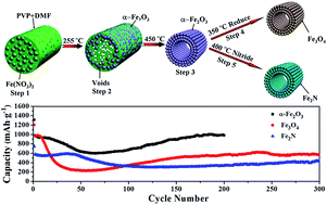
α-Fe2O3 hollow nanofibers were synthesized via a facile electrospinning process followed by a post-calcination process, and for the first time, Fe3O4 and Fe2N hollow nanofibers were successfully obtained via reduction and nitridation of the prepared α-Fe2O3 hollow nanofibers in the presence of NH3 atmosphere at 350 °C and 400 °C, respectively. The crystal structure, morphology and compositions of the α-Fe2O3, Fe3O4 and Fe2N hollow nanofibers were investigated by X-ray diffraction (XRD), scanning electron microscopy (SEM), transmission electron microscopy (TEM), and energy dispersive spectrometry (EDS). Electrochemical measurements show that the α-Fe2O3 and Fe3O4 hollow nanofibers electrodes deliver a high specific initial discharge capacity of 1314 and 1210 mA h g−1, respectively, and a stable cycling performance (980 mA h g−1 for α-Fe2O3 after 200 cycles and 572 mA h g−1 for Fe3O4 after 300 cycles) at a current density of 100 mA g−1. The Fe2N hollow nanofibers electrode demonstrates a high initial discharge capacity, good cycling stability (438 mA h g−1 at the 300th cycle with a current density of 100 mA g−1), high coulombic efficiency, and excellent rate capability. The superior electrochemical performances are attributed to the unique one-dimensional hollow nanostructure of the materials. The prepared hollow nanofibers are candidate anode materials for Li-ion batteries.
To improve the durability of Fe2O3, Fe3O4 and Fe2N electrodes, various types of nano-architectures have been employed as anode materials for LIBs, such as nanospheres,20 nanofibers,21 nanorods,22 nanotubes,23 and some other more complex ones.24 The results indicate that the initial discharge capacities increase obviously, and the capacity retention ratios are improved. Among the reported Fe2O3, Fe3O4 and Fe2N nanostructures, one-dimensional (1D) nanomaterials are expected to have high performance in energy storage systems because they facilitate the electron transport along the long dimension and the two short dimensions ensure fast Li-ion insertion/extraction.25 Electrospinning is an outstanding technique to process viscous solutions or melts into continuous fibers or ribbons with 1D nanostructure.26,27 Electrospinning is widely used to fabricate solid nanofibers and hollow nanofibers. In our early work, we have fabricated LiFePO4/C composite nanofibers and LiFePO4/C/Ag composite hollow nanofibers by electrospinning technology and used the composites as the cathode materials for LIBs.28,29 When the hollow-structure nanomaterials are explored as the electrode materials for LIBs, the underlying principle is that the void space within these hollow nanostructures could efficiently enhance their lithium storage properties by shortening the diffusion distance for Li-ion and reversibly accommodating large volume changes.30 Jeong et al.20 reported that hierarchical Fe2O3@PANI hollow spheres were prepared by a template-free sonochemical method, which exhibited high reversible capacity, excellent cycle stability and rate capability. Chen et al.31 reported that Fe3O4 beads exhibited superior performances with large reversible capacity and good cycling property benefiting from the unique hollow structures. Lei et al.32 synthesized coaxial Fe3O4@C hollow particles by confined nanospace pyrolysis, presenting enhanced electrochemical properties. All these works prove that hollow structure is desirable for achieving high capacity and cycling stability of electrode materials. However, it is still a great challenge to fabricate novel and well-defined hollow nanostructures of Fe2O3, Fe3O4 and Fe2N with high lithium storage capacity and excellent cycling stability.
Herein, we employed electrospinning followed by thermal treatment to fabricate α-Fe2O3 hollow nanofibers, and then cleverly used the reduction and nitridation of NH3 atmosphere for preparation of Fe3O4 and Fe2N hollow nanofibers by the control of the calcination temperature, and then they were used as anode materials for LIBs. The morphology, structure and electrochemical performances of the obtained hollow nanofibers were systematically studied. Some new insights into the design and synthesis of electrodes have been proposed, which will be very helpful for the development of future superior performance electrodes.
![]() | ||
Fig. 1 TG and DSC curves of Fe(NO3)3/PVP composite nanofibers (a); XRD patterns and PDF standard cards of the obtained α-Fe2O3 (b), Fe3O4 (c) and Fe2N (d) hollow nanofibers. |
Crystal structure and phase composition of α-Fe2O3 hollow nanofibers were confirmed by XRD measurements. As observed in Fig. 1b, all diffraction peaks are sharp and well-defined, implying that the sample is highly crystallized. It is seen that all the diffraction peaks are indexed as a pure rhombohedral phase of α-Fe2O3, which match well with those of the JCPDS standard card (PDF#33-0664), and no characteristic peaks are observed for other impurities, demonstrating the high purity of the prepared α-Fe2O3.
Typical XRD patterns of the obtained Fe3O4 hollow nanofibers are demonstrated in Fig. 1c. All diffraction peaks confirm that the obtained sample can be identified as cubic Fe3O4 with space group of Fmm, in accordance with the JCPDS standard card (PDF#19-0629). Strong and sharp diffraction peaks are situated at 2θ values of 18.31°, 30.12°, 35.48°, 37.12°, 43.12°, 53.50°, 57.03° and 62.63°, corresponding to (111), (220), (311), (222), (400), (422), (511) and (440) crystallographic planes, indicating the formation of pure cubic crystalline Fe3O4.
The XRD patterns of sample obtained via nitriding α-Fe2O3 hollow nanofibers at 400 °C are shown in Fig. 1d. All diffraction peaks are well indexed to pure Fe2N (PDF#76-0090) with orthorhombic structure, and the diffraction peaks are situated at 2θ values of 37.48°, 40.83°, 42.90°, 56.63°, 67.66° and 75.70°, corresponding to (110), (002), (−1−11), (−1−12), (300) and (−1−13) crystallographic planes of the orthorhombic Fe2N, respectively. Notably, no other impurity phase can be detected, implying that the sample has high purity.
SEM was employed to investigate the morphologies. Fig. 2a demonstrates the SEM image of Fe(NO3)3/PVP composite nanofibers. It is observed that the fibers have smooth surface and fibrous morphology. SEM image of α-Fe2O3 hollow nanofibers is shown in Fig. 2b. One can see that the sample retains the morphology of fibers after calcination at 450 °C. The shrink of diameter of α-Fe2O3 hollow nanofibers after calcination is due to the decomposition of PVP and inorganic salts. Inset of Fig. 2b illustrates that α-Fe2O3 nanofibers possess hollow and well-defined tubular nanostructure. SEM image of the obtained Fe3O4 hollow nanofibers reduced at 350 °C is shown in Fig. 2c. After being reduced at 350 °C, the sample retains the morphology of hollow fibers. Inset of Fig. 2c indicates that Fe3O4 nanofibers possess hollow nanostructure. SEM image of the obtained Fe2N hollow nanofibers nitrided at 400 °C is indicated in Fig. 2d. After being nitrided at 400 °C, the hollow nanofibers are broken, but still remain short fibrous structure.
![]() | ||
Fig. 2 SEM images of electrospun Fe(NO3)3/PVP composite nanofibers (a), α-Fe2O3 hollow nanofibers (b), Fe3O4 hollow nanofibers (c) and Fe2N hollow nanofibers (d). |
Under the 95% confidence level, these nanofibers analyzed by Shapiro–Wilk method are normal distribution. Histograms of diameters distribution of the nanofibers are indicated in Fig. 3. The diameters of Fe(NO3)3/PVP composite nanofibers, α-Fe2O3 hollow nanofibers, Fe3O4 hollow nanofibers and Fe2N hollow nanofibers are 236.81 ± 2.08 nm, 130.57 ± 0.90 nm, 129.18 ± 1.05 and 121.03 ± 1.39 nm, respectively.
![]() | ||
Fig. 3 Histograms of diameters distribution of Fe(NO3)3/PVP composite nanofibers (a), α-Fe2O3 hollow nanofibers (b), Fe3O4 hollow nanofibers (c) and Fe2N hollow nanofibers (d). |
The elementary compositions of Fe(NO3)3/PVP composite nanofibers, α-Fe2O3 hollow nanofibers, Fe3O4 hollow nanofibers and Fe2N hollow nanofibers were further confirmed by EDS, as revealed in Fig. 4. EDS spectra show that C, N, O, Fe are main elements in composite nanofibers, O and Fe elements exist in α-Fe2O3 and Fe3O4 hollow nanofibers. EDS spectrum reveals the presence of Fe and N elements in Fe2N hollow nanofibers. The element C, in α-Fe2O3, Fe3O4 and Fe2N hollow nanofibers, comes from the used conductive tape. The Pt peaks in the spectra come from Pt conductive film plated on the surface of the samples for SEM observation. No other elements are found in the samples, implying that the samples have high purity.
![]() | ||
Fig. 4 EDS spectra of Fe(NO3)3/PVP composite nanofibers (a), α-Fe2O3 hollow nanofibers (b), Fe3O4 hollow nanofibers (c) and Fe2N hollow nanofibers (d). |
To provide further insights into the morphology and structure of α-Fe2O3, Fe3O4 and Fe2N hollow nanofibers, TEM investigations were carried out. Fig. 5a, c and e manifest the typical TEM images of α-Fe2O3, Fe3O4 and Fe2N hollow nanofibers, inset of Fig. 5a, c and e demonstrate the corresponding selected-area electron diffraction (SAED) patterns. TEM images in Fig. 5a and c clearly indicate that α-Fe2O3 and Fe3O4 nanofibers are hollow structure, which agree with SEM observations. Fig. 5e shows the TEM image of Fe2N hollow nanofibers, which indicates that the sample is broken hollow nanofibers. Fig. 5b shows the HRTEM image of α-Fe2O3 hollow nanofibers. It is clearly demonstrated the well-textured and crystalline lattice with a distance of 0.27 nm matching very well with the lattice distance of (104) crystallographic plane of α-Fe2O3. The HRTEM image (Fig. 5d) of Fe3O4 hollow nanofibers displays clear crystal lattice with a spacing of 0.30 nm corresponding to the (220) crystallographic plane. The HRTEM image (Fig. 5f) exhibits clear lattice fringes with spacing of 0.21 nm, which correspond to the (−1−11) crystallographic plane of orthorhombic Fe2N. Inset of Fig. 5a and c are the SAED patterns of α-Fe2O3 and Fe3O4 hollow nanofibers, they exhibit clear diffraction dots, demonstrating the single crystalline in nature. The SAED pattern (inset of Fig. 5e) of Fe2N hollow nanofibers demonstrating the polycrystalline structure.
![]() | ||
Fig. 5 TEM images (a, c and e) and HRTEM images (b, d and f) of α-Fe2O3 (a and b), Fe3O4 (c and d) and Fe2N (e and f), the inset of (a, c and e) are the corresponding SAED patterns. |
Based on above analyses, we propose a possible formation mechanism for α-Fe2O3, Fe3O4 and Fe2N hollow nanofibers, as shown in Fig. 6. Firstly, PVP, residual DMF and Fe(NO3)3 were evenly dispersed in Fe(NO3)3/PVP composite nanofibers (step 1). During calcination process, nitrate was decomposed and formed the α-Fe2O3 crystallites, many crystallites were combined into nanoparticles, α-Fe2O3 nanoparticles moved to the surface of the composite fibers with evaporation of solvent DMF, then some nanoparticles were mutually connected to generate hollow-centered α-Fe2O3/PVP composite nanofibers (step 2). As the annealing temperature further increased, PVP was removed from the composite nanofibers via combustion. At the same time, α-Fe2O3 located near the surface of the composite nanofibers, and α-Fe2O3 hollow nanofibers were obtained (step 3). Then, α-Fe2O3 hollow nanofibers were heated to 350 °C in a quartz boat under a flow of NH3 atmosphere, Fe3O4 hollow nanofibers were obtained (step 4). When nitridation temperature was 400 °C, α-Fe2O3 hollow nanofibers turned to Fe2N hollow nanofibers, and Fe2N hollow nanofibers were obtained (step 5).
Samples | Saturation magnetization (emu g−1) | Remanence (emu g−1) | Coercivity (Oe) |
---|---|---|---|
α-Fe2O3 | 4.34 | — | — |
Fe3O4 | 82.99 | 39.27 | 400.45 |
Fe2N | 2.07 | — | — |
![]() | ||
Fig. 8 Typical discharge and charge profiles (a), cycling performance and coulombic efficiency (b) of α-Fe2O3 hollow nanofibers electrode at a current density of 100 mA g−1. |
The cycling performance and coulombic efficiency of α-Fe2O3 hollow nanofibers electrode at a current density of 100 mA g−1 are demonstrated in Fig. 8b. It is observed that the capacity of 980 mA h g−1 at 100 mA g−1 is retained after 200 cycles, indicating an excellent cycling stability is obtained. The superior electrochemical performances are attributed to the superfine hollow nanofibrous morphology which could enhance the contact between the electrolyte and electrode, facilitate the impregnation of the electrolyte into the electrode, shorten Li-ion diffusion path, the hollow core can buffer against the local volume change during charge–discharge and provide extra space for the storage of Li+, and hollow nanomaterials have big contacting area and fast diffusion rates along many grain boundaries existing in them.38 More interestingly, accompanied with the cycle number increasing, the discharge capacity increases after 63 cycles and then gradually levels off. It is likely that Li-ion diffusion is activated and stabilized gradually during cycling.39 The coulombic efficiency of the first cycle is poor as a result of the high irreversible capacity observed during the first discharge process. Nevertheless, after the first cycle, the coulombic efficiency of every cycle is more than 95.02%, implying a high capacity and charge–discharge reversibility of α-Fe2O3 hollow nanofibers electrode.
Fig. 9a reveals galvanostatic charge–discharge curves of Fe3O4 hollow nanofibers electrode in the 1st, 2nd and 300th cycles at a current density of 100 mA g−1. In the first discharge step, the voltage drops to 1.15 V, subsequently, the profile manifests a long voltage plateau at about 0.86 V corresponding to the transformation of Fe3O4 to Fe, followed by a sloping curve down to the cut-off voltage of 0.01 V, which are similar to literature descriptions for Fe3O4-based anodes.40,41 The overall capacity at the end of the first discharge and charge cycles are 1210 mA h g−1 and 915 mA h g−1, respectively. There is an irreversible capacity loss of 295 mA h g−1 for the first cycle, which can be mainly attributed to the formation of the SEI, formation of gel-type polymeric layer and also partially due to the reduction of the solvent in the electrolyte.37 For the second discharge step, the discharge curve shows two slopes at 1.58 V and 0.80 V, and then drops in a smooth curve till the cut-off at 0.01 V. The total capacity at the end of the second discharge and charge cycles are 969 mA h g−1 and 918 mA h g−1, respectively. In the 300th charge–discharge curves, discharge and charge capacities are 572 mA h g−1 and 562 mA h g−1, respectively, demonstrating high cycling stability. Fig. 9b reveals the cycling performance and coulombic efficiency of Fe3O4 hollow nanofibers electrode at a current density of 100 mA g−1. It is noticeable that a high reversible capacity of about 572 mA h g−1 is achieved after 300 cycles at 100 mA g−1, implying an excellent cycling stability is obtained. Noticeably, the capacity increases gradually after the 60 cycle and then gradually levels off. This phenomenon has also been observed previously for metal oxide electrodes in long time cycles.42 For this phenomenon, we propose two possibilities as follows. (i) The decrement of capacity in the initial stage is accompanied with irreversible reactions that generate Fe nanoparticles. The metallic particles will increase the overall conductivity of the electrodes. As a result, the charge transfer kinetics will also be improved, resulting in the increment of the capacity in the following cycles. (ii) The decomposition of the electrolyte forms an gel-type polymeric SEI layer on the electrode surface. This SEI layer could improve the mechanical cohesion of the active materials without hindering the ions transfer. The SEI layer could also provide excess lithium ions storage sites by a so-called “pseudocapacitance-type” behavior, especially in the low potential region.42 The coulombic efficiency of the first cycle is poor as a result of the high irreversible capacity observed during the first discharge process. Nevertheless, after the first cycle, the coulombic efficiency is more than 89.82%, indicating a high capacity and charge–discharge reversibility of Fe3O4 hollow nanofibers.
![]() | ||
Fig. 9 Typical discharge and charge profiles (a), cycling performance and coulombic efficiency (b) of Fe3O4 hollow nanofibers electrode at a current density of 100 mA g−1. |
The charge–discharge profiles of Fe2N hollow nanofibers electrode at a current density of 100 mA g−1 are manifested in Fig. 10a. In the first discharge curve, the sample exhibits long voltage plateau followed by a sloping curve down to 0.01 V, the long voltage plateau around 0.85 V. It is implied that the initial discharge and charge capacities are 750 mA h g−1 and 563 mA h g−1, respectively. There is an irreversible capacity loss of 187 mA h g−1 for the first cycle, which can be mainly attributed to the formation of the SEI. The second discharge cycle has a different profile, the discharge curve shows two slopes at 1.53 V and 0.82 V, and then drops in a smooth curve till the cut-off at 0.01 V. The total capacity at the end of the second discharge and charge cycles are 580 mA h g−1 and 553 mA h g−1, respectively. In the 300th charge–discharge curves, discharge and charge capacities are 438 mA h g−1 and 437 mA h g−1, respectively, demonstrating high cycling stability. Fig. 10b shows the cycling performance and coulombic efficiency of Fe2N hollow nanofibers electrode at a current density of 100 mA g−1. It can still remains above 438 mA h g−1 after 300 cycles at a current density of 100 mA g−1, indicating a good capacity retention is obtained. The coulombic efficiency of the first cycle is poor as a result of the high irreversible capacity observed during the first discharge process. Nevertheless, after the first cycle, the coulombic efficiency is more than 95.25%, implying a high capacity and charge–discharge reversibility of Fe2N hollow nanofibers electrode.
![]() | ||
Fig. 10 Typical discharge and charge profiles (a), cycling performance and coulombic efficiency (b) of Fe2N hollow nanofibers electrode at a current density of 100 mA g−1. |
The electrochemical performances of α-Fe2O3, Fe3O4 and Fe2N hollow nanofibers electrodes are compared with Fe2O3, Fe3O4 and Fe2N materials reported in the literatures,17,18,31,34,43–49 and summarized in Table 2. The as-prepared α-Fe2O3, Fe3O4 and Fe2N hollow nanofibers electrodes demonstrate comparable or even better Li storage performance, which could be ascribed to their hollow nanostructure.
Samples | Initial capacity (mA h g−1) | Current density (mA g−1) | Capacity retention (mA h g−1)/(cycles) | References |
---|---|---|---|---|
α-Fe2O3 hollow nanofibers | 1314 | 100 | 980 (200) | This work |
α-Fe2O3 nanoparticles | 1061 | 100 | 800 (70) | 43 |
α-Fe2O3 nanoparticles | 1500 | 200 | 115 (12) | 44 |
Porous flower-like α-Fe2O3 nanostructures | 1156 | 50 | 1069 (25) | 45 |
Mesostructured 3D Fe2O3 | 1620 | 200 | 400 (100) | 46 |
α-Fe2O3 nanorods | 1515 | 50 | 1095 (50) | 34 |
α-Fe2O3 submicron rods | 1115 | 150 | 415 (30) | 47 |
Fe3O4 hollow nanofibers | 1210 | 100 | 572 (300) | This work |
Fe3O4 nanoparticles | 920 | 200 | 300 (80) | 48 |
Fe3O4 hollow beads | 1250 | 100 | 500 (50) | 31 |
Fe3O4 mesoporous microspheres | 1307 | 185 | 450 (110) | 49 |
Fe2N hollow nanofibers | 750 | 100 | 438 (300) | This work |
Fe2N nanostructures | 515 | 100 | 80 (100) | 17 |
Fe2N power | 240 | 6000 | 59 (300) | 18 |
CV is a complementary technique to galvanostatic cycling and helps in understanding the nature and potentials of various structural transformations and redox reactions occurring during discharge–charge cycles.34 Fig. 11a reveals the CV curves of α-Fe2O3 hollow nanofibers in the potential window of 0.01–3 V at the scan rate of 0.1 mV s−1. It is clearly observed from the CV curves that there is substantial difference between the first and the subsequent cycles. During the first cycle, three cathodic peaks are observed at 1.63 V, 1.02 V and 0.72 V. The peaks at 1.63 V and 1.02 V correspond to formation of hexagonal α-LixFe2O3 and cubic LixFe2O3, without any crystal structure destruction.25 The large peak at 0.72 V is due to the decomposition of LixFe2O3 and the crystal structure destruction accompanied by the complete reduction of Fe2+/3+ to Fe0.34 During the anodic scan, two peaks are located at 1.64 V and 1.85 V, corresponding to the oxidation of Fe0 to Fe2+ and further oxidation to Fe3+. The subsequent cycles are significantly different from that of the first cycle except a new cathodic peak appears at 0.82 V with decreasing intensity, while the anodic curve only showed one broad peak with small decrease in the intensity. The difference in the first and second cathodic curves is due to irreversible phase transformation during lithium insertion and extraction in the initial cycle. After the first cycle, the intensity of all the peaks remains almost the same, implying enhanced stability during lithiation and delithiation processes.
Fig. 11b shows the first three CV curves of Fe3O4 hollow nanofibers electrode between 0.01 V and 3.0 V at a scan rate of 0.1 mV s−1. In agreement with literatures,50,51 it is clear that the CV curve of the first cycle is quite different from those of subsequent cycles, especially for the discharge branch. In the first cathodic process, there is one intense peak located at around 0.65 V, corresponding to the initial reduction of Fe3O4 to Fe accompanying with the electrochemical formation of amorphous Li2O. In the first anodic process, two peaks are located at around 1.65 and 1.87 V, which can be ascribed to the oxidation of Fe to Fe3O4 and the decomposition of Li2O. The lithium storage mechanism of Fe3O4 electrode can be described by the electrochemical conversion reaction: .52 In the second cycle, both the cathodic and anodic peaks shift positively, which is ascribed to the polarization of the electrode materials in the first cycle. In comparison, the distinct peak appear at 0.70 V during discharge and at 1.92 V during charge from the second cycle onward. It is noteworthy that after the first cycle, the CV curves almost overlapped, suggesting a high reversibility of lithium storage.
The CV curves of Fe2N hollow nanofibers electrode at a scan rate of 0.1 mV s−1 are shown in Fig. 11c. During the first cathodic scan, the peaks observed at around 0.16 V and 0.67 V represent the lithiation reactions of Fe2N, and another weak peak at around 0.45 V, corresponding to the formation of partially irreversible SEI layer. During the first anodic scan (Li extraction), the CV manifests two peaks at 1.63 V and 1.85 V. These peaks are the indication of decomposition of Li3−xFexN and formation of Fe2N. The lithium storage mechanism of Fe2N electrode can be described by the electrochemical conversion reaction: .17 During the subsequent cycles, the main reduction peaks are shifted to 0.16 V, 0.81 V and 0.97 V, peak intensity drops significantly in the second cycle, indicating the occurrence of some irreversible processes in the electrode material in the first cycle. On the other hand, the oxidation peak at around 1.85 V in the anodic scan exhibits little change in the first three cycles, demonstrating a good reversibility of the electrochemical reaction.
In addition, Fe2N hollow nanofibers electrode exhibits much better rate capability compared to α-Fe2O3 and Fe3O4 hollow nanofibers electrodes operated at various current density between 100 mA g−1 and 1000 mA g−1 (Fig. 12c), it can be found that the discharge remains stable and decreases regularly with an increased current density. After each 7 cycles at a specifical current density, the reversible capacities at 100, 200, 400, 600, 800 and 1000 mA g−1 are about 567, 518, 478, 442, 410 and 395 mA h g−1, implying that the rate cycling stability of Fe2N hollow nanofibers electrode is excellent. Remarkably, when the current density is again reduced back to 100 mA g−1, the discharge capacity can be recovered (even a little higher than the original capacity at 100 mA g−1) (Fig. 12c). Whereas, the reversible capacities of α-Fe2O3 and Fe3O4 nanofibers electrodes rapidly drop with an increase current density, as implied in Fig. 12a and b.
![]() | ||
Fig. 12 Rate capability of α-Fe2O3 (a), Fe3O4 (b) and Fe2N (c) hollow nanofibers electrodes at various current densities. |
To further understand the superior electrochemical performances of α-Fe2O3, Fe3O4 and Fe2N hollow nanofibers electrodes, an EIS measurement was performed with α-Fe2O3, Fe3O4 and Fe2N hollow nanofibers electrodes. EIS technique is one of the widely used and powerful analytical techniques to investigate charge transfer and Li-ion diffusion kinetics in various electrode materials.53 Fig. 13 indicates the Nyquist plots in the frequency range from 1 MHz to 0.01 Hz, with an amplitude of 10 mV. All of the measured values have similar Nyquist plots consisting of a semicircle at high frequency region, representing the charge transfer resistance between the electrode and electrolyte.41,54 Apparently, the charge transfer resistance of Fe2N (43.21 Ω) hollow nanofibers electrode is much lower than that of α-Fe2O3 (138.46 Ω) and Fe3O4 (91.63 Ω) hollow nanofibers electrodes, which can lead to rapid electron transport during the electrochemical lithium insertion/extraction reaction and thus result in significant improvement on the rate performance.
This journal is © The Royal Society of Chemistry 2016 |