DOI:
10.1039/C6RA23616B
(Paper)
RSC Adv., 2016,
6, 105550-105557
Amino acid-based ionic liquid surface modification of magnetic nanoparticles for the magnetic solid-phase extraction of heme proteins†
Received
22nd September 2016
, Accepted 20th October 2016
First published on 20th October 2016
Abstract
In this study, magnetic nanoparticles with an amino acid-based ionic liquid directly modified onto their surface were fabricated as a new magnetic adsorbent (Fe3O4@DIH–EMIMLpro) for the magnetic solid phase extraction of protein. The synthesized Fe3O4@DIH–EMIMLpro nanoparticles were characterized using Fourier transform infrared spectrometry, transmission electron microscopy, vibrating sample magnetometry, X-ray diffraction and thermogravimetric analysis. Hemoglobin was chosen as a model protein to investigate the extraction performance of the prepared materials. After extraction, the concentration of protein in the supernatant was determined by UV-vis spectrophotometry. The adsorbed hemoglobin could be eluted from the Fe3O4@DIH–EMIMLpro nanoparticles by sodium dodecyl sulfate solution. The Fe3O4@DIH–EMIMLpro nanoparticles could be used for rapid, efficient, and selective adsorption of hemoglobin with a binding capacity as high as 1.58 mg mg−1. Moreover, the extraction process of protein from human whole blood by Fe3O4@DIH–EMIMLpro has been performed to evaluate the practical application of the proposed method.
1. Introduction
The selective separation and detection of protein targets from complex biological samples is one of the most intensively researched subjects owing to its significant applications in diagnostics, proteomics and protein purification.1,2 Several analytical methods such as two aqueous phase extraction,3 solid phase extraction,4 immobilized metal ion affinity chromatography,5,6 electrophoresis7 and molecular imprinting8,9 have been reported for the separation and purification of proteins from cell debris or other complex mixtures. To the best of our knowledge, although these approaches have shown a broad range of bioapplications in the field of protein separation, some limitations including time consuming processes and high consumption of solvents, as well as complicated pre-treatment of samples, still exist. Therefore, it is highly necessary to develop a new alternative for the separation and purification of proteins.
In recent years, magnetic solid phase extraction (MSPE), as an improved solid phase extraction (SPE) that is based on the application of magnetic or magnetically modified adsorbents, has emerged as an alternative method.10,11 In MSPE procedures, the magnetic adsorbents are dispersed into the sample solution to capture the analytes and then directly separated from the sample matrix by an external magnetic field, which greatly simplifies the SPE procedure and enhances the extraction efficiency.12 Thus, some efforts have been made in developing various magnetic adsorbents, and further exploiting their potential applications in MSPE.13–16 For example, Ji’s group reported the use of amino acid-based polymer functionalized magnetic nanocomposites (MNPs) as adsorbents for the analysis of plant growth regulators in bean sprouts.17 Wu et al. evaluated graphene-based MNPs for the MSPE of carbamate pesticides from environmental water samples.18 Gao et al. prepared magnetic amino-functionalized polyacrylic acid–nanomagnetite (Fe3O4@PAA@NH2) for hexavalent chromium removal from polluted water.19 Although substantial progress has been made, new magnetic adsorbents with simple preparation processes, low costs and high adsorption efficiencies are still highly desirable.
Ionic liquids (ILs) are a kind of organic salt that possess unique chemical and physical properties, such as high thermal and chemical stability, negligible vapor pressure, electroconductivity and non-inflammability.20 These excellent properties make them promising solvents and functional materials for sample pre-treatment, including liquid–liquid extraction,21 liquid phase microextraction,22 and solid phase microextraction.23 For example, Pino’s group applied the ionic liquid 1-hexadecyl-3-methylimidazolium bromide in a microwave-assisted liquid–liquid extraction system for polycyclic aromatic hydrocarbons (PAHs) in sediments.24 Aliyari et al. investigated modified surface-active ionic liquid-coated magnetic graphene oxide as a new magnetic solid phase extraction sorbent for preconcentration of trace nickel.25 However, ILs directly used for protein separation can lead not only to changes of protein conformation, but also to loss of activity due to long term effects. Recycling and reuse of ILs can also be difficult. The immobilization of ILs on the surface of magnetic nanoparticles (ILs–MNPs) as a new magnetic adsorbent can solve this problem very well. Up to now, ILs–MNPs have been used in many fields, for example in the separation of materials such as natural products26 and environmental contamination.27 It is an inviting prospect that a target protein, specifically adsorbed on magnetic nanoparticles modified by ionic liquids, could be isolated with the help of a magnet, which favours recycling and reusing multiple times.
As is well known, amino acid-based ionic liquids are a kind of functional materials, which have good biocompatibility and are easy available.28 Moreover, this kind of ionic liquid contains multiple functional groups, such as imidazolium cations, carboxylic acid roots, etc. Therefore, we assume that amino acid-based ionic liquid functionalized magnetic nanoparticles should have great potential in the extraction of proteins via multiple interactions.
In this work, an L-proline-based ionic liquid (1-ethyl-3-methyl-imidazolium L-proline, EMIMLpro) was introduced as a fabricated MSPE adsorbent. EMIMLpro-functionalized magnetic nanoparticles Fe3O4@DIH–EMIMLpro were synthesized via a crosslinking reaction in which 1,6-diisocyanatohexane is a crosslinking agent, and were further used as MSPE adsorbents for the separation of heme proteins which have an iron-containing heme group (such as hemoglobin). The resultant EMIMLpro-functionalized nanoparticles show a good adsorption capacity and unique selectivity due to covalent coordination between the ferrous atoms and the imidazolium cations in the ionic liquid,29 and electrostatic forces. Moreover, the proposed nanoparticles were further applied in separating hemoglobin from a human blood sample.
2. Material and methods
2.1 Reagents and chemicals
All reagents used in this work were of analytical grade and commercially available: iron(III) chloride hexahydrate (FeCl3·6H2O) and sodium dodecyl sulfatepolyacrylamide (SDS) were purchased from Tianjin Fuchen Chemical Reagent Factory. Sodium chloride (NaCl), potassium dihydrogen phosphate (KH2PO4), potassium hydrogen phosphate anhydrous (K2HPO4·3H2O), propylene glycol (PG), succinic acid, urea, anhydrous ethanol, aqueous ammonia solution (25%), concentrated hydrochloric acid (37.5%) and dimethyl sulfoxide (DMSO) were bought from Beijing Chemical Factory. Tris(hydroxymethyl) aminomethane (Tris) was purchased from Amresco Company. 1,6-Diisocyanatohexane (DIH) was purchased from J&K Company. 1-Ethyl-3-methyl-imidazolium L-proline (EMIMLpro) was obtained from Shanghai Cheng Jie Chemical Co., Ltd. Hemoglobin (Hb, H8020, pI = 6.9) and lysozyme from chicken egg white (Lys, L8120, pI = 11) were purchased from Solarbio. The human whole blood sample was provided by the Beijing Maternity Hospital. Solvents used in the preparation process were of analytical grade.
2.2 Characterization and analytical conditions
The size and morphology of the prepared magnetic nanoparticles were observed by transmission electron microscopy (TEM Philips Tecnai 20, Netherlands). X-ray diffractometry (XRD) was performed on a RIGAKU diffractometer. Fourier transform infrared (FT-IR) spectra were obtained using a Thermo Electron Nexus 8700 FTIR spectrometer. Thermogravimetric analysis (TGA) was performed under a nitrogen atmosphere using STARTe System (TGA/DSC1 SF1100 of METTLER TOLEDO). An elemental analyzer (Elementar, Vario EL cube, Germany) was utilized to estimate the relative percentages of the elements C, H and N in Fe3O4@DIH–EMIMLpro. Room temperature magnetization isotherms of the magnetic nanoparticles were carried out using a vibrating sample magnetometer JDM-13 (vibrating sample magnetometer, VSM, Lake Shore 7410, USA). A water ultrasonicator KH-100E (Kunshan Ultrasonic Instrument, China) could be used to disperse the prepared nanoparticles in solutions. Moreover, a magnet, a mechanical stirrer RZR-2041 (Heidolph, Germany) and a vacuum drying oven DZG-6020 (Shanghai Senxin equipment, China) were used. A UV-vis spectrophotometer U-3010 (Hitachi, Japan) was used to measure the absorbance of the proteins. The maximum absorption wavelength of hemoglobin and Lys were 408 and 280 nm, respectively. All measurements were performed at ambient temperature.
2.3 Synthesis of Fe3O4 nanoparticles
A solvent-thermal method was used to synthesize Fe3O4 nanoparticles in our previous work.30 In a typical procedure, 0.81 g (3 mmol) of FeCl3·6H2O, 0.12 g (1 mmol) of succinic acid and 1.8 g (30 mmol) of urea were completely dissolved in 30 mL of propylene glycol by ultrasonication to obtain a homogeneous yellow solution. Then, the resulting mixture was transferred to a Teflon-lined stainless steel autoclave, sealed and maintained at 200 °C for 8 h. After being cooled down to room temperature, the black precipitate that formed was washed five times with ethanol and deionized water with the help of a magnet. Finally, it was dried at 60 °C for 24 h to obtain the freshly prepared magnetic nanoparticles.
2.4 Synthesis of Fe3O4@DIH–EMIMLpro nanoparticles
The above obtained Fe3O4 nanoparticles (0.2 g) were subsequently dispersed in 30 mL of DMSO and ultrasonicated for 10 minutes. 3 mL of EMIMLpro ionic liquid was then added to the solution and the mixture was stirred at 80 °C for 4 h. After stirring, 1 mL of DIH was added in batches as a coupling agent, and the resulting mixture was mechanically stirred and reacted for 48 h. As the reaction was completed, the synthesized magnetic nanoparticles were cooled to room temperature, then separated from the solvent using a magnet and washed five times with methanol and acetone to remove excess EMIMLpro ionic liquid and by-products. Finally, the product Fe3O4@DIH–EMIMLpro was dried under vacuum at 70 °C for 24 h.
2.5 Magnetic solid phase extraction of hemoglobin
All experiments were performed in 10 mL glass vials. To conduct extraction, 2 mL of the phosphate buffered protein solution or blood samples were added to the bottle, and then a certain amount of Fe3O4@DIH–EMIMLpro nanoparticles was homogeneously dispersed under ultrasonication for 5 min. The extraction was subsequently performed under vortex for 10 min to attain equilibrium. After extraction, an external magnet was placed beside the glass bottle to collect the adsorbents. After removing the phosphate buffered solution, 2 mL of Tris buffer solution containing SDS and NaCl was added to the glass vials as an eluent under vortex for 10 min, and then the magnetic nanoparticles were collected with the help of a magnet. The concentrations of proteins in the supernatant were determined by a UV-vis spectroscopy (the absorbance for hemoglobin being at 408 nm and for Lys at 280 nm).
2.5.1 Hemoglobin extraction efficiency at different pH. To explore the extraction performance in different solutions, hemoglobin was dissolved in phosphate buffer solution (1/15 mol L−1) of pH varying from 5 to 8. In a typical extraction experiment, the prepared magnetic nanoparticles were first washed three times with a buffer solution, and collected with a magnet. Then, the Fe3O4@DIH–EMIMLpro nanoparticles (3 mg) produced were added to 2 mL of phosphate buffer solution of corresponding pH containing 0.1 mg mL−1 hemoglobin. After ultrasonic vibration for 5 min, they were subjected to vortex mixing for 10 minutes, allowed to adsorb statically for 5 min and finally, the supernatant was collected using a permanent magnet for 30 s. Before and after extraction, hemoglobin concentration in the phosphate buffer solution was determined using UV-vis spectroscopy to measure the absorbance at its maximum absorption wavelength (408 nm for hemoglobin) which is a simple and effective technique to determine the concentration change and complex formation. Finally, the extraction efficiency of hemoglobin at different pH was obtained based on hemoglobin concentrations in the phosphate buffer solution before and after extraction.
2.5.2 The extraction capacity for hemoglobin. The extraction capacity for hemoglobin at optimal pH on Fe3O4@DIH–EMIMLpro magnetic nanoparticles was determined in 1/15 mol L−1 phosphate buffer solution at room temperature. As the standard curve of hemoglobin was obtained, 3 mg of the product was added to different initial concentrations of hemoglobin (2 mL), ranging from 0 to 30 mg mL−1. After the extraction procedure, the extraction capacity for hemoglobin on the prepared materials was calculated from the concentration change of hemoglobin in solution using the spectrophotometric method at 408 nm. The extraction quantity of hemoglobin on the magnetic nanoparticles was calculated using the following equation:
where C0 and V are the added concentration and volume in prepared buffer protein solution, respectively. Ce stands for the equilibrium concentration of protein in the supernatant after magnetic separation. m is the amount of Fe3O4@DIH–EMIMLpro added.
2.5.3 Selective recognition performance. To investigate the selective recognition performance, a dual protein compound consisting of hemoglobin (0.1 mg mL−1) and Lys (0.2 mg mL−1) was tested with the Fe3O4@DIH–EMIMLpro nanoparticles. 5 mg of the synthesized product was added to the binary protein mixture at the appropriate pH. After processing using the above method, the supernatant was gathered with a magnet. Then, the protein-bonded magnetic nanoparticles were cleaned with a solution of the appropriate pH three times before protein desorption. The original binary protein solution, the residual protein solution after adsorption and the desorbed protein solution were detected using UV-vis spectroscopy.
2.5.4 Recyclability. To study the recycling abilities of Fe3O4@DIH–EMIMLpro nanoparticles, the adsorbent was washed using 2 mL of ethanol by ultrasonication for 5 min and vortexed for 10 min, then washed using 2 mL of deionized water by ultrasonication and vortexed for the same time before continuous MSPE application. Finally, 2 mL of phosphate buffer solution was used to activate the magnetic adsorbent.
2.5.5 Analysis of actual samples. 5 mg of Fe3O4@DIH–EMIMLpro was dispersed in 2 mL of human whole blood sample diluted 500-fold with 1/15 mol L−1 phosphate buffer (pH 7.2) under ultrasonication for 5 min. Then, the sample solution was vortexed for 10 min, placed at 25 °C for 5 min and the adsorbed protein materials were collected using an external magnetic field. Finally, Tris–HCl buffer with SDS and NaCl solution was applied to elute the adsorbed protein and the eluate was analyzed by SDS-PAGE.
3. Results and discussion
3.1 Synthesis and characterization of Fe3O4@DIH–EMIMLpro nanoparticles
The synthetic route for Fe3O4@DIH–EMIMLpro nanoparticles is shown in Scheme 1. This preparation procedure contains two parts: the first step was the synthesis of Fe3O4 nanoparticles via a solvothermal method; the second step gives EMIMLpro ionic liquid modified magnetic nanoparticles (named as Fe3O4@DIH–EMIMLpro).
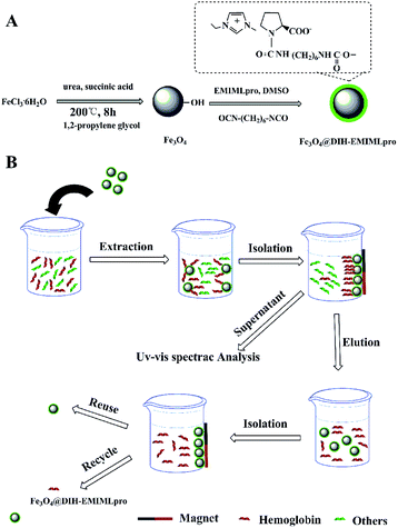 |
| | Scheme 1 Synthesis of Fe3O4@DIH–EMIMLpro (A) and its application for the MSPE of protein (B). | |
3.1.1 FT-IR. FT-IR spectra were used to confirm the successful modification of EMIMLpro onto the surface of Fe3O4 nanoparticles (Fig. 1). In the spectrum of Fe3O4@DIH–EMIMLpro, characteristic peaks are observed at 3030, 1621 and 1575 cm−1 corresponding to the stretching vibration of the unsaturated C–H bond of the imidazolium ring, the stretching vibration of C![[double bond, length as m-dash]](https://www.rsc.org/images/entities/char_e001.gif) O and the bending vibration of N–H, respectively. The band located at 1126 cm−1 belongs to the stretching vibration of C–N. The characteristic band of Fe–O at 600 cm−1 appears in the spectrum of Fe3O4@DIH–EMIMLpro, indicating the successful combination of EMIMLpro with Fe3O4 nanoparticles. Besides, as shown in Fig. S1,† the maximum absorption wavelengths of Fe3O4@DIH–EMIMLpro and EMIMLpro in the ultraviolet spectrum are 268 nm and 230 nm, respectively. This result further confirms the connection between EMIMLpro and the Fe3O4 nanoparticles due to the apparent red shift of the wavelength of Fe3O4@DIH–EMIMLpro nanoparticles.
O and the bending vibration of N–H, respectively. The band located at 1126 cm−1 belongs to the stretching vibration of C–N. The characteristic band of Fe–O at 600 cm−1 appears in the spectrum of Fe3O4@DIH–EMIMLpro, indicating the successful combination of EMIMLpro with Fe3O4 nanoparticles. Besides, as shown in Fig. S1,† the maximum absorption wavelengths of Fe3O4@DIH–EMIMLpro and EMIMLpro in the ultraviolet spectrum are 268 nm and 230 nm, respectively. This result further confirms the connection between EMIMLpro and the Fe3O4 nanoparticles due to the apparent red shift of the wavelength of Fe3O4@DIH–EMIMLpro nanoparticles.
 |
| | Fig. 1 FT-IR of Fe3O4 and Fe3O4@DIH–EMIMLpro. DIH is 1,6-diisocyanatohexane; EMIMLpro is 1-ethyl-3-methyl-imidazolium L-proline. | |
3.1.2 VSM. The magnetic properties of the prepared magnetic nanoparticles were investigated using VSM at room temperature. Seen from the characterization results shown in Fig. 2, it was found that the saturated magnetization values of Fe3O4 and Fe3O4@DIH–EMIMLpro are 72.4 and 26.7 emu g−1, respectively. Compared with Fe3O4 nanoparticles, the magnetization per gram of Fe3O4@DIH–EMIMLpro is obviously decreased due to the thick, non-magnetic, organic EMIMLpro layer coated onto the surface of Fe3O4 nanoparticles. Though the magnetization value of Fe3O4@DIH–EMIMLpro is reduced to 26.7 emu g−1, a strong magnetic response to an external magnetic field is exhibited, along with complete separation from solution within 30 s. The inset picture in Fig. 2 depicts that these nanoparticles could be uniformly dispersed in sample solution by ultrasonication or vortex mixing and collected from the suspension within 30 s, leaving a plain supernatant. The above results prove that the prepared microspheres could be applied in magnetic separation.
 |
| | Fig. 2 Magnetization curves of Fe3O4 and Fe3O4@DIH–EMIMLpro. | |
3.1.3 XRD. Fig. 3 shows the XRD pattern of Fe3O4@DIH–EMIMLpro. Six obvious diffraction peaks appear at 2θ = 30.11°, 35.53°, 43.28°, 53.66°, 57.17° and 62.80°, which correspond to (220), (311), (400), (422), (511) and (440), respectively.31 The results are consistent with the characteristic diffraction peaks of Fe3O4 (PDF 00-011-0614), which suggests that the crystal structure of Fe3O4 was not destroyed during the synthesis process of Fe3O4@DIH–EMIMLpro.
 |
| | Fig. 3 X-ray diffraction pattern of Fe3O4@DIH–EMIMLpro. | |
3.1.4 TGA. In order to measure the relative content of the organic shell on the Fe3O4@DIH–EMIMLpro nanoparticles, TGA was performed. As shown in Fig. 4, two mass loss profiles occur at 200 °C and 380 °C in the TGA curve of the Fe3O4@DIH–EMIMLpro nanoparticles. The first evident mass loss of about 41% is caused by the pyrolysis of EMIMLpro and the second small weight loss of about 13.47% is due to the removal of DIH onto the surface of Fe3O4.32 The organic compounds completely decompose at a temperature above 550 °C. Elemental analysis reflects the composition of Fe3O4@DIH–EMIMLpro. The data were as follows: C 35.41%, H 5.49% and N 11.2%.
 |
| | Fig. 4 Weight loss curves of Fe3O4 and Fe3O4@DIH–EMIMLpro. | |
3.1.5 TEM. To acquire a better understanding of the morphological structure of the as-synthesized magnetic nanoparticles, the magnetic nanoparticles were characterized using TEM. Seen in Fig. 5A, the magnetic nanoparticles are spherical and have a mean diameter of about 70 nm; containing some nanoparticles with an average size of about 13.31 nm calculated using the Scherrer equation from the X-ray diffraction results. The TEM image shown in Fig. 5B illustrates that core/shell structured EMIMLpro coated magnetic nanoparticles with an EMIMLpro layer with a thickness of about 20 nm were successfully prepared.
 |
| | Fig. 5 TEM images of Fe3O4 (A) and Fe3O4@DIH–EMIMLpro (B). | |
3.2 MSPE procedure
3.2.1 Optimization of pH. The optimization of the pH has a remarkable effect on the MSPE process of hemoglobin because the pH of sample solution influences the surface charge of the adsorbent and the existing state of analytes. Fig. 6 shows the extraction efficiency for a single protein at different pH. The extraction efficiency was calculated by the change of the absorbance of hemoglobin with UV-vis spectra before and after extraction by the as-synthesized materials. Seen in Fig. 6, the maximum extraction efficiency of hemoglobin was obtained at pH 7.2. The extraction efficiency of hemoglobin decreased at pH values lower or higher than the maximum extraction efficiency pH because of electrostatic repulsion between the protein and adsorbent and the different ionization state of hemoglobin.33
 |
| | Fig. 6 pH effect on the extraction efficiency of Fe3O4@DIH–EMIMLpro for hemoglobin. | |
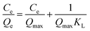
3.2.2 Extraction capacity for hemoglobin. To evaluate the extraction capacity of Fe3O4@DIH–EMIMLpro for hemoglobin, adsorption isotherms with different initial concentrations of hemoglobin from 0.1 to 30 mg mL−1 were obtained. As shown in Fig. 7, the adsorption capacity of Fe3O4@DIH–EMIMLpro for hemoglobin increased rapidly along with increasing the initial concentration and reached equilibrium over 5 mg mL−1. The maximum adsorption capacity (Qmax) of the prepared magnetic nanoparticles for hemoglobin was 1.58 mg mg−1, which was due to the contribution of modified magnetic nanoparticles whose imidazole functional groups were coordinated with Fe2+ contained in hemoglobin and the efficient utilization of imidazole groups because of lower space steric effects and higher curvature. The Langmuir and Freundlich isothermal models were applied to analyze the adsorption data. The equations are expressed as follows:| |
 | (1) |
| |
log![[thin space (1/6-em)]](https://www.rsc.org/images/entities/char_2009.gif) Qe = 1/nlogCe + logKF Qe = 1/nlogCe + logKF
| (2) |
where Qe (mg mg−1) is the adsorption quantity of hemoglobin bound to Fe3O4@DIH–EMIMLpro at equilibrium in different initial concentration, Qmax (mg g−1) is the maximum adsorption capacity, Ce (mg mL−1) is the equilibrium concentration of proteins, KL (mL mg−1) is the Langmuir constant, and KF (mg mL−1) and m are the Freundlich constants which represent the adsorption capacity and heterogeneity of the system, respectively.
 |
| | Fig. 7 Adsorption isotherm for the adsorption of hemoglobin on Fe3O4@DIH–EMIMLpro in pH 7.2 phosphate buffer (1/15 mol L−1). Qe is the equilibrium adsorption amount of the proteins (mg mg−1) and Ce is the protein equilibrium concentration in solution (mg mL−1) after adsorption. | |
Compared with the linear correlation coefficients of the two isothermal models in Table 1, we found that the Langmuir isotherm model (r = 0.9981) is more suitable for describing the adsorption process of hemoglobin onto Fe3O4@DIH–EMIMLpro than the Freundlich model (r = 0.5721). This result indicates that the adsorption process of proteins onto Fe3O4@DIH–EMIMLpro follows monolayer adsorption. Moreover, the maximum adsorption capacity of hemoglobin obtained from experiments (1.528 mg mg−1) is also close to the apparent maximum adsorption capacity of hemoglobin (1.581 mg g−1). Therefore, the binding of hemoglobin onto the prepared magnetic nanoparticles follows monolayer adsorption. In addition, the adsorption capacity of our work is much higher than some other reported work, shown in Table S1.†
Table 1 Langmuir and Freundlich isotherm constants of Fe3O4@DHI–EMIMLpro
| Langmuir model |
Freundlich model |
| Qe (mg mg−1) |
KL (mL mg−1) |
R2 |
KF (mg mg−1) |
1/n |
R2 |
| 1.528 |
5.606 |
0.981 |
0.948 |
0.233 |
0.572 |
3.2.3 Selective recognition of hemoglobin. The high specific recognition performance of the prepared material was also indispensable for obtaining a single biological sample from complex biological samples and for the recovery of products. A standard protein mixture of hemoglobin and Lys as a model was investigated to discuss the selectivity of the Fe3O4@DIH–EMIMLpro. Fig. 8 shows the UV-vis absorption spectra of a binary protein before and after treatment with the prepared adsorbent materials as well as the spectra of the solution eluted with the desorption solvent. As we expected, the maximum absorption wavelength of the curve is consistent with the characteristic absorption peaks that appear for a binary protein, and the recoveries of hemoglobin are satisfactory. The absorbance of hemoglobin in the residual solution sharply decreases after absorption by the Fe3O4@DIH–EMIMLpro. More than 90% of hemoglobin was adsorbed, while only about 10% of Lys was removed from the binary solution. In addition, the absorbance of hemoglobin after desorption with the eluent was high enough. The above results confirm that Fe3O4@DIH–EMIMLpro as a magnetic absorbent has wide prospects for application in the separation and purification of hemoglobin.
 |
| | Fig. 8 (A) UV-vis spectra for standard solutions of Hb and Lys; (B) UV-vis spectra of a mixture of Hb and Lys in 2 mL of pH 7.0 phosphate solution before and after adsorption by 5 mg Fe3O4@DIH–EMIMLpro; without adsorption; remaining protein after adsorption of Fe3O4@DIH–EMIMLpro; and desorbed protein solution. | |
3.2.4 Recyclability. As a matter of fact, recyclability of prepared functional magnetic nanoparticles is usually considered to have a positive effect on cost for future applications. In order to evaluate recyclability, the hemoglobin adsorption–desorption cycle was repeated eight times using the same Fe3O4@DIH–EMIMLpro nanoparticles. As shown in Fig. 9, the adsorption efficiency of Fe3O4@DIH–EMIMLpro was found to still be above 80% for hemoglobin after eight cycles. The change range of adsorption efficiency calculated from the eight cycles was not great, which illustrates that the adsorption capacity of the EMIMLpro decorated magnetic nanoparticles did not significantly decrease after using for eight cycles. This manifests that the as-synthesized nanomaterial has a favorable stability for the separation of hemoglobin.
 |
| | Fig. 9 The use of recycled Fe3O4@DIH–EMIMLpro for Hb adsorption. | |
3.3 Practical isolation of hemoglobin from human whole blood
The practical applicability of the prepared magnetic nanoparticles was investigated through the selective separation of hemoglobin from diluted human whole blood. In this work, SDS-PAGE was used to analyze the removal of proteins by the magnetic absorbent. As seen in Fig. 10, the intensity of the hemoglobin band (B14 kDa) is significantly weaker (Fig. 10, lane 3) after adsorption by the Fe3O4@DIH–EMIMLpro nanoparticles and the band of hemoglobin appeared again as the adsorbent was eluted with a desorption solution (Fig. 10, lane 4). In conclusion, these results prove the practical applicability of the EMIMLpro-modified magnetic nanomaterial for the effective isolation of hemoglobin from complex biological samples.
 |
| | Fig. 10 Standard SDS-PAGE of proteins. The samples were subjected to electrophoresis on a 30% acrylamide gel at 120 V. Lane 0, protein molecular weight marker; lane 1, pure hemoglobin solution; lane 2, 500-fold diluted human whole blood; lane 3, 500-fold diluted human whole blood after pre-treatment by the Fe3O4@DIH–EMIMLpro nanoparticles; lane 4, hemoglobin isolated from human whole blood using the present procedure. | |
4. Conclusions
In this work, the prepared Fe3O4@DIH–EMIMLpro combines the superiorities of amino acid-based ILs and Fe3O4 nanoparticles, revealing many fascinating properties which could not be achieved by either component alone. An environmentally friendly amino acid-based IL was utilized to directly decorate the Fe3O4 nanoparticles with 1,6-diisocyanatohexane to synthesize Fe3O4@DIH–EMIMLpro. In an MSPE process, several key elements such as solution pH, extraction capacity, selectivity recognition, recyclability and practical application were investigated. High extraction capacity, short extraction time, reusability and selective extraction were the advantages of Fe3O4@DIH–EMIMLpro as an adsorbent compared with traditional extraction materials. This result suggests that it has attractive potential to offer new possibilities in the extraction of proteins and proves that Fe3O4@DIH–EMIMLpro would be an important tool in bioseparation technology, as well as in other biotechnological applications.
Acknowledgements
This work was supported by the National Natural Science Foundation of China (NSFC, Grant No. 21075007), and the Program for New Century Excellent Talents in University (NCET-11-0563) and Beijing Nova program Interdisciplinary Cooperation Project are gratefully acknowledged.
Notes and references
- B. Alvarez-Sanchez, F. Priego-Capote and M. D. Castro, TrAC, Trends Anal. Chem., 2010, 29, 120–127 CrossRef CAS.
- H. M. Chen, D. W. Qi, C. H. Deng, P. Y. Yang and X. M. Zhang, Proteomics, 2009, 9, 380–387 CrossRef CAS PubMed.
- N. Li, Y. Z. Wang, K. J. Xu, Y. H. Huang, Q. Wen and X. Ding, Talanta, 2016, 152, 23–32 CrossRef CAS PubMed.
- M. V. Barberan, M. J. L. García, E. F. S. Alfonso and J. M. H. Martínez, Anal. Chim. Acta, 2016, 917, 37–43 CrossRef PubMed.
- J. L. Cao, X. H. Zhang, X. W. He, L. X. Chen and Y. K. Zhang, J. Mater. Chem. B, 2013, 1, 3625–3632 RSC.
- C. Ding, X. D. Ma, X. Yao and L. Jia, J. Chromatogr. A, 2015, 1424, 18–26 CrossRef CAS PubMed.
- B. Yu, M. Chi, Y. X. Han, H. L. Cong, J. B. Tang and Q. H. Peng, Talanta, 2016, 152, 76–81 CrossRef CAS PubMed.
- Y. T. Liu, Y. X. Gu, M. L. Li and Y. Wei, New J. Chem., 2014, 38, 6064–6072 RSC.
- H. M. Duan, X. J. Wang, Y. H. Wang, J. B. Li and C. N. Luo, RSC Adv., 2015, 5, 88492–88499 RSC.
- Q. Liu, J. B. Shi, M. T. Cheng, G. L. Li, D. Cao and G. B. Jiang, Chem. Commun., 2012, 48, 1874–1876 RSC.
- J. Wang, Z. Y. Chen, Z. M. Li and Y. L. Yang, Food Chem., 2016, 204, 135–140 CrossRef CAS PubMed.
- H. Parham and F. Khoshnam, Talanta, 2013, 114, 90–94 CrossRef CAS PubMed.
- R. M. Frizzarin, C. P. Cabello, M. M. Bauza, L. A. Portugal, F. Maya, V. Cerda, J. M. Estela and G. T. Palomino, Anal. Chem., 2016, 88, 6990–6995 CrossRef CAS PubMed.
- M. A. Habila, Z. A. ALOthman, A. M. El-Toni, J. P. Labis and M. Soylak, Talanta, 2016, 154, 539–547 CrossRef CAS PubMed.
- S. L. Zhang, W. X. Yao, J. B. Ying and H. T. Zhao, J. Chromatogr. A, 2016, 1452, 18–26 CrossRef CAS PubMed.
- J. H. Zhu, S. Y. Wei, H. B. Gu, S. B. Rapole, Q. Wang, Z. P. Luo, N. Haldolaarachige, D. P. Young and Z. H. Guo, Environ. Sci. Technol., 2012, 46, 977–985 CrossRef CAS PubMed.
- S. L. Ji, L. Qi, N. Li and M. L. Wang, Talanta, 2016, 158, 229–234 CrossRef CAS PubMed.
- Q. H. Wu, G. Y. Zhao, C. Feng, C. Wang and Z. Wang, J. Chromatogr. A, 2011, 1218, 7936–7942 CrossRef CAS PubMed.
- F. Gao, H. B. Gu, H. W. Wang, X. F. Wang, B. Xiang and Z. H. Guo, RSC Adv., 2015, 5, 60208–60219 RSC.
- N. V. Plechkova and K. R. Seddon, Chem. Soc. Rev., 2008, 37, 123–150 RSC.
- C. C. Chang and S. D. Huang, Anal. Chim. Acta, 2010, 662, 39–43 CrossRef CAS PubMed.
- H. H. Bai, Q. X. Zhou, G. H. Xie and J. P. Xiao, Talanta, 2010, 80, 1638–1642 CrossRef CAS PubMed.
- X. Zhou, P. F. Xie, J. Wang, B. B. Zhang, M. M. Liu, H. L. Liu and X. H. Feng, J. Chromatogr. A, 2011, 1218, 3571–3580 CrossRef CAS PubMed.
- V. Pino, J. L. Anderson, J. H. Ayala, V. González and A. M. Afonso, J. Chromatogr. A, 2008, 1182, 145–152 CrossRef CAS PubMed.
- E. Aliyari, M. Alvand and F. Shemirani, RSC Adv., 2016, 6, 64193–64202 RSC.
- H. He, D. H. Yuan, Z. Q. Gao, D. L. Xiao, H. He, H. Dai, J. Peng and N. Li, J. Chromatogr. A, 2014, 1324, 78–85 CrossRef CAS PubMed.
- E. Yilmaz and M. Soylak, Talanta, 2013, 116, 882–886 CrossRef CAS PubMed.
- D. M. Kroupa, C. J. Brown, L. M. Heckman and T. A. Hopkins, J. Phys. Chem. B, 2012, 116, 4952–4958 CrossRef CAS PubMed.
- Y. Wei, Y. Li, A. L. Tian, Y. T. Fan and X. Wang, J. Mater. Chem. B, 2013, 1, 2066–2071 RSC.
- Y. T. Liu, Y. Li and Y. Wei, J. Sep. Sci., 2014, 37, 3745–3752 CrossRef CAS PubMed.
- Y. H. Huang, Y. Z. Wang, Y. Wang, Q. Pan, X. Q. Ding, K. J. Xu, N. Li and Q. Wen, RSC Adv., 2016, 6, 5718–5728 RSC.
- Y. T. Liu, A. L. Tian, X. Wang, J. Qi, F. K. Wang, Y. Ito and Y. Wei, J. Chromatogr. A, 2015, 1400, 40–46 CrossRef CAS PubMed.
- X. Xue, B. H. Wang, X. J. Xi, Q. Chu and Y. Wei, New J. Chem., 2015, 39, 5735–5742 RSC.
Footnote |
| † Electronic supplementary information (ESI) available. See DOI: 10.1039/c6ra23616b |
|
| This journal is © The Royal Society of Chemistry 2016 |
Click here to see how this site uses Cookies. View our privacy policy here. 
 *a
*a


