DOI:
10.1039/C6RA21736B
(Review Article)
RSC Adv., 2016,
6, 114587-114592
Potential roles of annexin A7 GTPase in autophagy, senescence and apoptosis
Received
30th August 2016
, Accepted 2nd December 2016
First published on 2nd December 2016
Abstract
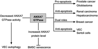
Annexin A7 (ANXA7) is a Ca2+-dependent phospholipid binding protein with intrinsic GTPase activity, belonging to the annexin superfamily. Accumulating research reveals that ANXA7 participates in cell autophagy, senescence and apoptosis in various cell types including vein endothelial cells (VECs), bone marrow-derived mesenchymal stem cells (BMSCs), Sertoli cells and cancer cells. This implies essential roles for ANXA7 in the physiological regulation of mammalian cells. Decreased ANXA7 GTPase activity induced by 6-amino-2,3-dihydro-3-hydroxymethyl-1,4-benzoxazine (ABO) promoted autophagy either by reducing the phosphorylation of ANXA7-interacting proteins or by inhibiting phosphatidylcholine-specific phospholipase C (PC-PLC) activity. ANXA7 inhibited senescence in BMSCs and protected cells from apoptosis in VECs and Sertoli cells. In cancer cells, ANXA7 had differing effects: it had a pro-apoptotic effect on prostate cancer and glioblastoma cells but an anti-apoptotic effect on renal cancer and hepatocarcinoma cells, and it played dual roles in breast cancer. The roles of ANXA7 GTPase activity in autophagy, senescence and apoptosis as well as its potential action mechanisms are summarized and discussed in the present review. An understanding of the global architecture of ANXA7 among autophagy, senescence and apoptosis interactive networks would be instrumental in providing novel therapeutic strategies for various human diseases.
1. Introduction
Annexins are a family of 13 Ca2+- and phospholipid-binding proteins. Mammalian annexins are classified as group A annexins. They have a highly conserved C-terminal region composed of 20–200 amino acid residues and a more variable N-terminal region. Four repeats of approximately 70 amino acids constitute the core domain. All annexins differ significantly in their N-terminal domains and this bestows different properties on each member. The sequence length of the N-terminal region is variable, but is typically short, consisting of 10 to 30 amino acids. This region regulates the annexin-membrane association and mediates interactions with protein ligands.1 Annexins participate in a host of cellular processes, including anti-coagulation and anti-inflammatory action, endocytosis and exocytosis, signal transduction in cell proliferation, differentiation, apoptosis and metastasis.2–4
Annexin A7 (ANXA7) is an evolutionarily conserved protein with a bipartite structure, a variable N-terminal domain and a conserved C-terminal domain (Fig. 1). Different from other annexins, ANXA7 has a special N-terminus which is extraordinarily long and highly hydrophobic and consists of more than 100 amino acids.5 It is believed that the N-terminus of ANXA7 has important sites implicated in interactions with other proteins.5–7 The ability of ANXA7 to participate in membrane fusion stems from its C-terminal architecture via binding of Ca2+ and phospholipid.3,8 Alternative splicing gives rise to two isoforms of 47 and 51 kDa, differing in their N-terminal domains. These different isoforms present different expression patterns. Most tissues harbor only the 47 kDa isoform, while the larger isoform exclusively exists in mature skeletal muscle, heart and brain.9–11 ANXA7 predominantly localizes in cytoplasm, but is partly found in the plasma membrane, nucleus, vesicular structures and t-tubule system.10,12,13 The Ca2+ concentration has an enormous impact on the ANXA7 sub-cellular localization. When the intracellular Ca2+ level increases, ANXA7 translocates to membranes, and it subsequently redistributes to the nuclear membrane and the plasma as well as to intracellular vesicles as the Ca2+ concentration returns back to a baseline level.
 |
| Fig. 1 Schematic diagram depicting the domain organization of ANXA7. Human ANXA7 contains 488 amino acid residues. It has two isoforms of 51 kDa and 47 kDa. Compared with the larger isoform, the smaller one lacks residues 141–163 of the ANXA7 N-terminus. The N-terminal domain is extraordinarily long (more than 100 amino acids) and highly hydrophobic. The proline (Pro)-rich region contains the majority of protein-binding sites. The four repeating sequence domains may also be implicated in protein interactions. The C-terminal domain can bind Ca2+ and phospholipid which confers the exocytosis and secretion functions of ANXA7. | |
ANXA7 is involved in multiple physical and pathological modulatory processes. A remarkable feature of ANXA7 is its Ca2+-dependent GTPase activity that is activated when protein kinase C increases the phosphorylation of ANXA7.8 In exocytotic secretory processes, ANXA7 GTPase activity is increased and ANXA7 forms a complex with Ca2+, protein kinase C and GTP. The concerted contribution of GTP and Ca2+ provides a huge boost to membrane fusion and secretion.8,14 In addition, it has been reported that certain small molecules can modulate annexins, such as withaferin A for annexin II.15 Based on this idea, we synthesized and identified two small molecules, (S)-ethyl 1-(3-(4-chlorophenoxy)-2-hydroxypropyl)-3-(4-methoxyphenyl)-1H-pyrazole-5-carboxylate (SEC) as an ANXA7 GTPase activator, and 6-amino-2,3-dihydro-3-hydroxymethyl-1,4-benzoxazine (ABO) as an inhibitor.16,17 Using the two modulators, we revealed that decreased ANXA7 GTPase activity led to enhanced autophagy in vein endothelial cells (VECs), that elevated ANXA7 protein levels caused suppression of senescence in cultured bone marrow-derived mesenchymal stem cells (BMSCs), and that increased ANXA7 GTPase activity promoted apoptosis in prostate cancer cells.
Homozygous ANXA7 (−/−) mice showed a lethal phenotype due to cerebral hemorrhage at embryonic day 10,18 indicating that ANXA7 is of vital importance in physiological control. In the current review, we summarize and discuss the current knowledge of the roles of ANXA7 in autophagy, senescence and apoptosis.
2. Decreased ANXA7 GTPase activity promoted autophagy
Autophagy is an indispensable cellular process for protein and organelle quality control. Autophagy can perform a pro-survival function, or it may lead to cell death. The sign that ANXA7 has a hand in regulating autophagy was first shown in ANXA7 gene knockout mice. Srivastava et al. reported that a reduced level of ANXA7 in heterozygous ANXA7 (+/−) mice caused deficient Ca2+ signal transduction due to the reduction of inositol 1,4,5-trisphosphate receptors (IP3Rs).19 IP3R is reported to invoke autophagy via activating calcium/calmodulin-dependent protein kinase kinase-beta (CaMKKβ) and to inhibit autophagy by forming a complex with BECN1/beclin 1.20,21 It is possible that ANXA7, acting as a mediator of calcium action, increased autophagy by upregulating IP3R and activating CaMKKβ. On the other hand, it has been reported that Ca2+ influx increased BECN1 phosphorylation and suppressed autophagy.22 We presume that ANXA7 might block autophagy initiation by activating Ca2+ signaling and promoting BECN1 phosphorylation.
In our previous study we identified ABO as an ANXA7 GTPase inhibitor that acts by inhibiting ANXA7 T286 phosphorylation.16 In addition, this small chemical compound significantly induces autophagy in an ANXA7-dependent manner in VECs.23 In this scenario, the ANXA7 level was elevated both in vivo24 and in vitro.23 ABO triggered the co-localization of ANXA7 with LC3. The accumulation of autophagic microtubule-associated protein 1A/1B-light chain 3 (LC3-II) was restrained in ANXA7 knockdown cells. Overexpression of ANXA7 potently stimulated autophagy due to LC3-II accumulation. Though ABO decreased ANXA7 GTPase activity, ANXA7 is indispensable in ABO-induced autophagy. However, the exact mechanisms are still ambiguous. Significant advances in the understanding of the function of ANXA7 in autophagy mainly stem from the characterization of its interacting proteins, including grancalcin (GCA), integrin β4 (ITGB4), T-cell intracellular antigen 1 (TIA1) and phosphatidylcholine-specific phospholipase C (PC-PLC).
2.1 Decreased ANXA7 GTPase activity inhibited GCA phosphorylation
GCA, calpain, sorcin, peflin, and ALG-2 belong to the penta-EF-hand subfamily whose members have the ability to bind to Ca2+.25 Two members of this family, sorcin6 and ALG-2,7 were demonstrated to interact with ANXA7 in a previous study. Our recent data showed that GCA can also bind to ANXA7 and function as an ANXA7-interacting partner in the ANXA7-dependent autophagy induced by ABO.16 The decreased ANXA7 GTPase activity caused by ABO led to reduced phosphorylation of GCA at serine residues. Moreover, ANXA7 knockdown almost totally inhibited GCA phosphorylation, and resulted in ineffectiveness in inducing autophagy. Therefore, in ABO-induced autophagy, the reduced ANXA7 GTPase activity decreased GCA phosphorylation to a low but not excessive level. In addition, as the ANXA7 GTPase activity was inhibited by ABO, the intracellular Ca2+ level was reduced. A further study showed that GCA knockdown dramatically elevated the intracellular Ca2+ concentration, and in these circumstances, ABO had no effect on the intracellular free Ca2+ level and failed to induce autophagy. These results indicate that ANXA7 might modulate calcium levels via GCA and suggest a potential mechanism: ANXA7 increases autophagy via binding to GCA and inhibiting GCA phosphorylation. GCA could therefore be an attractive target for further investigations into the regulatory effect of ANXA7 on intracellular free Ca2+ levels.
2.2 Decreased ANXA7 GTPase activity inhibited ITGB4 phosphorylation
ITGB4 is an integral membrane protein and has been shown to participate in VEC autophagy.26 Interestingly, we found that ANXA7 binds to ITGB4 in VECs.27 ABO promoted the binding at 3 h, while this interaction was inhibited with long-term ABO treatment for 24 h. ABO cannot induce autophagy in VECs with ITGB4 knockdown or in HEK293 cells with little ITGB4 expression. Knocking down ANXA7 caused the elevation of the ITGB4 protein level, and ABO could not trigger autophagy under this condition. The phosphorylation of ITGB4 Y1494 was inhibited with ABO treatment which was consistent with the results that ABO inhibited ANXA7 GTPase activity and further decreased ANXA7-interacting protein phosphorylation. Therefore, both ITGB4 and ANXA7 are of critical importance to ABO-induced autophagy and ITGB4 phosphorylation may negatively regulate autophagy.
2.3 Decreased ANXA7 GTPase activity inhibited TIA1 phosphorylation
TIA1 is a DNA/RNA binding protein and functions in RNA splicing and translation. TIA1 has been identified as a new ANXA7-interacting protein, and is implicated in the regulation of ANXA7-mediated autophagy induced by ABO.28 ABO promoted the interaction of ANXA7 and TIA1 and, in accordance with that result, TIA1 phosphorylation was inhibited with ABO treatment. It has been reported that TIA1 is responsible for the processing of a long non-coding RNA FLJ11812 and that elevated TIA1 phosphorylation decreased the FLJ11812 level. FLJ11812 can sequester the microRNA MIR4459 that targets autophagy related protein 13 (ATG13).29 The decreased ANXA7 GTPase activity caused by ABO treatment inhibited TIA1 phosphorylation; unphosphorylated TIA1 facilitated the production of FLJ11812 and promoted autophagy by decreasing MIR4459 and so increasing the ATG13 level. The results were also certified in vivo. In the endothelium of apoE−/− mice, ABO promoted the co-localization of ANXA7 and TIA1, and elevated the ATG13 level.
2.4 Decreased ANXA7 GTPase activity suppressed PC-PLC activity
Further evidence revealed that, under proatherogenic conditions, ABO promoted the co-localization of ANXA7 and PC-PLC, and that ANXA7 is an endogenous regulator of PC-PLC.24 PC-PLC is an important member of the phospholipase family. Its activity is implicated in a host of cellular processes.30 PC-PLC activity was sharply reduced in Cd2+-induced autophagy31 and sphingosylphosphorylcholine-induced autophagy.32 ABO suppressed oxidized low-density lipoprotein (oxLDL)-induced PC-PLC activity and triggered autophagy, both of which were impaired in the absence of ANXA7.24 Taken together, these data all indicate that ABO inhibited oxLDL-induced PC-PLC activity and promoted autophagy via triggering the co-localization of ANXA7 and PC-PLC in an ANXA7-dependent manner.
Collectively, the above evidence has fueled speculation that ANXA7 plays a key role in autophagy control through its GTPase activity. Reduced ANXA7 GTPase activity promoted autophagy and inhibited phosphorylation of its interacting proteins. The GTPase activity of ANXA7 was inhibited by ABO, but the ANXA7 level was elevated. These paradoxical attributes might be explained if ABO decreases ANXA7 GTPase activity to a low but not excessive point. The homeostasis is of vital importance in autophagy control. Exploring the crosstalk among these ANXA7-interacting proteins is interesting and valuable in elucidating a more accurate and comprehensive mechanism by which ANXA7 regulates autophagy.
3. ANXA7 GTPase inhibited senescence
Evidence connecting ANXA7 with senescence is sparse. Caterino et al. observed that ANXA7 was present in reticulocytes but was lost with cell maturation. The lower ANXA7 level in “old” red cells than “young” ones was possibly due to the degradation of ANXA7 during the aging or remodeling of cells. The cytoskeleton may have a significant influence on the turnover of ANXA7 during erythrocyte maturation, because ANXA7 was present in both mature and immature red cell membranes in band 3-deficient samples, in which the cytoskeleton was abnormal.33
Our group illustrated that ANXA7 inhibited senescence in BMSCs. In the process of BMSC senescence, ANXA7 was downregulated, and ABO treatment dramatically elevated the level of ANXA7 and mitigated senescence. ABO could not decrease senescence-associated beta-galactosidase (SA-β-gal) activity in ANXA7 knockdown BMSCs. On the one hand, ANXA7 played a protective role in BMSCs treated with bafilomycin-A1 (Baf-A1), a recognized inhibitor of the vacuolar H+-ATPase (V-ATPase) located in lysosomal membranes. Upon ABO treatment, a marked potentiation between ANXA7 and Lysotracker was shown, suggesting that ABO dramatically elevated the level of ANXA7 and subsequently triggered its translocation to the lysosomal membranes of BMSCs. ANXA7 might enhance lysosomal activity to antagonise the cell senescence process. On the other hand, Hmbox1, a transcription factor belonging to the hepatocyte nuclear factors in the homeobox family, was also increased upon ABO challenge. Knockdown of ANXA7 significantly decreased the Hmbox1 protein, but there was no significant difference in the Hmbox1 mRNA level, suggesting that ANXA7 might serve as an upstream regulator of Hmbox1 at the post-transcriptional level. Hmbox1 silencing suppressed the anti-aging effect of ABO, and Hmbox1 overexpression reduced SA-β-gal positively stained cells.34 An important question that remains to be clarified is whether or not the GTPase activity of ANXA7 is involved in this process.
Autophagy has profound implications for senescence. Lysosomal dysfunction impaired autophagy flux and triggered senescence.35 Hmbox1 could function as an autophagy inducer.36 In view of the above data, it seems plausible to suspect that ANXA7 enhances autophagy activity by sustaining the lysosomal function and upregulating the Hmbox1 level, collectively contributing to senescence suppression. Since the decreased ANXA7 GTPase activity induced by ABO was found to elevate autophagy, another possibility is that the inhibitory effect of ANXA7 on senescence can be attributed to decreased GTPase activity.
4. ANXA7 GTPase in apoptosis
Most previous research has demonstrated that ANXA7 is closely associated with tumor growth by serving either as a tumor suppressor or as a promoter. ANXA7 had a pro-apoptotic effect on prostate cancer and glioblastoma as well as an anti-apoptotic effect on renal cancer and hepatocarcinoma. In breast cancer ANXA7 played dual roles. Recently, emerging evidence has shown that ANXA7 had an apoptosis-suppression influence on normal mammalian cells such as VECs and testicular Sertoli cells.
4.1 ANXA7 GTPase mediates pro-apoptotic and anti-apoptotic actions in cancer cells
ANXA7 demonstrated a tumor suppressor function in prostate cancer.37 In androgen-sensitive LNCaP cells, wild-type (WT)-ANXA7 promoted the upregulation of the retinoblastoma (RB1)-binding transcription factor E2F1 and activated its downstream apoptosis signal-stimulating targets such as ASPP2 and ASK1. In contrast, dominant-negative (DN)-ANXA7 had no effect on apoptosis due to the activation of pro-survival ERK5. In androgen-resistant PC3 and DU145 cells, WT-ANXA7 mainly functioned by surpassing PTEN deficiency and forcing the PTEN and RB1-deficient cells into apoptosis.38 In addition, Cai et al. reported that ANXA7 was the target of miRNA-155 and that inhibition of miRNA-155 promoted apoptosis by elevating ANXA7 in LNCaP and PC3 prostate cancer cells.39 In our recent study, we found that the small molecule SEC promoted PC3 cell apoptosis via increasing ANXA7 GTPase activity. Activated ANXA7, bound to ITGB4, promoted the Y1494 phosphorylation of ITGB4 and induced its nuclear translocation. Nuclear ITGB4 activated ATF3 transcription and stimulated the expression of downstream pro-apoptotic genes, leading to apoptosis. The phenomenon was destroyed by the ANXA7 GTPase inhibitor ABO.17 Thus the GTPase activity of ANXA7 potentially plays an apoptosis-promoting role in prostate cancer.
A genetic landscape study in gliomas identified ANXA7 as one of the survival-associated landscape genes and suggested that an alteration of its function might contribute to gliomagenesis.40 In U87 and U373 glioma cells, inhibition of GSK3β activity by the inhibitor AR-A 014418 caused the downregulation of splicing factors, and led to a decrease in the ANXA7 variant and an increase in WT-ANXA7. Meanwhile, the anti-apoptotic genes were reduced, such as BAL2, BCLxL, survivin and MCL1.41 Therefore, ANXA7 could play a pro-apoptotic role in glioblastoma. Whether ANXA7 undergoes small deletions or site mutations in gliomas should be explored further.
ANXA7 has both inhibitory and promoting effects on breast cancer development. An ethanol extract of Chinese propolis (EECP) induced apoptosis in MCF-7 (human breast cancer ER+) and MDA-MB-231 (human breast cancer ER−) cells, which was accompanied by elevated ANXA7 expression. Meanwhile, the nuclear translocation of p65 was suppressed. The level of p53 was increased in MCF-7 but decreased in MDA-MB-231 cells.42 The coordinating roles of ANXA7, p65 and p53 in the regulation of apoptosis remain to be further investigated. In contrast, in breast cancer cell line BT549, ANXA7 interacted with galectin-3 and facilitated its translocation to perinuclear membranes. This prevented mitochondrial damage, suppressed caspase activity and inhibited apoptosis. ANXA7 also downregulated abrogated galectin-3 anti-apoptotic activity.43
The apoptosis-suppressing function of ANXA7 via its regulation of galectin-3 translocation also exists in renal carcinoma. Following arsenic trioxide (ATO) treatment, ANXA7 was co-translocated with galectin-3 from the nucleus to the cytoplasm in renal carcinoma cell lines, which inhibited ATO-induced apoptosis.44 These results suggest that the localization of ANXA7 could be functionally related to apoptosis.
ANXA7 plays a tumor-promotion role in hepatocellular carcinoma. It was reported that suppressing the expression of ANXA7 could increase the apoptosis capacity in the mouse hepatocarcinoma cell line Hca-P by decreasing the expression of Bcl2 and increasing the expression of cytochrome C by the mitochondrial pathway.45,46 Further study revealed that ANXA7 could form a complex with BAG4 that is a negative regulator of apoptosis. Hsp70 and Bcl2 are BAG-interacting proteins and inhibit cytochrome C-dependent caspase 3 activation.47 A recent study demonstrated that the 47 kDa isoform of ANXA7 was expressed in both the cytoplasm and the mitochondria and formed a complex with Bcl2. Decreased expression of the ANXA7 47 kDa isoform downregulated Bcl2 levels and impaired the mitochondrial membrane potential, contributing to apoptosis.48 Therefore, ANXA7 functions in mitochondrial apoptosis in hepatocellular carcinoma by modulating BAG, Bcl2 and their interacting partners.
4.2 Anti-apoptotic action of ANXA7 GTPase in VECs
Oxidized low-density lipoprotein (oxLDL) is believed to cause endothelial injury and trigger the development of atherosclerosis. In vitro, propolis attenuated oxLDL-induced VEC apoptosis by upregulating the ANXA7 level; meanwhile the PC-PLC level and activity were downregulated.49 In vivo, ABO (targeting ANXA7) reduced the oxLDL-induced increase in PC-PLC activity, decreased endothelium apoptosis and restricted atherosclerosis development in apoE−/− mice.24 Therefore, the anti-apoptotic role of ANXA7 in VECs is closely related to its regulation of PC-PLC levels and activity.
4.3 Anti-apoptotic action of ANXA7 GTPase in Sertoli cells
Nonylphenol (NP) is known to impair the reproductive system. NP could induce apoptosis in testicular Sertoli cells and decrease ANXA7 levels, resulting in dysregulation of spermatogenesis.50 Three other proteins that were identified by two-dimensional gel electrophoresis (2DE) and mass spectrometry were all upregulated, namely RKIP, ERp57 and PRDX6. ERp57, located mainly in the endoplasmic reticulum (ER), modulates the redox state of the ER in a Ca2+-dependent manner.51 The downregulation of ANXA7 may result in an imbalance of cellular Ca2+, which probably disrupts the ERp57 function and thus induces apoptosis.
5. Conclusions
The ANXA7 GTPase orchestrates autophagy, senescence and apoptosis interactive networks (Fig. 2), to ensure that cell progress proceeds normally. A decrease in the GTPase activity of ANXA7 triggered autophagy. The ANXA7 GTPase activity is also of considerable interest in the regulation of senescence and apoptosis. The mechanisms by which ANXA7 contributes to physiological and pathological processes have been discussed in this review. The function of ANXA7 is mainly derived from its functional and physical interactions with other partners (Table 1). The decreased ANXA7 GTPase activity caused by ABO treatment inhibited the phosphorylation of ANXA7-binding proteins including GCA, ITGB4 and TIA1. SEC works as an ANXA7 GTPase activator and probably inverts the pathway by activating ANXA7 GTPase. Among ANXA7 binding proteins, GCA, sorcin and ALG2 all bind Ca2+. It is tempting to speculate that ANXA7 may interact with its Ca2+-binding partners to fine-tune intracellular Ca2+ levels, thus regulating various Ca2+-dependent cellular processes, such as autophagy and apoptosis. ANXA7-mediated autophagy could be a promising way to suppress senescence. The interweaving roles of ABO and SEC in senescence regulation are useful. The two small molecules are distinct from each other but also interrelated. Since the ANXA7 GTPase activity participates in multiple biological functions, it is inadequate to focus merely on protein level changes in order to clarify specific mechanisms; the inhibitor ABO and activator SEC are powerful tools which can be used to explore a comprehensive understanding of ANXA7 action. The elucidation of ANXA7-mediated physiological events will open up new avenues for research and provide potential opportunities for clinical diagnosis of, and therapeutic interventions in, a variety of disorders.
 |
| Fig. 2 ANXA7 in the regulation of autophagy, senescence and apoptosis. (a) With reduced GTPase activity by ABO, ANXA7 mediates autophagy by inhibiting PC-PLC activity and reducing the phosphorylation of GCA, ITGB4 and TIA1. SEC, as an ANXA7 GTPase activator, probably inverts the pathway, although this remains to be examined. (b) With elevated protein levels by ABO, ANXA7 suppresses senescence by upregulating lysosomal activity and Hmbox1 levels. ANXA7 might also enhance autophagy activity by sustaining the lysosomal function and upregulating Hmbox1 levels, which collectively contribute to senescence suppression. (c) ANXA7 functions in apoptosis in both cancer cells and normal mammalian cells. ANXA7 promotes apoptosis in prostate cancer and glioblastoma, and suppresses apoptosis in renal cancer and hepatocarcinoma. In breast cancer cells, ANXA7 has roles in both inhibiting and promoting apoptosis. ANXA7 exerts an anti-apoptosis effect on normal mammalian cells, such as VECs and testicular Sertoli cells. | |
Table 1 Summary of ANXA7 interacting proteins
ANXA7-binding proteins |
ANXA7-binding motif |
Cellular activity |
ALG2 |
N-Terminal domain |
Anti-apoptosis and Ca2+ binding |
Sorcin |
N-Terminal domain |
Ca2+ binding |
GCA |
Unknown |
Ca2+ binding |
ITGB4 |
Unknown |
Hemidesmosome and signal transduction |
TIA1 |
Unknown |
Cellular metabolism |
Galectin-3 |
Unknown |
Anti-apoptosis |
BAG4 |
Unknown |
Anti-apoptosis |
Bcl2 |
Unknown |
Anti-apoptosis |
Acknowledgements
This work was supported by the National Natural Science Foundation of China (no. 31270877, 91313303, 31570834, 20972088, 31070735) and the Key Technology Special Project of Shandong Province (no. 2015ZDJS04001 and 2015ZDJS04003).
Notes and references
- V. Gerke and S. E. Moss, Physiol. Rev., 2002, 82, 331–371 CrossRef CAS PubMed.
- K. Monastyrskaya, E. B. Babiychuk and A. Draeger, Cell. Mol. Life Sci., 2009, 66, 2623–2642 CrossRef CAS PubMed.
- C. Guo, S. Liu, F. Greenaway and M. Z. Sun, Clin. Chim. Acta, 2013, 423, 83–89 CrossRef CAS PubMed.
- S. Mussunoor and G. I. Murray, J. Pathol., 2008, 216, 131–140 CrossRef CAS PubMed.
- C. E. Creutz, Gen. Physiol. Biophys., 2009, 28, F7–F13 Search PubMed.
- A. M. Brownawell and C. E. Creutz, J. Biol. Chem., 1997, 272, 22182–22190 CrossRef CAS PubMed.
- H. Satoh, Y. Nakano, H. Shibata and M. Maki, Biochim. Biophys. Acta, 2002, 1600, 61–67 CrossRef CAS.
- H. Caohuy and H. B. Pollard, J. Biol. Chem., 2002, 277, 25217–25225 CrossRef CAS PubMed.
- C. Herr, C. S. Clemen, G. Lehnert, R. Kutschkow, S. M. Picker, B. S. Gathof, C. Zamparelli, M. Schleicher and A. A. Noegel, BMC Biochem., 2003, 4, 8 CrossRef PubMed.
- S. Selbert, P. Fischer, D. Pongratz, M. Stewart and A. A. Noegel, J. Cell Sci., 1995, 108(1), 85–95 CAS.
- C. S. Clemen, A. Hofmann, C. Zamparelli and A. A. Noegel, J. Muscle Res. Cell Motil., 1999, 20, 669–679 CrossRef CAS PubMed.
- C. S. Clemen, C. Herr, A. A. Lie, A. A. Noegel and R. Schroder, NeuroReport, 2001, 12, 1139–1144 CrossRef CAS PubMed.
- M. Rick, S. I. Ramos Garrido, C. Herr, D. R. Thal, A. A. Noegel and C. S. Clemen, BMC Neurosci., 2005, 6, 25 CrossRef PubMed.
- H. Caohuy, M. Srivastava and H. B. Pollard, Proc. Natl. Acad. Sci. U. S. A., 1996, 93, 10797–10802 CrossRef CAS.
- G. Ozorowski, C. M. Ryan, J. P. Whitelegge and H. Luecke, Biol. Chem., 2012, 393, 1151–1163 CrossRef CAS PubMed.
- H. Li, N. Liu, S. Wang, L. Wang, J. Zhao, L. Su, Y. Zhang, S. Zhang, Z. Xu, B. Zhao and J. Miao, Biochim. Biophys. Acta, 2013, 1833, 2092–2099 CrossRef CAS PubMed.
- S. Y. Liu, D. Ge, L. N. Chen, J. Zhao, L. Su, S. L. Zhang, J. Y. Miao and B. X. Zhao, Oncotarget, 2016, 7, 16282–16296 Search PubMed.
- M. Srivastava, C. Montagna, X. Leighton, M. Glasman, S. Naga, O. Eidelman, T. Ried and H. B. Pollard, Proc. Natl. Acad. Sci. U. S. A., 2003, 100, 14287–14292 CrossRef CAS PubMed.
- M. Srivastava, I. Atwater, M. Glasman, X. Leighton, G. Goping, H. Caohuy, G. Miller, J. Pichel, H. Westphal, D. Mears, E. Rojas and H. B. Pollard, Proc. Natl. Acad. Sci. U. S. A., 1999, 96, 13783–13788 CrossRef CAS.
- Y. Gu, B. Qi, Y. Zhou, X. Jiang, X. Zhang, X. Li and W. Fang, Viruses, 2016, 8, E135 CrossRef PubMed.
- J. M. Vicencio, C. Ortiz, A. Criollo, A. W. Jones, O. Kepp, L. Galluzzi, N. Joza, I. Vitale, E. Morselli, M. Tailler, M. Castedo, M. C. Maiuri, J. Molgo, G. Szabadkai, S. Lavandero and G. Kroemer, Cell Death Differ., 2009, 16, 1006–1017 CrossRef CAS PubMed.
- Q. Wang, W. Guo, B. Hao, X. Shi, Y. Lu, C. W. Wong, V. W. Ma, T. T. Yip, J. S. Au, Q. Hao, K. H. Cheung, W. Wu, G. R. Li and J. Yue, Autophagy, 2016, 12, 1340–1354 CrossRef CAS PubMed.
- L. Wang, Z. Dong, B. Huang, B. Zhao, H. Wang, J. Zhao, H. Kung, S. Zhang and J. Miao, Autophagy, 2010, 6, 1115–1124 CrossRef CAS PubMed.
- H. Li, S. Huang, S. Wang, J. Zhao, L. Su, B. Zhao, Y. Zhang, S. Zhang and J. Miao, Cell Death Dis., 2013, 4, e806 CrossRef CAS PubMed.
- K. Lollike, A. H. Johnsen, I. Durussel, N. Borregaard and J. A. Cox, J. Biol. Chem., 2001, 276, 17762–17769 CrossRef CAS PubMed.
- L. Wang, Z. Dong, Y. Zhang and J. Miao, J. Cell. Physiol., 2012, 227, 474–478 CrossRef CAS PubMed.
- H. Li, S. Huang, S. Wang, L. Wang, L. Qi, Y. Zhang, S. Zhang, B. Zhao and J. Miao, Int. J. Biochem. Cell Biol., 2013, 45, 2605–2611 CrossRef CAS PubMed.
- S. Huang, N. Liu, H. Li, J. Zhao, L. Su, Y. Zhang, S. Zhang, B. Zhao and J. Miao, Int. J. Biochem. Cell Biol., 2014, 57, 115–122 CrossRef CAS PubMed.
- D. Ge, L. Han, S. Huang, N. Peng, P. Wang, Z. Jiang, J. Zhao, L. Su, S. Zhang, Y. Zhang, H. Kung, B. Zhao and J. Miao, Autophagy, 2014, 10, 957–971 CrossRef CAS PubMed.
- H. Li, L. Zhang, D. Yin, Y. Zhang and J. Miao, Trends Cardiovasc. Med., 2010, 20, 172–176 CrossRef CAS PubMed.
- Z. Dong, L. Wang, J. Xu, Y. Li, Y. Zhang, S. Zhang and J. Miao, Toxicol. In Vitro, 2009, 23, 105–110 CrossRef CAS PubMed.
- D. Ge, Q. Jing, N. Meng, L. Su, Y. Zhang, S. Zhang, J. Miao and J. Zhao, J. Cell. Physiol., 2011, 226, 2827–2833 CrossRef CAS PubMed.
- M. Caterino, M. Ruoppolo, S. Orru, M. Savoia, S. Perrotta, L. Del Vecchio, F. Salvatore, G. W. Stewart and A. Iolascon, FEBS Lett., 2006, 580, 6527–6532 CrossRef CAS PubMed.
- F.-W. Wang, F. Zhao, X.-Y. Qian, Z.-Z. Yu, J. Zhao, L. Su, Y. Zhang, S.-L. Zhang, B.-X. Zhao and J.-Y. Miao, RSC Adv., 2014, 4, 56722–56730 RSC.
- H. Tai, Z. Wang, H. Gong, X. Han, J. Zhou, X. Wang, X. Wei, Y. Ding, N. Huang, J. Qin, J. Zhang, S. Wang, F. Gao, Z. M. Chrzanowska-Lightowlers, R. Xiang and H. Xiao, Autophagy, 2016, 1–15 CrossRef CAS PubMed.
- H. Ma, L. Su, H. Yue, X. Yin, J. Zhao, S. Zhang, H. Kung, Z. Xu and J. Miao, Sci. Rep., 2015, 5, 15121 CrossRef CAS PubMed.
- M. Srivastava, L. Bubendorf, L. Nolan, M. Glasman, X. Leighton, G. Miller, W. Fehrle, M. Raffeld, O. Eidelman, O. P. Kallioniemi, S. Srivastava and H. B. Pollard, Dis. Markers, 2001, 17, 115–120 CrossRef CAS PubMed.
- Y. Torosyan, O. Simakova, S. Naga, K. Mezhevaya, X. Leighton, J. Diaz, W. Huang, H. Pollard and M. Srivastava, Int. J. Cancer, 2009, 125, 2528–2539 CrossRef CAS PubMed.
- Z. K. Cai, Q. Chen, Y. B. Chen, M. Gu, D. C. Zheng, J. Zhou and Z. Wang, Mol. Med. Rep., 2015, 11, 533–538 CAS.
- M. Bredel, D. M. Scholtens, G. R. Harsh, C. Bredel, J. P. Chandler, J. J. Renfrow, A. K. Yadav, H. Vogel, A. C. Scheck, R. Tibshirani and B. I. Sikic, J. Am. Med. Assoc., 2009, 302, 261–275 CrossRef CAS PubMed.
- A. K. Yadav, V. Vashishta, N. Joshi and P. Taneja, J. Oncol., 2014, 2014, 695325 Search PubMed.
- H. Xuan, Z. Li, H. Yan, Q. Sang, K. Wang, Q. He, Y. Wang and F. Hu, J. Evidence-Based Complementary Altern. Med., 2014, 2014, 280120 Search PubMed.
- F. Yu, R. L. Finley Jr, A. Raz and H. R. Kim, J. Biol. Chem., 2002, 277, 15819–15827 CrossRef CAS PubMed.
- Y. Xu, X. Gu, M. Gong, G. Guo, K. Han and R. An, Cancer Biol. Ther., 2013, 14, 897–906 CrossRef CAS PubMed.
- Y. Huang, Y. Du, X. Zhang, L. Bai, M. Mibrahim, J. Zhang, Y. Wei, C. Li, S. Fan, H. Wang, Z. Zhao and J. Tang, Biomed. Pharmacother., 2015, 70, 146–150 CrossRef CAS PubMed.
- Y. Huang, Q. Wang, Y. Du, L. Bai, F. Jin, J. Zhang, S. Fan, H. Wang, L. Song, Y. Gao, X. Wang and J. Tang, Biomed. Pharmacother., 2014, 68, 819–824 CrossRef CAS PubMed.
- Y. Du, Y. Huang, Y. Gao, B. Song, J. Mao, L. Chen, L. Bai and J. Tang, Biomed. Pharmacother., 2015, 74, 30–34 CrossRef CAS PubMed.
- L. Bai, Y. Guo, Y. Du, H. Wang, Z. Zhao, Y. Huang and J. Tang, Biomed. Pharmacother., 2016, 83, 1127–1131 CrossRef CAS PubMed.
- H. Xuan, Z. Li, J. Wang, K. Wang, C. Fu, J. Yuan and F. Hu, J. Evidence-Based Complementary Altern. Med., 2014, 2014, 465383 Search PubMed.
- J. Wu, F. Wang, Y. Gong, D. Li, J. Sha, X. Huang and X. Han, Chem. Res. Toxicol., 2009, 22, 668–675 CrossRef CAS PubMed.
- J. S. Kim-Han and K. L. O'Malley, Antioxid. Redox Signaling, 2007, 9, 2255–2264 CrossRef CAS PubMed.
|
This journal is © The Royal Society of Chemistry 2016 |
Click here to see how this site uses Cookies. View our privacy policy here.