Fatemeh Zarei baygia,
Nafiseh Farhadian*a,
Bizhan Malaekeh-Nikoueib and
Zahra Maghsouda
aChemical Engineering Department, Faculty of Engineering, Ferdowsi University of Mashhad, Mashhad 9177948974, Iran. E-mail: n.farhadian@um.ac.ir
bNanotechnology Research Center, School of Pharmacy, Mashhad University of Medical Sciences, I. R. Iran
First published on 10th November 2016
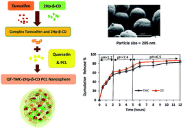
The aim of this study is to prepare a nanosphere containing the insoluble anti-cancer drug called tamoxifen citrate. The synthesized nanosphere is composed of tamoxifen citrate (TMC), 2-hydroxy propyl-β-cyclodextrin (2HP-β-CD), quercetin (QT) and polycaprolactone (PCL), which was prepared using water in oil in water emulsion solvent extraction method. 2HP-β-CD and QT were applied to enhance the solubility and bioavailability of the drug, respectively. The response surface method was used to optimize the operating variables such as PCL polymer mass, stabilizer percentage, continuous water volume and ratio based on the central composite design. Quadratic models were developed for two responses: the drug encapsulation efficiency and the nanosphere size. According to the results, the optimum conditions to achieve a nanosphere of a small size and high drug entrapment efficiency included 30 mg PCL, 2% (w/v) PVA stabilizer, 80 ml water and
equivalent ratio of 0.5. According to the UV analysis based on the second derivative spectrophotometric technique, the estimated encapsulation efficiency of TMC was 53% ± 2. Scanning electron microscopy (SEM) showed that the mean particle size of the synthesized nanospheres was 205 nm, which is appropriate for oral usage. In addition, the in vitro release of TMC and QT in the simulated body environment (SGF and SIF) was performed at 37 °C. The in vitro release of TMC at pH = 7.4 showed that TMC and QT had a slow and simultaneous release behaviour. This confirms the controllable release of TMC in the new synthesized nanoparticle form.
Tamoxifen citrate is a selective oestrogen receptor antagonist oral drug.5,6 In patients with breast cancer, the anti-oestrogen effects of the breast tissue have a simultaneous effect on other tissues such as bone,7 liver,8 endometrial,9 and cardiovascular system.10 As a result, there is a competition between tamoxifen and estradiol for the oestrogen receptor, which prevents the binding of estradiol to the receptor. This mechanism can treat breast cancer.11 Moreover, after primary treatment, tamoxifen can be used to reduce the risk of an invasive breast cancer for 10 years.12 The long-time usage of this oral drug can produce a number of serious side effects, such as hot flashes, fatigue, toxic hepatitis, multifocal hepatic fatty infiltration, sub massive hepatic necrosis and cirrhosis as well as increased risk of blood clots and oxidative stress mediated hepatotoxicity.13 Furthermore, tamoxifen increases the risk of endometrial cancer, which is mainly dependent upon the treatment duration and dose accumulation.9 Thus, lower drug doses with higher selective adsorption would be more effective.8 One of the best strategies to enhance drug adsorption efficiency is to reduce the size of drug carrier as it increases the contact surface area of the drug with the surrounding environment. This enhances the therapeutic effect of drug and thus lowers the dose usage. It has been shown that some of the side effects of this drug can be reduced.14
In addition to general side effects of chemotherapeutic drugs, each drug may have some specific problems in relation to its consumption. For TMC, two main problems have been reported, including its low solubility in the hydrophilic environment of body due to its hydrophobic nature, and its low bioavailability and insufficient adsorption in the gastrointestinal tract and removal by the first-pass liver metabolism.8 To overcome the first problem, some compounds with hydrophobic–hydrophilic nature have been suggested. In this regard, 2-hydroxy propyl-β-cyclodextrin (2HP-β-CD) is one of the best candidates owing to its high solubility in aqueous environments and low toxicity. Also, producing a complex containing TMC and 2HP-β-CD is rather simple.15,16 Shukla et al., formulated and characterized a water-soluble complex of TMC-2HP-β-CD to enhance TMC solubility. They found that TMC-2HP-β-CD nanoassembly infiltrated the tissue due to the enhanced permeability and retention property of tumour tissue. This produced excellent anticancer effect in the ovarian cancer cell line.17 In another study, 2HP-β-CD was applied to enhance bioavailability and solubility of tamoxifen citrate. Results showed the low level of synthesized nanoparticles in plasma and liver in comparison to oral TMC suspension.18
The second problem of TMC consumption as low bioavailability is related to the aromatase enzyme function. Aromatase is the enzyme that catalyzes the conversion of androstenedione and testosterone to oestrogen and estradiol.19 Tamoxifen citrate, inhibiting the function of this enzyme, reduces the amount of estradiol.17 Applying aromatase inhibitors to the TMC formulation may decrease drug resistance in the breast cancer and enhance bioavailability.8 The application of flavonoids, due to special phenolic structure, can prevent injury caused by free radicals, which may lead to cell damage,20 aging and stroke.21 Quercetin (QT) is a flavonoid with high therapeutic effects, which has been used with TMC in literature.8,18 Caltagirone et al. showed that TMC and quercetin were effective in inhibiting in vitro. Also, TMC was found to compete with quercetin in whole-cell assays for type-II binding, thus inhibiting cell proliferation in a dose-dependent manner in a multidrug-resistant, ER-negative MCF-7 breast cancer cell line.22 In another study, a combination of quercetin and tamoxifen was shown to be more effective in reducing cell viability than tamoxifen and quercetin separately. In addition, TMC-QT complex led to a significant inhibition of cell viability by decreasing Bcl-2 protein expression and inducing apoptosis.23 Jain et al. showed that free TMC and QT had 2% (ref. 8) oral bioavailability in humans, which is a low value. The co-administration of quercetin enhanced the oral bioavailability of TMC by 5.31-fold.8
A review of the literature suggests that both 2HP-β-CD and QT have been applied to TMC formulation separately. However, the co-administration of TMC with both TMC and QT has not been reported. The main goal of this study is to provide a co-encapsulation of TMC with 2HP-β-CD and QT in a nanostructure form. To do so, nanosphere was selected as an appropriate form of drug carrier at nanometre scale. In oral delivery, polyester-based NPs could protect the drug against the gastrointestinal (GI) harsh environment and enhance the oral adsorption of drug with systemic targeting by avoiding the intravenous route.24 Thus, this type of carrier was adopted in this study. Also, polycaprolactone (PCL) was chosen as the appropriate polymer. PCL has a slow degradation rate in comparison to other polymers such as poly(lactic-co-glycolic acid) (PLGA),25 polylactic acid (PLA) and polyglycolide (PGA)26 is slow. Hence, PCL nanosphere could be a suitable carrier for long-term drug release systems.27,28 According to previous research, the oral bioavailability of tamoxifen citrate was significantly enhanced by its encapsulation in PCL NPs, leading to specific immunosuppression without increased adverse effects.24 Finding the best nanosphere structure with the appropriate size, drug loading and drug entrapment efficiency is a critical consideration in the design of drug delivery systems. In this study, the design expert method using response surface methodology was adopted to optimize the preparation parameters. Then, the best structure of the drug release was examined in experimental environments at pH = 7.4 and simulated gastric and intestinal fluid. Designing a best structure for slow and simultaneous release of TMC and QT is the main novelty of this research.
Tamoxifen citrate (3.54 mM) and 2HP-β-CD (3.54, 7.1, 10.62 mM) were mixed in 2 ml of distilled water, stirred (14000 rpm for 24 h) at 25 °C and filtered through a 0.45 μm membrane filter to remove undissolved material. Then, the TMC content was determined by UV spectroscopy (UV-FP-6200, JASCO Instruments, Japan) at a wavelength of λ = 237 nm. The inclusion efficiency (%) was calculated using the following formula.17,29
![]() | (1) |
Furthermore, the extinction coefficient of drug was calculated using eqn (2) as follows:
A = εCL | (2) |
Finally, the nanospheres were separated from the bulk suspension, and the free drug was removed by centrifugation. The supernatant (liquid layer above the sediment) was maintained for drug assay with the sedimented nanospheres at the bottom of falcon tube being washed three times with deionized water in successive centrifugation/redispersion cycles for 20 min at a temperature of 25 °C. Finally, nanoparticles were freeze-dried.30,31 All experiments were repeated 3 times to confirm the observed trends. In Scheme 1, the schematic of applied procedure for nanosphere synthesis has been shown.
A first and second derivative spectrum of each component were subsequently recorded (see ESI file, Fig. S1†). As shown in Fig. S1,† in the second derivative curve, the simultaneous determination of TMC can be performed at a wavelength of 215 nm. This new wavelength was selected to calculate TMC concentration in the nanosphere form. The calibration curves of TMC and QT are presented in the ESI file (Fig. S2–S4†). It should be noted that by using UV visible analysis, it is possible to measure TMC content in the free form not in the encapsulated form by CD. Moreover, at the applied wavelength for TMC measurement, other components have the zero contribution.
Dependent variables | Constraints |
---|---|
Y1 = particle size (nm) | Minimum |
Y2 = entrapment efficiency of TMC (%) | Maximum |
![]() | (3) |
The particles suspension was gently stirred at 37 °C in a shaker incubator (100 rpm). The final suspension was withdrawn at appropriate intervals (0.5, 1, 1.5, 2, 3, 4, 5, 6, 8, 10 and 12 h) with each sample being centrifuged at 22000 rpm for 10 min. The filtrate was replaced by 2 ml of fresh buffer. The content of tamoxifen citrate and quercetin in the release medium were determined by UV Vis analysis. It should be noted that all experiments were repeated three times at each stage to achieve optimal evaluation.
![]() | ||
Fig. 2 The inclusion efficiencies of TMC and 2HP-β-CD mixture at different molar ratios. Each data point represents mean ± SD (n = 3). |
Moreover, the extinction coefficient of drug before and after encapsulation with β-CD indicated that the ratio of εafter encapsulation with β-CD/εbefore encapsulation with β-CD was 0.34. This demonstrates a reduction in the extinction coefficient of drug, which can affect the release rate of TMC from nanosphere. It will be discussed in details later.
Run | Independent variable | Y1 (nm) | Y2 (%) | |||
---|---|---|---|---|---|---|
X1 (mg) | X2 (%) | X3 (ml) | X4 (—) | |||
1 | 40.00 | 1.00 | 80.00 | 0.75 | 250 | 50 |
2 | 50.00 | 1.00 | 60.00 | 0.50 | 300 | 60 |
3 | 60.00 | 2.00 | 80.00 | 0.75 | 350 | 70 |
4 | 40.00 | 2.00 | 80.00 | 0.75 | 270 | 53 |
5 | 30.00 | 1.00 | 60.00 | 1.00 | 180 | 40 |
6 | 50.00 | 3.00 | 100.00 | 0.50 | 310 | 64 |
7 | 40.00 | 2.00 | 40.00 | 0.75 | 340 | 52 |
8 | 50.00 | 3.00 | 60.00 | 1.00 | 336 | 58 |
9 | 50.00 | 3.00 | 100.00 | 1.00 | 300 | 55 |
10 | 30.00 | 1.00 | 100.00 | 1.00 | 150 | 44 |
11 | 50.00 | 1.00 | 100.00 | 1.00 | 310 | 65 |
12 | 50.00 | 1.00 | 100.00 | 0.50 | 280 | 63 |
13 | 50.00 | 3.00 | 60.00 | 0.50 | 326 | 64 |
14 | 30.00 | 1.00 | 100.00 | 0.50 | 145 | 43 |
15 | 40.00 | 2.00 | 80.00 | 1.25 | 295 | 54 |
16 | 30.00 | 3.00 | 100.00 | 1.00 | 190 | 50 |
17 | 40.00 | 2.00 | 120.00 | 0.75 | 274 | 53 |
18 | 30.00 | 1.00 | 60.00 | 0.50 | 165 | 47 |
19 | 30.00 | 2.00 | 80.00 | 0.50 | 205 | 53 |
20 | 40.00 | 2.00 | 80.00 | 0.25 | 220 | 59 |
21 | 40.00 | 4.00 | 80.00 | 0.75 | 296 | 60 |
22 | 20.00 | 2.00 | 80.00 | 0.75 | 120 | 43 |
23 | 30.00 | 3.00 | 60.00 | 0.50 | 185 | 52 |
24 | 50.00 | 1.00 | 60.00 | 1.00 | 320 | 60 |
25 | 30.00 | 3.00 | 100.00 | 0.50 | 176 | 48 |
![]() | ||
Fig. 3 Response surface plot showing the effect of (a) PCL mass (X1) and % PVA (X2), (b) % PVA (X2), and continues phase volume (X3), (c) ![]() |
![]() | ||
Fig. 4 Response surface plot showing the effect of (a) PCL mass (X1) and % PVA (X2), (b) % PVA (X2), and continues phase volume (X3), (c) ![]() |
Also, the particle size is increased as a result of reduction in the continuous phase volume. With a reduction in the volume of continuous phase molecules, drugs stick together and produce larger nanosphere. An increase in the tension force between dispersed and continuous phases increases the particle size (Fig. 3b) and encapsulation efficiency. However any change in the volume of continues phase exerts a significant effect on the particle size in comparison to encapsulation efficiency (Fig. 4b).43 Furthermore, an increase in the concentration of surfactant (% PVA) can reduce the size of nanospheres.
Applying surfactant as a stabilizer in the emulsification stage diminishes the surface tension of the continuous phase, resulting in a significant hike in the net shear stress at a constant energy density. This promotes the formation of small-size emulsion droplets and thus decreases the mean diameter of nanospheres.44
According to our observations, there is an optimum percentage for the surfactant concentrations. Applying 1 to 2% of the PVA leads to a significant decrease in the nanosphere size. However, increasing PVA concentration from 2 to 3% (p < 0.05) produces a plateau trend in the particle size curve. This may be related to the saturation of the particle surfaces by emulsifiers. This confirms that an increase of more than 2% in the emulsifier concentration will not have any effect on the particle size (Fig. 3a and d), with a decrease in the particle size from 1 to 2% PVA reducing the extent of encapsulation efficiency (Fig. 4a).
According to Fig. 3(c), no significant relationship was observed between particle size and TMC to QT ratio.
The statistical analysis of the experimental data was used to identify the best fitting models for the independent variables. The quadratic model for particle size (R2 = 0.981) and the entrapment efficiency of TMC (R2 = 0.982) and QT (R2 = 0.979) was established. The fitting equations obtained for the particle size and encapsulation efficiency were as follows:
Particle size = 284.8 + 63.92X1 + 15.57X2 − 12.5X3 + 10.5X4 − 15.42X12 − 11.41X22 − 9.79X42 | (4) |
Encapsulation efficiency = 52.40 + 7.33X1 + 2.1X2 + 0.083X3 − 0.33X4 − 1.12X1X2 + 1.38X1X3 + 0.63X3X4 + 1.03X12 + 0.79X22 + 1.03X24 | (5) |
Finally, the optimum nanosphere size to achieve the small particle size and high encapsulation efficiency simultaneously was formulated as containing 30 mg PCL, 80 ml of continuous phase volume (w2), 2% PVA and a TMC to QT ratio of 0.5. Under optimum conditions, the particle size is estimated as 205 with a TMC encapsulation and entrapment of 53%.
![]() | (6) |
![]() | (7) |
Using eqn (6) and (7), the encapsulation efficiency was estimated as 53 ± 2% for TMC and 55 ± 2% for QT. In addition, the estimated drug loading was 10.6 ± 0.1% for TMC and 11 ± 0.1% for QT.
This observation confirms the long-term delivery of drugs from nanosphere. Moreover, TMC and QT were released from the nanosphere at a similar rate. This can improve the bioavailability of TMC and decrease some side effects of TMC tablets, which is consistent with the findings of Jain8 and Diab.24
This controllable release of drugs from nanosphere can be attributed to the slow application of degradable PCL in the polymeric matrix.25 The drug release from biodegradable polymers is controlled by three mechanisms: Fickian diffusion through the polymer matrix, diffusion through pores in the matrix, and drug liberation by polymer erosion.39 The slow PCL degradation compared to poly glycolic acid and other polymers makes it suitable for long-term delivery greater than a year.45,46 Given the slow degradation of PCL in aqueous environments due to its semi-crystalline and hydrophobic nature, the drug release from synthesized NP may be due to diffusion mechanism no the polymer degradation.47
This slow release of TMC from nanosphere structure is in agreement with previous studies. Jain K. et al. prepared a nanosphere structure containing TMC drug in a polymeric matrix using PLGA instead of PCL in the absence of β-CD usage.8 The release of TMC from their NP (TMC–PLGA) lasted for 20 days, which is a long term delivery for the drug. It should be noted that the difference between the structure proposed by Jain K et al. and our nanosphere release time can be attributed to different nature of applied polymers (PLGA and PCL).8 Moreover, applying β-CD to our structure can enhance drug release in aqueous environments.48 In other words, water penetration into the PCL polymer matrix in the present of β-CD is easier than other corresponding structures.
The sustained release of TMC at pH = 7 shows that this drug can be a good candidate for injectable usage compared to orally administrated drugs, but the convenient and cost effective route of drug usage is the oral route. In some cases, this drug is prescribed in a dosage of 20 mg per day for a period of at least 5 to 10 years. The oral administration for this long time period is the best method. In addition, since the main goal of this study was to improve the solubility and bioavailability of TMC in the commercial form (tablet), the injectable usage of this drug was not considered.
According to previous studies, the release rate of TMC in tablet form was 80% in SGF and 45% in SIF environments after 10 min.50 A comparison of the results of this study with previous results confirms that the release of TMC as a synthesized nanosphere in SGF and SIF environments was slower than its usual form. The slow release of TMC in the proposed form may enhance its bioavailability for long-term usage in patients with breast cancer.51
Similar to the PBS environment (pH = 7.4), the mechanism through which drug is released from nanosphere in SGF and SIF environments is diffusion. According to the previous studies,26 PCL microparticles are resistant to the simulated gastric fluid and can protect adjuvant and/or the antigen from proteolytic destruction in the stomach. This allows the entrapped drugs to pass into the intestine for the presentation of the gut-associated lymphoid system.
Moreover, as shown in Fig. 7 and 8, TMC is not released from the nanosphere completely. This may be due to the reduced extinction coefficient of TMC caused by encapsulation in β-CD. As discussed above, the calculated values of TMC by UV visible analysis is related to the free form of TMC not encapsulation form. In the other word, some TMC content remains in β-CD and could not be released in the buffer medium due to strong interactions between TMC and CD surface.
Footnote |
† Electronic supplementary information (ESI) available. See DOI: 10.1039/c6ra21166f |
This journal is © The Royal Society of Chemistry 2016 |