DOI:
10.1039/C6RA21062G
(Paper)
RSC Adv., 2016,
6, 92996-93002
Design of boronic acid-attributed carbon dots on inhibits HIV-1 entry†
Received
22nd August 2016
, Accepted 12th September 2016
First published on 12th September 2016
Abstract
The development of gp120 targeted human immunodeficiency virus (HIV) drug has improved antiretroviral therapies owing to its effects on attachment to target cells. Some currently available antiretroviral therapies that act by inhibiting viral infection still are limited on the toxicity issues; therefore, novel approaches to treat and prevent HIV infections are still needed. Herein, we introduce carbon dot nanoparticles as a new strategy for preventing HIV-1 infection via interaction with gp120 and subsequent elimination of target cell interaction. Carbon dots (diameter: ∼2 nm) exhibiting a graphene-like structure were prepared by pyrolysis of citric acid and further associated with boronic acid-containing molecules. Specific peaks from Fourier transform infrared spectroscopy and X-ray photoelectron spectroscopy analysis indicated successful modification of the carbon dots by boronic acid. The lower cytotoxic effects of this carbon-based material were evaluated using WST-1 assays. The existence of boronic acid moieties on the edge of carbon dots enhanced the inhibitory activity by suppressing syncytium formation. These findings provide a basis for further studies of carbon dot-based applications in HIV prevention and therapy.
Introduction
Human immunodeficiency virus (HIV) is a widely investigated virus that remains a serious challenge for the pharmaceutical, medical, and biotechnological fields due to its association with high mortality rates.1,2 In recent decades, considerable efforts have been made in an attempt to develop medicines and vaccines to minimize the harmful effects of HIV infection. Notably, several therapeutic strategies, including combination antiretroviral therapy (cART) and highly active antiretroviral therapy (HAART), have been developed to prevent further spread of HIV.3–5 However, despite improvements in antiviral therapy, in common cases, the disease has not yet been completely controlled.6
Rapid interactions with the current host and spread to new hosts are major concerns associated with the handling of HIV.7 Moreover, other major challenges, such as drug resistance, toxicity, poor access to reservoirs, complicating side effects of multidrug treatment and the limited role of the drug in HIV prevention, have restricted the development of innovative approaches to the treatment of HIV infection.8,9 Nonspecific interactions and suboptimal efficacy have also led to the failure of several antiviral drugs during clinical trials.10,11 Thus, novel approaches are needed to treat and study HIV.
The viral infection cycle encompasses several important steps; the initial step of HIV infection is binding of the gp120 receptor on the viral envelope to a surface protein on the target cell. This receptor is heavily glycosylated, containing complex-type oligosaccharides and high mannose-type structures. Therefore, researchers have focused on the development of antiviral drugs based on the concept of gp120-targeted entry inhibition by presenting particular molecules as gp120-binding agents.12–15 For example, the boronic acid moieties, which may selectively and reversibly react with either 1,2- or 1,3-cis diols to produce a boronate diester complex such as those on gp120 and saccharides, have emphasized the applicability of boronic acid materials as antiretroviral agents that may effectively inactivate HIV infections. Some studies in the field of medicinal chemistry have demonstrated that boronic acid as a crucial part on carbohydrate-based sensors and catalysis of reactions involving carbohydrate derivatives due to the strong affinity of boronic acid for carbohydrates,16–18 and this may apply in prevention or treatment of HIV infection.
Antiviral drugs have generally been designed to specifically target either viral protein or cellular protein, and a major aim of antiviral drug discovery is minimizing problems with drug administration. In order to fulfill this need, several studies have begun focusing on the use of nanoparticles.19,20 Utilization of drug-conjugated nanoparticles has been informed to be more favorable than directly active drug owing to the attraction of the drug to the target due to the presence of multiple active sites on the nanoparticle.21,22 To date, several types of nanoparticles have been developed for use as drug delivering agents for HIV therapeutics based on noble metals,23–25 polymers,26–28 composite materials,29–32 and lipids.33,34 However, these nanoparticles have shown limited effects because of the inertness of the starting material, toxicity, colloidal stability, and complicated fabrication methods. Thus, utilization of carbon based nanoparticles, like carbon dots (Cdots), have received considerable attention as potential nanoparticles for drug delivery in clinical applications. Cdots on forming graphene-like nanostructure has been shown to have promising results in the fields of sensing, catalysts, bio-imaging, solar energy, optoelectronics, fluorescent ink, and drug delivery.35–41 Moreover, Cdots also exhibit excellent photostability and tunable photoluminescence and are colloidally stable, cost effective, eco-friendly, nontoxic, biocompatible and chemically inert, which makes them more reliable than conventional nanostructures.
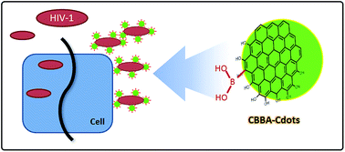
To date, several studies have reported the use of carbon-based materials for HIV treatment.42–45 Zhang et al. recently reported the potency of both graphene and graphene oxide, which induced conformational changes and aggregation of fragments of the viral R protein.44 Unfortunately, the validation of Cdots potency to inhibit viral entry has not been approved as a therapeutic strategy for the treatment or prevention of HIV infections. Therefore, in this study, we report a preparation method for Cdots as an entry inhibitor to prevent HIV infection. Preparation of Cdots was carried out by pyrolysis of citric acid (CA) to produce a graphene-like structure with an abundance of hydroxyl and carboxylate sites on the surface. These sites were then further modified with boronic acid by conjugating Cdots with carboxyl phenylboronic acid (CBBA) to block viral entry. These findings provide important information regarding the characteristics, toxicity, and inhibitory activity of Cdots for further clinical applications.
Experimental
Materials
4-Dimethylaminopyridine (DMAP), 1-ethyl-3-(3-dimethylaminopropyl) carbodiimide (EDC) and 4-carboxy-3-chlorobenzeneboronic acid (CBBA, 97%) were purchased from Sigma-Aldrich (Milwaukee, WI, USA). Sodium hydroxide and citric acid anhydrous (CA) were purchased from Showa Kako Corp. (Osaka, Japan). All chemicals were used directly without further purification.
Synthesis of carbon dots (Cdots)
Cdots were prepared by following previous work with some modifications.39 Experimentally, 200 mg of CA was calcined at 250 °C for 30 min. This process produces the CA orange indicating carbonization process to form graphitic nanostructures or Cdots. Once cooled to room temperature, Cdots were added with NaOH solution (0.5 M) until the pH was neutral. Then, the solution was further dialyzed on the membrane with a molecular-weight cut off (MWCO) of 1000 Da to narrow the Cdots size distribution and remove small molecules.
Preparation of CBBA-Attributed Cdots (CBBA–Cdots). 10 mL of CBBA solution (0.3 mg mL−1) was added with 2.4 mg of DMAP under magnetic stirring until clearly dissolved and then 3.1 mg of EDC was added. After mixing for 30 minutes, the solution was mixed with 5 mL of Cdots and the stirring was continued for 24 h. The mixture was then purified by a dialysis process for 24 h.
Cells
The Human acute T lymphoblastic leukemia cell lines (MT-4 and MOLT-4) were cultured in RPMI-1640 medium (GIBCO, USA) supplemented with 10% heat-inactivated fetal bovine serum (Sigma), 1% sodium bicarbonate, 100 U mL−1 penicillin G, and 100 μg mL−1 streptomycin (culture medium) and incubated for three days in a CO2 incubator. The virus persistently infected the MT-4 cells (MT-4/HIV-1), which were obtained from co-culture of HIV-1 infected peripheral blood mononuclear cells (PBMCs) with uninfected PBMCs using stimulated phytohemagglutinin (PHA, 10 μg mL−1). The co-culture was incubated in growth RPMI-1640 medium containing T cell growth factor cytokine, IL-2 for three days in a CO2 incubator. The virus stock was produced from supernatants of persistently infected MT-4 cells with the HIV-1 isolates from Surabaya, Indonesia.
Cytotoxicity assessment
Cell viability was quantified using the colorimetric WST-1 Cell Proliferation Reagent (Roche Applied Science, Switzerland). This assessment was based on the reduction of 2-(4-iodophenyl)-3-(4-nitrophenyl)-5-(2,4-disulfophenyl)-2H-tetrazolium sodium salt (WST-1) to water-soluble formazan from living cells. In the first step, the as-prepared Cdots and CBBA samples were dissolved in 1 mL of the RPMI-160 medium, then, added to MOLT-4 cells cultured in a 96 well plate with density of 2 × 10⁵ cells per μL in 100 μL of sample dissolved medium. 10 μL of WST-1 reagents was subsequently added to each well and incubated for 24 h at 37 °C in a CO2 incubator. After the incubation, the viable cells were measured by the absorbance of each sample using a microplate reader at a wavelength of 450 nm. The cytotoxicity effect of Cdots and CBBA–Cdots were also evaluated at half the cytotoxic concentration (CC50) and were analyzed with Origin software.
Antiviral assay (syncytia formation)
Syncytia formation assay evaluated the antiviral activity of the as-prepared Cdots and CBBA samples. Herein, MT-4/HIV-1 cells and MOLT-4 cells were cultured in RPMI-1640 medium. The cells were cultured at 37 °C for 5 days. After the incubation, the number of cells was counted with a hemocytometer. The MT4/HIV-1 cells (2 × 10⁴ cells per μL) and MOLT-4 cells (4 × 10⁵ cells per μL) were suspended in culture medium and placed in 96 well plates. Samples (Cdots or CBBA–Cdots) at specific concentrations were first mixed with MT4/HIV-1 cells and incubated for 30 minutes in a CO2 incubator. The mixed sample was subsequently added with 4 × 10⁵ cells per μL following incubation at 37 °C for 24 h in a CO2 incubator using a mixed sample on cultured MOLT-4 as a negative control (NC). After incubation, the amount of produced syncytia was microscopically counted. The data were analysed based on the percent of inhibition compared with the negative control as follows:
Inhibiting activity was also evaluated on half the concentration of inhibition (IC50) value counted by the Origin software.
Statistical analysis
The statistical analysis, including the cytotoxic concentration decreasing 50% of cell viability (CC50) and inhibition concentration inducing 50% of cell viability, were carried out using dose response mode on non-linear fitting curve of Origin®software (version 8.0724, OriginLab Inc., Northampton, MA). All data were performed in triplicate and the means were compared using a paired-sample t-test.
Other characterizations
Physicochemical analysis obtaining dynamic light scattering (DLS) and zeta potential (ζ) were collected using a Zetasizer Nanoseries 3000 HS from Malvern instruments. Transmission electron microscopy (TEM) images of samples were prepared by layering a Cdots solution onto copper grids (200-mesh) coated with a thin Formvar-carbon film and removing the solvent by drying at room temperature. High-resolution TEM images (HR-TEM) were acquired using a Tecnai G2 F20 instrument (Philips, Holland). X-ray diffraction (XRD) patterns were obtained using a Rigaku 18 kW rotating anode source X-ray diffractometer with the Cu Kα1 line (l = 1.54 Å). Atomic Force Microscopy (AFM) images were acquired with a scanning probe SPM-9600 (Shimadzu Co., Japan) at ambient temperature, X-ray photoelectron (XPS) spectra were obtained using a VG ESCA scientific theta probe spectrometer equipped with an Al Kα (1486.6 eV) X-ray source and 28 eV pass energy. Fourier transform infrared (FTIR) spectra were collected with an FTIR spectrometer (Shimadzu, Japan).
Results and discussion
Structural and morphological characterization of Cdots
The Cdots morphologies and particle sizes were first investigated by electron microscopy. The TEM images shown in Fig. 1a demonstrated that Cdots were nearly spherical, with particle sizes less than 5 nm. These data were further confirmed by atomic force microscopy (AFM); (Fig. S1 in the ESI†). A histogram of the AFM data showed that Cdots were typically about 2 nm in size. Moreover, HR-TEM analysis (Fig. 1b) showed that the Cdots exhibited a lattice finger structure, indicating the presence of carbon crystals surrounding the amorphous phase. Further physicochemical analysis (Tabel 1) using DLS also showed that the hydrodynamic diameter of Cdots was 2.8 nm, similar to the data obtained from AFM.
 |
| Fig. 1 TEM image of as-prepared Cdots (a) and its high magnification image performed with high-resolution TEM (b). White arrays show lattice finger of graphene like structure on Cdots. | |
Table 1 Physicochemical properties of prepared nanoparticlesa
Type |
Hydrodynamic diameter (nm) |
Polydispersity index |
ζ (mV) |
Each data presented as mean ± SD (n = 3). |
Cdots |
2.8 ± 1.1 |
1.4 ± 0.08 |
−55.4 ± 8.2 |
CBBA–Cdots |
6.2 ± 0.8 |
0.9 ± 0.02 |
−90.9 ± 7.01 |
During Cdots synthesis, the pyrolysis of CA induced carbonization, producing a crystal phase in the form of the observed graphene-like structure. Further high-magnification HR-TEM images allowed us to determine the distance between the carbon atoms in the lattice (0.34 nm). Moreover, XRD analysis (Fig. 2a) showed that the diffractogram pattern of Cdots had a broad peak at 2θ (range: 20–30°), confirming the graphitic structure based on JCPDS26-1076, with d-spacing at a higher intensity (006) of 0.335 nm. This result was consistent with the above HR-TEM images and also similar to the results of some previous studies on identifying the crystal structure of Cdots.37,46,47 These findings were further supported by Raman measurements (Fig. 2b), where the G-band peak significantly appeared at 1582 cm−1, representing the formation of sp2-hybridized carbon atoms. Moreover, a D-band peak was also recorded at 1332 cm−1, which is associated with vibration of the sp3-hybridized carbon atoms at the edge of the crystal. This peak indicated that the Cdots formed a crystal graphene-like structure. The higher intensity D-band indicated the formation of bonds at the edge, yielding the amorphous phase, or bonds at the terminal end of the carbon plane, which would contribute to the water solubility of the Cdots.
 |
| Fig. 2 (a) Diffractogram pattern of Cdots. (b) its Raman spectra of Cdots (green line) and citric acid (yellow line). | |
Surface modification of Cdots
Based on the results of previous studies, boronic acid containing-material has positive effects on the selectivity of glycopeptides and on glycoprotein enrichment. Therefore, introducing CBBA onto Cdots is critical and guarantees a specific interaction between Cdots and HIV, which is covered abundantly by glycoprotein. As illustrated in Scheme 1, introducing CBBA onto Cdots was accelerated via the EDCI/DMAP reagents, which activated the carboxylate moiety of CBBA before reacting specifically with the hydroxyl sites presented on Cdots.
 |
| Scheme 1 Schematic of conjugating CBBA onto Cdots and further mechanism on inhibition entry. | |
The obtained CBBA–Cdots were then investigated further using X-ray photoelectron spectroscopy (XPS; Fig. 3a–d), which revealed the existence of atoms, namely carbon (around 286 eV), oxygen (around 531 eV), and boron (around 193 eV). Deconvolution of high-resolution C1 XPS spectra yielded peaks at 285.4 (i) and 286.5 eV (ii), which indicated C–C/C
C (graphite) and C–O species, respectively, on Cdots. The carbon spectrum also showed a peak at 288.2 eV (iii) for –C
O (ester) on boronic acid-conjugated Cdots.48 Further deconvolution of O1 XPS spectra yielded peaks at 531.3 (iv), 532.7 (v), and 533 eV (vi), which were assigned to H–O, C–O, and B–O on CBBA–Cdots, respectively.49–51 Moreover, the appearance of a peak at 193.7 eV for B–O species strongly indicated successful conjugation of CBBA onto Cdots.52 Moreover, the appearance of a peak at 193.3 eV for the B–O species strongly indicated successful conjugation of CBBA onto Cdots.53 Functional group analyses using Fourier transform infrared (FTIR) spectroscopy were carried out to further support our characterization of CBBA–Cdots. When FTIR spectra of CBBA–Cdots were compared with bare Cdots (Fig. 3e), the presence of CBBA conjugated onto Cdots was demonstrated by specific peaks at 1572 (1), 1080 (2), 758 (3), and 680 cm−1 (4), which were assigned to the stretching vibrations of the B–OH, B–C, C–H, and C–C bonds of CBBA, respectively, which is consistent with previous studies.53,54 However, the FTIR data also yielded several peaks, including peaks at 3448.84 (5), 2852.81 (6), and 1414.93 (7), assigned to O–H, C–H, and C
O, respectively, for Cdots. Lastly, we obtained the zeta potential values for Cdots and CBBA–Cdots from our physicochemical analysis (Table 1) to determine whether CBBA was successfully conjugated onto the Cdots. The existence of boronic acid sites from CBBA onto the surface of nanoparticles was observed by the decrease in the zeta potential value to −90.9 eV.
 |
| Fig. 3 XPS spectrum of Cdots on (a) full scan range, (b) the C1s spectrum, (c) the O1s spectrum, and (d) the B1s spectrum. (e) FTIR spectra of Cdots (green line) and CBBA–Cdots (red line). | |
Cytotoxicity assay
In order to evaluate the potential utility of CBBA–Cdots in biological applications, we assessed the toxicity of the obtained CBBA–Cdots in MOLT-4 human leukemia cells using WST-1 assays, as shown in Fig. 4a. After 24 h incubation with both Cdots and CBBA–Cdots, MOLT-4 cells proliferated as rapidly as untreated cells (control), even when the concentrations were raised up to 300 μg mL−1. The figure also shows that average percent cell viability at each concentration was greater than 80%, confirming the absence of cytotoxic effects for both Cdots and CBBA–Cdots in human cells. Further analysis on proving low toxicity of this nanomaterial was determined by CC50 values, where both Cdots and CBBA–Cdots had high CC50 values of 2901.2 and 1991.9 μg mL−1, respectively (Fig. S2 in the ESI†). These high CC50 values indicate that both Cdots and CBBA–Cdots could be applied safely under the conditions used in this study.
 |
| Fig. 4 Cell viability evaluation using WST-1 assay of MOLT-4 cells after 24 h treatment with Cdots (red) and CBBA–Cdots (green) compared negative control. All data perform in the format mean ± SD (n = 3). | |
Evaluation of anti-HIV efficacy in vitro
During HIV-1 infection, HIV entry into target cells is initiated through binding of gp120 overexpressed on the virus with the surface receptor CD4 and the co-receptors CXCR4 or CCR5 on the cell surface. Insertion of the virus onto the cell is affected by the viral protein and cell killing is induced by syncytium production. As a common condition, the spread of HIV can take on virus-to-cell and cell-to-cell pathways. Unlike the former pathway, transitions of the virus from the infected cell to the normal cell upon direct contact was confirmed as a more efficient way via a virological synapse process.55,56 However, both of the pathways include binding of gp120 to surface receptor of the targeted cell.
In this study, we examined the HIV inhibitory activity of Cdots and CBBA–Cdots by setting the infection in a cell-to-cell manner and counting the numbers of syncytia produced after a 24 h incubation of cultured MT4/HIV-1 and MOLT-4 cells with nanoparticles. Basically, syncytial formation is not equal to a cell-to-cell pathway, but it represents the interaction between gp120 and CD4/CXCR4 during the pathway. Therefore, syncytia formation assay is a useful method for the screening of inhibitors for viral cell-to-cell transmission. In the investigation, the Cdots were first incubated with MT4/HIV-1 cells to allow preliminary interaction with gp120 of the virus before the addition of MOLT-4 cells. Observation by microscopy showed that incubation with Cdots (300 μg mL−1) did not effectively prevent the formation of syncytia, similar to the results of the negative control (Fig. 5a–d). In contrast, CBBA–Cdots were found to efficiently bind to gp120 on the virus, thereby preventing the binding of MOLT-4 cells and blocking infection. Accordingly, when high concentrations of CBBA–Cdots were used, no syncytia were found on the cultured cells. Formation of tetravalent boronate diester cyclic complex from reaction between boronic acid sites of CBBA–Cdots to 1,2-cis diols sites on gp120 was predicted as being the factor responsible for this phenomena. We further analyzed the potency of Cdots on the inhibition of HIV infection by varying the Cdots concentration (Fig. 5e). Conjugation of CBBA onto Cdots significantly increased the inhibitory capacity of Cdots at high concentrations (greater than 75 μg mL−1), whereas the inhibitory effects were decreased at lower concentrations. However, Fig. 4b also proves that Cdots, itself, has an ability to prevent HIV infection, even at low concentrations. This phenomena was caused by the existence of hydroxyl and carboxylate functional groups on the edges of Cdots that may facilitate the occurrence of non-covalent interactions, such hydrogen bonding, with molecules expressed on the viral envelope, including gp120, gp41, and transmembrane subunits, thus conferring Cdots with their observed inhibitory activity. This finding also confirms that increasing inhibitor ability was mainly caused by the existence of boronic acid site on CBBA–Cdots. Further improving inhibitor ability was shown by the IC50 values, where both Cdots and CBBA–Cdots showed very different values, namely 9506.3 and 26.7 μg mL−1, respectively (Fig. S3 in the ESI†); thus, CBBA–Cdots had a significantly lower IC50 than Cdots (with probability value p < 0.05). These findings supported that the conjugated boronic acid functioned synergistically to prevent HIV infection.
 |
| Fig. 5 Microscopic images consisting mix of cultured MOLT-4 cell and Cdots as positive control (a); mix of cultured MOLT-4 cell and MT-4/HIV as negative control (b); mix of cultured MOLT-4 cell, MT-4/HIV, and 300 μg mL−1 of Cdots (c); mix of cultured MOLT-4 cell, MT-4/HIV, and 300 μg mL−1 of CBBA–Cdots (d). All data performs after 24 h incubation. Blue arrays indicate position of obtained syncytia. (e) Inhibition intensity of Cdots (green) and CBBA–Cdots (blue) against MT4/HIV-1 cells over 24 h incubation. All data perform in the format mean ± SD (n = 3).*p < 0.05, **p < 0.01. | |
Conclusion
In this study, we investigated the antiviral effects of synthetic Cdots and CBBA–Cdots on HIV. Our findings showed that these synthetic nanoparticles, which formed graphene-like structures, inhibited HIV entry. In vitro analysis of the cytotoxicity of Cdots and CBBA–Cdots revealed that these nanoparticles could be safely applied, even at high concentration and after modification with CBBA. Furthermore, conjugation of Cdots with CBBA increased the inhibitory effects of the nanoparticles on HIV infection by preventing binding with the target cell. These data support the potential applications of Cdots and CBBA–Cdots in the clinical setting. The mechanism through which CBBA–Cdots prevent HIV infection may also be further developed to establish a dual-effect HIV drug for prevention and therapy by loading currently available HIV drugs onto the graphene scaffold of Cdots.
Competing interests
The authors have declared that no competing interest exists.
Acknowledgements
The authors thank the MANDAT research founding program of Airlangga University for financially supporting this research under Contract No. 401/UN3/2016.
References
- U. a. W. UNAIDS, Global AIDS response progress reporting 2015, 2015 Search PubMed.
- U. a. W. UNAIDS, Global AIDS response progress reporting 2014, Joint United Nations Programme on HIV/AIDS (UNAIDS), 2014 Search PubMed.
- A. Mocroft, A. N. Phillips, J. Gatell, B. Ledergerber, M. Fisher, N. Clumeck, M. Losso, A. Lazzarin, G. Fatkenheuer and J. Lundgren, Lancet, 2007, 370, 407–413 CrossRef CAS.
- R. K. Heaton, D. R. Franklin, R. J. Ellis, J. A. McCutchan, S. L. Letendre, S. LeBlanc, S. H. Corkran, N. A. Duarte, D. B. Clifford, S. P. Woods, A. C. Collier, C. M. Marra, M. R. Mindt and M. J. Tylor, J. Neurovirol., 2011, 17, 3–16 CrossRef CAS PubMed.
- M. J. Buzón, M. Massanella, J. M. Llibre, A. Esteve, V. Dahl, M. C. Puertas, J. M. Gatell, P. Domingo, R. Paredes, M. Sharkey, S. Palmer, M. Stevenson, B. Coltet, J. Blanco and J. Martinez-Picado, Nat. Med., 2010, 16, 460–465 CrossRef PubMed.
- N. Bulteel, L. Bansi-Matharu, D. Churchill, D. Dunn, D. Bibby, T. Hill, C. Sabin, M. Nelson, U. C. H. Cohort and U. H. D. R. D. U. H. Study, J. Infect., 2014, 68, 77–84 CrossRef PubMed.
- C. A. Derdeyn and G. Silvestri, Curr. Opin. Immunol., 2005, 17, 366–373 CrossRef CAS PubMed.
- P. A. Volberding and S. G. Deeks, Lancet, 2010, 376, 49–62 CrossRef.
- D. D. Richman, Nature, 2001, 410, 995–1001 CrossRef CAS PubMed.
- L. Van Damme, R. Govinden, F. M. Mirembe, F. Guédou, S. Solomon, M. L. Becker, B. Pradeep, A. Krishnan, M. Alary, B. Pande, G. Ramjee, J. Deese, T. Crucitti and D. Taylor, N. Engl. J. Med., 2008, 359, 463–472 CrossRef CAS PubMed.
- V. Pirrone, B. Wigdahl and F. C. Krebs, Antiviral Res., 2011, 90, 168–182 CrossRef CAS PubMed.
- J. Balzarini, Nat. Rev. Microbiol., 2007, 5, 583–597 CrossRef CAS PubMed.
- O. Francesconi, C. Nativi, G. Gabrielli, I. De Simone, S. Noppen, J. Balzarini, S. Liekens and S. Roelens, Chem.–Eur. J., 2015, 21, 10089–10093 CrossRef CAS PubMed.
- B. Iáiz, O. Martínez-Ávila, J. M. Falcon-Perez and S. Penadés, Bioconjugate Chem., 2012, 23, 814–825 CrossRef PubMed.
- T. J. Henrich and D. R. Kuritzkes, Curr. Opin. Virol., 2013, 3, 51–57 CrossRef CAS PubMed.
- D. Lee and M. S. Taylor, J. Am. Chem. Soc., 2011, 133, 3724–3727 CrossRef CAS PubMed.
- J. N. Cambre and B. S. Sumerlin, Polymer, 2011, 52, 4631–4643 CrossRef CAS.
- G. Springsteen and B. Wang, Chem. Commun., 2001, 1608–1609 RSC.
- D. Seth, A. Sarkar and D. Mitra, Nanosci. Nanoeng., 2014, 2, 1–9 CAS.
- I. Capek, Adv. Colloid Interface Sci., 2015, 222, 119–134 CrossRef CAS PubMed.
- G. Pastorin, W. Wu, S. Wieckowski, J.-P. Briand, K. Kostarelos, M. Prato and A. Bianco, Chem. Commun., 2006, 1182–1184 RSC.
- J. Kreuter, J. Nanosci. Nanotechnol., 2004, 4, 484–488 CrossRef CAS PubMed.
- B. Arnaiz, O. Martinez-Avila, J. M. Falcon-Perez and S. Penades, Bioconjugate Chem., 2012, 23, 814–825 CrossRef CAS PubMed.
- F. Chiodo, P. M. Enriquez-Navas, J. Angulo, M. Marradi and S. Penades, Carbohydr. Res., 2015, 405, 102–109 CrossRef CAS PubMed.
- C. Y. Shing, C. G. Whiteley and D.-J. Lee, J. Taiwan Inst. Chem. Eng., 2014, 45, 1140–1148 CrossRef CAS.
- E. M. Dahmane, M. Rhazi and M. Taourirte, Bull. Korean Chem. Soc., 2013, 34, 1333–1338 CrossRef CAS.
- J. L. Lenjisa, M. A. Woldu and G. D. Satessa, J. Nanobiotechnol., 2014, 12, 9 CrossRef PubMed.
- J. das Neves, F. Araujo, F. Andrade, J. Michiels, K. K. Arien, G. Vanham, M. Amiji, M. F. Bahia and B. Sarmento, Mol. Pharm., 2013, 10, 2793–2807 CrossRef CAS PubMed.
- L. M. Bimbo, O. V. Denisova, E. Mäkilä, M. Kaasalainen, J. K. De Brabander, J. Hirvonen, J. Salonen, L. Kakkola, D. Kainov and H. l. A. Santos, ACS Nano, 2013, 7, 6884–6893 CrossRef CAS PubMed.
- M. Nair, R. Guduru, P. Liang, J. Hong, V. Sagar and S. Khizroev, Nat. Commun., 2013, 4, 1707 CrossRef PubMed.
- J. P. Williams, P. Southern, A. Lissina, H. C. Christian, A. K. Sewell, R. Phillips, Q. Pankhurst and J. Frater, Int. J. Nanomed., 2013, 8, 2543–2554 CrossRef PubMed.
- J. Vonnemann, C. Sieben, C. Wolff, K. Ludwig, C. Bottcher, A. Herrmann and R. Haag, Nanoscale, 2014, 6, 2353–2360 RSC.
- D. Alukda, T. Sturgis and B. B. C. Youan, J. Pharm. Sci., 2011, 100, 3345–3356 CrossRef CAS PubMed.
- N. Chattopadhyay, J. Zastre, H.-L. Wong, X. Y. Wu and R. Bendayan, Pharm. Res., 2008, 25, 2262–2271 CrossRef CAS PubMed.
- D. Sun, R. Ban, P.-H. Zhang, G.-H. Wu, J.-R. Zhang and J.-J. Zhu, Carbon, 2013, 64, 424–434 CrossRef CAS.
- L. Zhu, Y. Yin, C.-F. Wang and S. Chen, J. Mater. Chem. C, 2013, 1, 4925 RSC.
- S. Gao, Y. Chen, H. Fan, X. Wei, C. Hu, L. Wang and L. Qu, J. Mater. Chem. A, 2014, 2, 6320–6325 CAS.
- R. Purbia and S. Paria, Biosens. Bioelectron., 2016, 79, 467–475 CrossRef CAS PubMed.
- M. Z. Fahmi, J.-K. Chen, C.-C. Huang, Y.-C. Ling and J.-Y. Chang, J. Mater. Chem. B, 2015, 3, 5532–5543 RSC.
- Q.-Q. Shi, Y.-H. Li, Y. Xu, Y. Wang, X.-B. Yin, X.-W. He and Y.-K. Zhang, RSC Adv., 2014, 4, 1563–1566 RSC.
- S. Qu, X. Wang, Q. Lu, X. Liu and L. Wang, Angew. Chem., 2012, 124, 12381–12384 CrossRef.
- T. A. Strom, S. Durdagi, S. S. Ersoz, R. E. Salmas, C. T. Supuran and A. R. Barron, J. Pept. Sci., 2015, 21, 862–870 CrossRef CAS PubMed.
- S. H. Friedman, D. L. DeCamp, R. P. Sijbesma, G. Srdanov, F. Wudl and G. L. Kenyon, J. Am. Chem. Soc., 1993, 115, 6506–6509 CrossRef CAS.
- M. Zhang, X. Mao, C. Wang, W. Zeng, C. Zhang, Z. Li, Y. Fang, Y. Yang, W. Liang and C. Wang, Biomaterials, 2013, 34, 1383–1390 CrossRef CAS PubMed.
- K. A. Mahmoud and J. H. T. Luong, Anal. Chem., 2008, 80, 7056–7062 CrossRef CAS PubMed.
- L. Tian, D. Ghosh, W. Chen, S. Pradhan, X. Chang and S. Chen, Chem. Mater., 2009, 21, 2803–2809 CrossRef CAS.
- R. Zhang, Y. Liu, L. Yu, Z. Li and S. Sun, Nanotechnology, 2013, 24, 225601 CrossRef PubMed.
- H. Yu, Y. Zhao, C. Zhou, L. Shang, Y. Peng, Y. Cao, L.-Z. Wu, C.-H. Tung and T. Zhang, J. Mater. Chem. A, 2014, 2, 3344 CAS.
- S. Knipe, J. Mycroft, A. Pratt, H. Nesbitt and G. Bancroff, Geochim. Cosmochim. Acta, 1995, 59, 1079–1090 CrossRef CAS.
- D. Yang, A. Velamakanni, G. Bozoklu, S. Park, M. Stoller, R. D. Piner, S. Stankovich, I. Jung, D. A. Field, C. A. Ventrice and R. S. Ruoff, Carbon, 2009, 47, 145–152 CrossRef CAS.
- A. Hayashi, M. Nakai, M. Tatsumisago, T. Minami, Y. Himei, Y. Miura and M. Katada, J. Non-Cryst. Solids, 2002, 306, 227–237 CrossRef CAS.
- T. V. Khai, H. G. Na, D. S. Kwak, Y. J. Kwon, H. Ham, K. B. Shim and H. W. Kim, Chem. Eng. J., 2012, 211–212, 369–377 CrossRef CAS.
- M. K. Smith and B. H. Northrop, Chem. Mater., 2014, 26, 3781–3795 CrossRef CAS.
- J. Faniran and H. Shurvell, Can. J. Chem., 1968, 46, 2089–2095 CrossRef CAS.
- L. M. Agosto, P. D. Uchil and W. Mothes, Trends Microbiol., 2015, 23, 289–295 CrossRef CAS PubMed.
- K. Ohki, M. Kishi, Y. Nishino, M. Sumiya, T. Kimura, T. Goto, M. Nakai and K. Ikuta, J. Acquired Immune Defic. Syndr., 1991, 4, 1233–1240 CAS.
Footnote |
† Electronic supplementary information (ESI) available: AFM data, CC50 graph of Cdots and CBBA–Cdots, and IC50 graph of Cdots and CBBA–Cdots. See DOI: 10.1039/c6ra21062g |
|
This journal is © The Royal Society of Chemistry 2016 |
Click here to see how this site uses Cookies. View our privacy policy here.