Synthesis, optical properties, and acid–base indicating performance of novel ketene hydroxybenzylidene nopinone derivatives

Jinlai Yangabc, Xu Xuad, Yiqin Yange, Jian Ruia, Yan Zhanga, Hongbo Kuanga, Shifa Wang*ad and Liangru Wu*bc
aCollege of Chemical Engineering, Nanjing Forestry University, Nanjing, Jiangsu 210037, China. E-mail: wangshifa65@163.com; Fax: +86 25 85427812; Tel: +86 25 85427812
bChina National Bamboo Research Center, Hangzhou 310012, Zhejiang, China. E-mail: bamshoots@163.com
cKey Laboratory of High Efficient Processing of Bamboo of Zhejiang Province, Hangzhou 310012, Zhejiang, China
dJiangsu Key Lab of Biomass-based Green Fuels and Chemicals, Nanjing 210037, China
eInstitute of Light Industry Science and Engineering, Nanjing Forestry University, Nanjing, Jiangsu 210037, China

Received 18th August 2016 , Accepted 10th November 2016

First published on 10th November 2016


Abstract

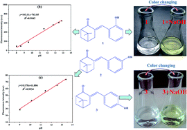
Three ketene nopinone derivatives (1–3) were successfully synthesized from β-pinene derivative nopinone and characterized by Fourier transform infrared spectroscope (FT-IR), nuclear magnetic resonance (NMR), mass spectrometry, and X-ray single crystal diffraction. Then, their optical properties were investigated by ultraviolet-visible and fluorescence spectroscopy. The ethanol solutions of compound 1 or 3 could change color from colorless to saffron yellow and Kelly, respectively, after adding NaOH solution. Moreover, the addition of potassium tert-butoxide could lead to a change in the UV-vis absorption and fluorescence spectra and enhance fluorescence, with compounds 1 and 3 being greatly affected by alkali. A good linear relationship between fluorescence intensity and pH values (9.01–13.38) was obtained for compound 2 (for detecting further pH value, y = 10.178x − 61.806, R2 = 0.9934). Compounds 1 and 3 were ideal acid–base indicators because of their high sensitivity, which was better than that of phenolphthalein. Therefore, these new nopinone derivatives may be candidates for future acid–base indicators.


1. Introduction

Acid–base indicators such as azo compound, nitrophenol and phenolphthalein are very useful chemical reagents. In recent years, some new acid–base indicators have been extensively described due to their applications in acid–base titration. One fluorescent pH indicator that exhibits a pH sensitivity in the 3.0–7.0 pH range is 4-[(p-N,N-dimethylamino)benzylidene]-2-phenyloxazole-5-one.1,2 Another indicator was 1,3-bisdicyanovinylindane, which displayed an strong blue color with a high extinction coefficient in the 3.8–5.2 pH range.3 It was found that (triphenylphosphoranylidene)cyclopenta-1,3-dienes display an extremely sharp, clear and reversible color change when they are used as pH indicators in the 10–12 range.4 In addition, 4[(4-dimethylamino-benzylidene)-amino]-benzene sulphonamide, which was synthesized via the condensation of sulfanilamide with p-dimethylaminobenzaldehyde, showed a yellow color at pH = 9.5 and this color disappeared at pH = 10.5.5 Finally, 7,8-dihydro-10-aryl-5H-indeno[1,2-b]quinoline-9,11-diones was a new pH indicator that showed excellent sensitivity in the 9.2–12 pH range, with an evident visual change from pale orange (pH = 9.2) to intense blue (pH = 12).6 As important pH indicators, some sulfonphthaleine dyes were fully investigated.7 There was also a colorimetric alginate-catechol hydrogel used as a spreadable pH indicator.8 More interestingly, a reversible photochromic dye (1-MEH) was investigated and the pH indicator dye could be used to photoswitch efficiently.9

Nopinone is an important derivative of β-pinene and has a C[double bond, length as m-dash]O group,10,11 which makes it easy to synthesize new derivatives. Thus, plenty of nopinone derivatives,12 such as (R)-(−)-cryptone,13 (1S,5S)-4-alkyl-6,6-dimethylbicyclo[3.1.1]hept-3-en-2-one,14 2-dialkylamino-6,6-dimethylbicyclo[3.1.1]heptan-3-ol,15 nopinone alkylation derivatives,16 chiral annulated indenes,17 nopinone-based triazole ketones,18 pinene-derived pyridines,19 chiral 1,3-aminoalcohols and 1,3-diols,20 have been synthesized. In addition, nopinone derivatives could also be used in fluorescent materials. Quinazolin-2-amine nopinone derivatives were successfully synthesized for use in cellular fluorescence imaging,21 while a novel high selective tetrahydroquinazolin-2-amine-based fluorescent sensor for Zn2+ was prepared from nopinone.22 However, to the best of our knowledge, there are no reported nopinone derivatives used as acid–base indicators. Moreover, we have been investigating β-pinene derivatives and their utility for new acid–base indicators synthesized from cheap β-pinene.

In this study, we report three novel ketene nopinone derivatives (1–3) that are easily synthesized from nopinone. Among them, compounds 1 and 3 could be used as acid–base indicators because of their good color indicating performance under alkaline conditions. Thus, compounds 1 and 3 were evaluated in base titration experiments in which they were compared with phenolphthalein.

2. Experimental

2.1. Materials and instruments

All the raw materials and solvents were purchased from commercial suppliers and used without further purification. 1H-NMR and 13C-NMR spectra were recorded on a Bruker 500 MHz FT-NMR spectrometer in DMSO or CDCl3 with TMS as the internal standard. Fourier-transform infrared (FT-IR) spectra were recorded on a Nicolet 380 FT-IR spectrophotometer. Melting points were measured with an X-6 microscopic melting point apparatus. UV-vis absorption spectra were obtained on a UV-2450 spectrophotometer (SHIMADZU) using a quartz cuvette with a 1 cm path length. Fluorescence emission spectra were acquired on a Perkin Elmer LS 55 fluorescence spectrophotometer at an excitation wavelength of 300 nm, scanning from 350 nm to 700 nm at a 100 nm min−1 scan speed. X-ray data were collected on a Bruker D8 Venture area diffractometer.

2.2. Synthesis of compounds 1–3

(+)-Nopinone with a purity of 95.6% (GC) was prepared from (−)-β-pinene (purchased from Deqing Forest Chemical Plant of China) by a reported method,23 and the specific rotation was also measured, [α]25D = +26.91 (c = 0.0016 g mL−1, CH3CH2OH).
2.2.1. 3-(2′-Hydroxybenzylidene)nopinone (1). A dried 50 mL three-necked flask equipped with a thermometer, stirrer and condenser was charged with nopinone (10 mmol), salicylaldehyde (12 mmol), and potassium tert-butoxide (27 mmol) in tert-butyl alcohol (30 mL). The resulting mixture was refluxed for 3 h until conversion reached over 95% (monitored by GC). The reacted mixture was extracted three times with 30 mL of ethyl acetate, and the combined organic layers were washed with saturated brine to neutrality, dried over Na2SO4, and concentrated to afford the crude product, which was purified twice by recrystallization in 15 mL of ethanol. Pale yellow crystals, 60% yield; mp: 238.8–238.9 °C; [α]25D = −89.80 (c = 0.0006 g mL−1, CH3CH2OH); FT-IR (KBr) ν (cm−1): 3109, 2962, 2930, 1662, 1591, 1489, 1451, 752; 1H NMR (DMSO-d6, 500 MHz): 0.82 (s, 3H), 1.31 (s, 3H), 1.34–1.37 (d, 1H), 2.26–2.27 (d, 1H), 2.51–2.53 (t, 1H), 2.54–2.58 (m, 1H), 2.85–2.86 (d, 2H), 6.83–6.85 (d, 1H), 6.86–6.94 (d, 1H), 7.18–7.22 (m, 1H), 7.55–7.56 (d, 1H), 7.94 (s, 1H), 9.99 (s, 1H); 13C NMR (DMSO-d6, 75 MHz), δ (ppm): 21.30, 25.81, 26.84, 30.20, 55.24, 115.62, 118.84, 122.17, 129.48, 129.55, 130.38, 131.25, 157.06, 201.88; EI-MS m/z (%): 242 (M+, 20), 201 (100), 199 (42), 131 (59), 77 (36), 55 (44); HRMS (m/z): [M + H]+ calcd for C16H18O2 + H+, 243.1379; found, 243.1368; anal. calcd for C16H18O2: C 79.31, H 7.49; found C 79.27; H 7.51.
2.2.2. 3-(3′-Hydroxybenzylidene)nopinone (2). A mixture of nopinone (10 mmol), 3-hydroxybenzaldehyde (12 mmol), potassium tert-butoxide (27 mmol) and tert-butyl alcohol (40 mL) was refluxed for 1.5 h. Other conditions were the same as those for compound 1. Faint yellow crystals, 80% yield; mp: 177.4–177.5 °C; [α]25D = −58.14 (c = 0.0009 g mL−1, CH3CH2OH); FT-IR (KBr) ν (cm−1): 3411, 2959, 2933, 1672, 1604, 1581, 1472, 1441, 874, 794, 685. 1H NMR (DMSO-d6, 500 MHz): 0.82 (s, 3H), 1.32 (s, 3H), 1.37–1.40 (d, 1H), 2.29–2.30 (d, 1H), 2.53–2.56 (t, 1H), 2.57–2.61 (m, 1H), 2.90–2.91 (d, 2H), 6.81–6.83 (d, 1H), 7.05–7.07 (d, 2H), 7.23–7.26 (t, 1H), 7.47 (s, 1H), 9.56 (s, 1H); 13C NMR (DMSO-d6, 75 MHz), δ (ppm): 21.25, 25.76, 26.84, 30.43, 55.19, 116.26, 116.98, 121.74, 129.59, 132.51, 134.63, 136.23, 157.41, 201.61; EI-MS m/z (%): 242 (M+, 100), 227 (30), 199 (50), 132 (30), 77 (22), 55 (31). HRMS (m/z): [M + H]+ calcd for C16H18O2 + H+, 243.1379; found, 243.1378; anal. calcd for C16H18O2: C 79.31H 7.49; found C 79.32, H 7.45.
2.2.3. 3-(4′-Hydroxybenzylidene)nopinone (3). A mixture of nopinone (10 mmol), p-hydroxybenzaldehyde (12 mmol), potassium tert-butoxide (27 mmol) and tert-butyl alcohol (30 mL) was refluxed for 8 h. Other conditions were the same as that for compound 1. Light yellow crystals, 69% yield; mp: 206.4–206.7 °C; [α]25D = −78.57 (c = 0.0004 g mL−1, CH3CH2OH); FT-IR (KBr) ν (cm−1): 3378, 2962, 2949, 2911, 1668, 1595, 1579, 1505, 1466, 836; 1H NMR (CDCl3, 300 MHz): 0.93 (s, 3H), 1.37 (s, 3H), 1.49–1.53 (d, 1H), 2.37 (s, 1H), 2.58–2.66 (m, 1H), 2.69–2.73 (t, 1H), 2.95 (s, 2H), 6.91–6.94 (d, 3H), 7.50–7.53 (d, 2H), 7.69 (s, 1H); 13C NMR (CDCl3, 75 MHz), δ (ppm): 21.62, 26.19, 27.60, 30.99, 39.48, 40.95, 55.88, 115.81, 128.04, 129.76, 132.97, 136.38, 157.31, 205.08; EI-MS m/z (%): 242 (M+, 100), 199 (37), 171 (39), 132 (41), 107 (40), 77 (24), 55 (27); HRMS (m/z): [M + H]+ calcd for C16H18O2 + H+, 243.1379; found, 243.1377; anal. calcd for C16H18O2: C 79.31, H 7.49; found C 79.23, H 7.48.

3. Results and discussion

3.1. Synthesis

Ketene nopinone derivatives (1–3) were successfully synthesized from nopinone according to the reaction shown in Scheme 1. The three compounds were characterized by FT-IR, NMR and MS. In addition, compound 1 was also characterized by X-ray single crystal diffraction (Fig. 1 and Table S1, ESI). These analyses confirmed 1 to be 3-(2′-hydroxybenzylidene)nopinone.
image file: c6ra20809f-s1.tif
Scheme 1 Synthesis of compounds 1–3.

image file: c6ra20809f-f1.tif
Fig. 1 X-ray crystal structure of compound 1.

3.2. Color changing properties

To investigate the color changes in ethanol after adding a certain amount of NaOH solution, 1.0 × 10⁻³ M solutions of compounds 1–3 were prepared. As shown in Fig. 2, the three solutions were colorless; however, the solutions of compounds 1 and 3 showed a clear color change to saffron yellow and Kelly, respectively, after adding two drops of 0.1 M NaOH. In contrast, compound 2 showed no significant change in color. Thus, compounds 1 and 3 were potentially suitable as acid–base indicators. Comparing the final color of compounds 1 and 3, it could be seen that the first color was more visible than the latter, and therefore compound 1 would be a better acid–base indicator.
image file: c6ra20809f-f2.tif
Fig. 2 Images showing the color changes for compounds 1–3.

3.3. Optical properties

3.3.1. UV-vis spectral changes. To further explore how the UV-vis absorption spectra was affected by the system's pH, trifluoroacetic acid (TFA) and potassium tert-butoxide were used to adjust the pH in ethanolic solutions of compounds 1–3 with a concentration of 1 × 10⁻⁴ mol L−1. Then, these solutions were analyzed by ultraviolet spectroscopy, as shown in Fig. 3 and S1 (ESI).
image file: c6ra20809f-f3.tif
Fig. 3 UV-vis absorption spectra of compounds 1 (a), 2 (b) and 3 (c) in different pH solutions.

Fig. 3a depicts the changes in the UV-vis absorption spectrum for 3-(2′-hydroxybenzylidene)nopinone (1) at different pH of the solutions. At a pH ≤ 9.55, the intensity of the 337 nm absorption peak decreased as the solution pH increased. This peak disappeared when the pH value reached 11.54; moreover, a new absorption peak appeared at 408 nm. When the pH was adjusted to 8.55, there was absorbance at 408 nm and the solution of compound 1 went from colorless to orange. This indicates that a chemical reaction occurred. When the pH was adjusted to 9.55, a clear absorption peak appeared at 408 nm and the solution exhibited a dark color. A clear absorption peak appeared at 408 nm when the pH value was above 11.54, and the intensity increased with an increasing pH value. It was evident that compound 1 had reacted under alkaline conditions. Therefore, compound 1 solution could change color when potassium tert-butoxide was added. Compound 1 has a 2′-OH group, which reacts easily with OH, leading to a change in the chemical structure from 2′-OH to 2′-O,24 with a color change to orange. Fig. S1a clearly shows the change in absorbance at 408 nm with an increased pH value. As potassium tert-butoxide was gradually added to the system, the rise in pH led to a continuous enhancement of the absorbance intensity.

As shown in Fig. 3b, compound 2 shows little change in the UV-vis absorption spectra, when compared with compound 1 (shown in Fig. 3a). The peak (292 nm) of compound 2 did not change at pH ≤ 10.12, but a red-shift appeared when the pH value increased to 11.43, and reached 306 nm at pH ≥ 12.54. Moreover, a new absorption peak emerged at 371 nm when the pH was above 11.43, however, the absorbance for this peak was weaker than that of compound 1. As shown in Fig. S1b, absorbance at 371 nm barely changed when pH was below 10, but increased markedly with continuous rising pH values. These results show that the 3′-OH group of compound 2 was stable at pH ≤ 10, and it could be affected by OH only when more potassium tert-butoxide was added to the solution. This could be the reason why compound 2 cannot be used as an acid–base indicator.

The results for compound 3 (Fig. 3c) were similar to those for compound 1. The UV-vis absorption spectrum of 3-(4′-hydroxybenzylidene)nopinone was also significantly affected by the solution's pH value. At pH values below 8.62, there was only one peak, at 324 nm, which barely changed with pH. However, two peaks were visible at a pH of 8.7, at 324 nm and 387 nm, and the solution's color turned to Kelly. This clearly indicated that a reaction occurred, which agreed with Fig. 2. As shown in Fig. S1c, the absorbance changed little at pH ≤ 8.0 and increased gradually to a maximum when potassium tert-butoxide was continuously added to the system. It indicated that the 4′-OH group of compound 3 also reacted easily with OH, leading to a change in color.

From the abovementioned research, it could be concluded that a 2′-OH or 4′-OH in ketene nopinone derivatives was easy to deprotonate under alkaline conditions24 and offered an apparent color changing process.25 Thus, 1 and 3 compounds could be further used as acid–base indicators.

3.3.2. Fluorescence spectral changes. In order to confirm the reason for the change in color, as with the UV-vis absorption spectra, fluorescence spectra of compounds 1–3 were recorded at different pH values (Fig. 4 and S2).
image file: c6ra20809f-f4.tif
Fig. 4 Fluorescence spectra of compounds 1 (a), 2 (b) and 3 (c) in different pH solutions, λex 300 nm.

Fig. 4a shows that the spectrum of compound 1 had two peaks at 436 and 603 nm and they barely changed (pH ≤ 8.55). When the pH increased to 9.55, the fluorescence intensity of the 436 nm peak decreased significantly, whereas the intensity of the 603 nm peak increased markedly. Moreover, a new peak appeared at 560 nm. This suggests that the structure of compound 1 had changed, which agreed with the results from the UV-Vis spectrum in Fig. 3a. As shown in Fig. S2a, there was a strong fluorescence intensity (568.73) at 560 nm when the pH was gradually raised to 11.54. At this point, the peak at 436 nm disappeared. At pH ≥ 13.45, the fluorescence intensity did not change. These results showed that the addition of potassium tert-butoxide could change the structure of compound 1 leading to a fluorescence enhancement. This indicates that the reason for the change in color was the change in the chemical structure of compound 1 when reacting with OH.

From Fig. 4b, it was clear that there was no remarkable fluorescence enhancement under strong alkaline condition, which means that the addition of potassium tert-butoxide had little effect on compound 2, which is in agreement with Fig. 3b. The relationship between the fluorescence intensity (601 nm) and pH value is shown in Fig. S2b. At a pH below 9.01, the intensity changed little, but it increased slightly when pH was adjusted to 10.12. The fluorescence intensity increased continuously when pH increased from 10.12 to 13.80.

As shown in Fig. 4c and Fig. S2c, there was little change in the fluorescence intensity at 601 nm for compound 3 at a pH below 8.62. Fluorescence for compound 3 increased after pH was raised to 8.70. After that, the fluorescence intensity increased gradually as pH increased, leading to a significant fluorescence enhancement. This indicates that the –OH group of compound 3 reacted easily with the OH originated from potassium tert-butoxide. The results showed that the addition of potassium tert-butoxide could enhance fluorescence.

As a result, the addition of potassium tert-butoxide could strongly enhance fluorescence for compounds 1 and 3, but led to only a small fluorescence enhancement for compound 2, which agreed well with the results for 2-acetyl-4-methyl-6-nitrophenol when affected by another alkali of SDS.26 It was concluded that the –OH group in compounds 1 and 3 reacted easily with OH, thus deprotonation occurred,24 leading to a fluorescence enhancement.

3.4. Linearity

To obtain a linear relationship for compounds 1–3 against different pH values, which would allow determining pH, data in Fig. S1 and S2 were treated by linear regression. As a result, the linear relationship between absorbance and fluorescence intensity of compound 1 and pH value of ethanol solution were obtained, y = 0.227x − 1.9938 (pH range 8.55–13.82), R2 = 0.9734 (Fig. 5a), y = 103.11x − 763.95 (pH range 8.01–13.82), R2 = 0.9842 (Fig. 5b). At the same time, another linear relationship between fluorescence intensity of compound 2 and pH was also obtained, y = 10.178x − 61.806 (pH range 9.01–13.82), R2 = 0.9934 (Fig. 5c). However, data for compound 3 showed no linear relationship. Therefore, compound 1 had the ability to measure the pH value of the ethanol solution using UV-vis absorption and fluorescence spectroscopy, and compound 2 could also be used to determine the pH of a solution by fluorescence spectroscopy. Thus, a new idea for confirming a solution's pH had been provided for future use.
image file: c6ra20809f-f5.tif
Fig. 5 Absorbance (a) and fluorescence intensity (b) of compound 1 vs. pH values; (c) fluorescence intensity of compound 2 vs. pH values.

3.5. Acid–base indicating performance

Compounds 1 and 3 showed a clear color change after adding some NaOH, indicating that they could be used as acid–base indicators. Therefore, their acid–base indicating performance was studied by comparing it with phenolphthalein. Compounds 1, 3 and phenolphthalein were separately dissolved in 95% ethanol to prepare three 0.2% solutions for use as indicators. Experimental samples were 0.2574 mol L−1 hydrochloric acid standard solution; then titration experiments were carried out using 0.1497 mol L−1 sodium hydroxide solution after adding two drops of one of the above indicators. The results were shown in Tables 1 and 2.
Table 1 Indicating performance of compound 1 and phenolphthalein
Type of indicator Measured HCl solution concentration (mol L−1) Average concentration (mol L−1) RSD (%) Indicating color
Phenolphthalein 0.2596 0.2610 0.454 Pink
0.2617
0.2616
Compound 1 0.2597 0.2596 0.089 Saffron yellow
0.2597
0.2593


Table 2 Indicating performance of compound 3 and phenolphthalein
Type of indicator Measured HCl solution concentration (mol L−1) Average concentration (mol L−1) RSD (%) Indicating color
Phenolphthalein 0.2601 0.2612 0.362 Pink
0.2619
0.2615
Compound 3 0.2598 0.2598 0.135 Kelly
0.2602
0.2595


The indicating color of compound 1 and 3 were saffron yellow and Kelly, respectively, while phenolphthalein turned pink. The measured HCl concentration using compound 1 and phenolphthalein as indicators were 0.2596 and 0.2610 mol L−1, respectively, with a relative standard deviation (RSD) of 0.089% and 0.454%, respectively. Compared to the hydrochloric acid standard concentration (0.2574 mol L−1), compound 1 as an indicator presented a high sensitivity, clear and reversible color and low RSD, and the performance was better than that of phenolphthalein. Thus, 3-(2′-hydroxybenzylidene)nopinone was an ideal acid–base indicator.

After that, the indicating performance of compound 3 was also investigated by comparing it with phenolphthalein (Table 2). Using compound 3 and phenolphthalein as indicators separately, the HCl solution concentration was measured by titration. With compound 3 as an indicator, a concentration of 0.2598 mol L−1 was obtained, whereas with phenolphthalein the measured concentration was 0.2612 mol L−1. The first concentration was closer to 0.2574 mol L−1 (standard concentration), thus, compound 3 as an indicator was better than phenolphthalein. It was concluded that 3-(4′-hydroxybenzylidene)nopinone was also a good acid–base indicator.

Among the three acid–base indicators, 3-(2′-hydroxybenzylidene)nopinone was the best one, followed by 3-(4′-hydroxybenzylidene)nopinone. In addition, the indicating color of the first was easier to see than the latter. Thus, 3-(2′-hydroxybenzylidene)nopinone is a potential acid–base indicator for future use.

4. Conclusion

Three ketene nopinone derivatives (1–3) were synthesized by aldol condensation from nopinone (β-pinene derivative). The color of compound 1 ethanol solution went from colorless to saffron yellow after adding two drops of NaOH solution, whereas another solution of compound 3 changed its color to Kelly under the same conditions. OH could easily react with the –OH group of compounds 1 and 3, leading to a significant change in the UV-vis absorption and fluorescence spectra and an enhanced fluorescence. The absorbance and fluorescence intensity of compound 1 had a linear relationship with the pH of the solution; y = 0.227x − 1.9938 (pH: 8.55–13.82), R2 = 0.9734; y = 103.11x − 763.95 (pH: 8.01–13.82), R2 = 0.9842. Compound 2, in the pH range of 9.01–13.38, also had a linear relationship between fluorescence intensity and pH, y = 10.178x − 61.806, R2 = 0.9934. The acid–base indicating performance of compounds 1 and 3 showed that they were better than phenolphthalein. Thus, they have a potential application as acid-base indicators, which increases the value of turpentine.

Acknowledgements

The authors gratefully acknowledge the project supported by the “Five-Year” plan of National Science and Technology for Chinese Rural Development Project (2015BAD15B04), the University Science Research Project of Jiangsu Province (14KJA220001), and the Committee of National Science Foundation of China (Grant No. 31470592).

Notes and references

  1. K. Ertekin, C. Karapire, S. Alp, B. Yenigül and S. Içli, Dyes Pigm., 2003, 56, 125–133 CrossRef.
  2. K. Ertekin, S. Cinar, T. Aydemir and S. Alp, Dyes Pigm., 2005, 67, 133–138 CrossRef.
  3. Y. A. Son and S. H. Kim, Dyes Pigm., 2005, 64, 153–155 CrossRef.
  4. F. Nourmohammadian, M. Davoodzadeh and A. A. Alizadeh, Dyes Pigm., 2007, 74, 741–743 CrossRef.
  5. R. A. Khalil, A. H. Jalil and A. Y. Abd-Alrazzak, J. Iran. Chem. Soc., 2009, 6, 345–352 CrossRef.
  6. F. Shirini, S. S. Beigbaghlou, S. V. Atghia and S. A. R. Mousazadeh, Dyes Pigm., 2013, 97, 19–25 CrossRef.
  7. T. De Meyer, K. Hemelsoet, V. Van Speybroeck and K. De Clerck, Dyes Pigm., 2014, 102, 241–250 CrossRef.
  8. Y. K. Lee and S. Y. Lee, Dyes Pigm., 2014, 108, 1–6 CrossRef.
  9. J. Vallet, J. C. Micheau and C. Coudret, Dyes Pigm., 2016, 125, 179–184 CrossRef.
  10. T. Szuppa, A. Stolle, B. Ondruschka and W. Hopfe, ChemSusChem, 2010, 3, 1181–1191 CrossRef PubMed.
  11. A. Stolle, Eur. J. Org. Chem., 2013, 44, 2265–2278 CrossRef.
  12. E. Mena, P. Van De Witte and J. Lub, Liq. Cryst., 2000, 27, 929–933 CrossRef.
  13. M. Kato, M. Watanabe, B. Vogler, Y. Tooyama and A. Yoshikoshi, J. Chem. Soc., Chem. Commun., 1990, 23, 1706–1707 RSC.
  14. M. Watanabe, B. Z. Awen and M. Kato, J. Org. Chem., 1993, 58, 3923–3927 CrossRef.
  15. C. T. Goralski, W. Chrisman, D. L. Hasha, L. W. Nicholson, P. R. Rudolf, D. Zakett and B. Singaram, Tetrahedron: Asymmetry, 1997, 8, 3863–3871 CrossRef.
  16. K. R. Campos, S. Lee, M. Journet, J. J. Kowal, D. Cai, R. D. Larsen and P. J. Reider, Tetrahedron Lett., 2002, 43, 6957–6959 CrossRef.
  17. R. L. Halterman and L. D. Crow, Tetrahedron Lett., 2003, 44, 2907–2909 CrossRef.
  18. R. C. da Silva Junior, V. F. Ferreira and S. Pinheiro, Tetrahedron: Asymmetry, 2004, 15, 3719–3722 CrossRef.
  19. A. V. Malkov, A. J. P. Stewart-Liddon, F. Teplý, L. Kobr, K. W. Muir, D. Haigh and P. Kočovský, Tetrahedron, 2008, 64, 4011–4025 CrossRef.
  20. Z. Szakonyi, T. Gonda, S. B. Oetvoes and F. Fueloep, Tetrahedron: Asymmetry, 2014, 25, 1138–1145 CrossRef.
  21. J. L. Yang, H. J. Xu, X. Xu, J. Rui, X. Y. Fang, X. Q. Cao and S. F. Wang, Dyes Pigm., 2016, 128, 75–83 CrossRef.
  22. J. L. Yang, H. Fang, X. Y. Fang, X. Xu, Y. Q. Yang, J. Rui, C. L. Wu, S. F. Wang and H. J. Xu, Tetrahedron, 2016, 72, 4503–4509 CrossRef.
  23. B. Liu, W. W. Ao, Y. Yang and S. F. Wang, J. Nanjing For. Univ., 2010, 34, 89–94 Search PubMed.
  24. A. G. Bracamonte and A. V. Veglia, J. Photochem. Photobiol., A, 2013, 261, 20–25 CrossRef.
  25. K. Yumura, H. Otani and J. Mizuguchi, Dyes Pigm., 1998, 38, 227–242 CrossRef.
  26. P. M. Gashnga, T. S. Singh and S. Mitra, J. Mol. Liq., 2016, 218, 549–557 CrossRef.

Footnote

Electronic supplementary information (ESI) available. CCDC 1437626. For ESI and crystallographic data in CIF or other electronic format see DOI: 10.1039/c6ra20809f

This journal is © The Royal Society of Chemistry 2016
Click here to see how this site uses Cookies. View our privacy policy here.