Cadmium(II) carboxyphosphonates based on mixed ligands: syntheses, crystal structures and recognition properties toward amino acids

Zhou Zhao, Dan Yang, Bo Xing, Chao Ma, Zhen-Gang Sun*, Yan-Yu Zhu, Huan-Yu Li and Jing Li
School of Chemistry and Chemical Engineering, Liaoning Normal University, Dalian 116029, P. R. China. E-mail: szg188@163.com

Received 13th August 2016 , Accepted 12th September 2016

First published on 12th September 2016


Abstract

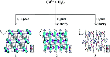
Three novel cadmium(II) carboxyphosphonates with three-dimensional (3D) frameworks and supramolecular structures, namely, Cd3[(L)2(1,10-phen)1.5]·H2O (1), Cd[(L′)(H2biim)] (2), and Cd[(L′)(H2biim)(H2O)]·H2O (3) (L = OOC–C6H4–CH2PO3, L′ = OOC–C6H4–CH2PO2(OC2H5), 1,10-phen = 1,10-phenanthroline, H2biim = 2,2′-biimidazole), were synthesized under hydrothermal conditions and were structurally characterized. For compound 1, the Cd(1)O6, Cd(2)O4N2, Cd(3)O2N2, Cd(4)O4 and CPO3 polyhedra were interconnected in a one-dimensional (1D) chain along the b-axis via corner- and edge-sharing. The adjacent chains were connected with each other by sharing the L3− anions, giving rise to a 3D framework structure. Compound 2 adopted a 3D supramolecular structure. The interconnection of two Cd(1)O4N2 polyhedra and two CPO3 tetrahedra via corner-sharing formed a structure unit, with such units further connected in the bc-plane by sharing the L′2− anions to exhibit a 2D layer, and then the neighboring layers were further assembled into a 3D supramolecular structure by π–π stacking interactions. In compound 3, the Cd(1)O4N2 and CPO3 polyhedra were interconnected to form a unit, and then the interconnection of multi-units were assembled into two kinds of 1D chains, whereby chains and other chains are further connected by π–π stacking interactions to present a 3D supramolecular structure. The luminescence properties of compounds 1–3 were investigated. Meanwhile, the excellent abilities of compounds 2 and 3 for the selective recognition of tryptophan (Trp) were demonstrated.

Introduction

Metal–organic frameworks (MOFs), as a new kind of functional material, have attracted considerable attention over the past few years because of their structural diversities and potential applications in the areas of adsorption,1 molecular recognition,2 ion recognition,3 catalysis,4 optics,5 and magnetism,6 and so on.

Compared with the traditional porous materials, MOFs have various advantages due to their type diversity, strong functions, and control of the pore size, etc. Metal phosphonates are an important class of MOF materials and have been widely studied due to their interesting structures and properties.7 Up to now, a great number of metal phosphonates have been successfully constructed by careful selection of phosphonic acids, metal ions, and the second organic ligand.8 To design a novel functional framework, an important and effective method is to modify phosphonic acids with additional organic functional groups, such as amines, hydroxyls, and carboxylate groups in the synthesis of metal phosphonates. Another important strategy for building new types of metal phosphonate with framework structures is to introduce a second organic ligand, such as an oxalate, carboxylic acid, 2,2′-bipyridine, 4,4′-bipyridine, or 1,10-phenanthroline, into the structure of the metal phosphonate. In recent years, a series of metal phosphonates with mixed ligands have been obtained by our group and other groups.9,10 The results achieved indicate that the introduction of a second organic ligand has a significant effect on the framework structures. To gain a deeper understanding of the properties and structures obtained with metal phosphonates, we have continued our work by carrying out reactions with the multifunctional organic phosphonate monoester ligand, HOOC–C6H4–CH2PO–(OH)(OC2H5) (H2L′), because we believe it may lead to a variety of interesting structures by adopting various kinds of coordination modes. Although phosphonic acid has been widely employed as an organic linker in the synthesis of metal phosphonates, organic phosphonate monoesters are an underexplored class of ligands.11 As for the phosphonate monoester ligands, a hydrophobic alkyl group (–R) tethered on the ligand might function as a steric shield to protect the M–O bonds and thus could enhance the stability of the resultant MOFs. In this paper, by selecting HOOC–C6H4–CH2PO–(OH)(OC2H5) (H2L′) as the organic phosphonate ligand and 1,10-phen and H2biim as the second metal linker, we successfully obtained three novel cadmium(II) carboxyphosphonates with three-dimensional (3D) frameworks and supramolecular structures, namely, Cd3[(L)2(1,10-phen)1.5]·H2O (1), Cd[(L′)(H2biim)] (2), and Cd[(L′)(H2biim)(H2O)]·H2O (3). Herein, we report their syntheses, crystal structures, and recognition properties for amino acids. To the best of our knowledge, research on the properties of metal phosphonates is mainly focused on the magnetism, luminescence, proton conductivity, and ion exchange etc., and there are no reports about amino acids' recognition properties of these materials. In recent years, due to the biological importance of amino acids, the design and development of optical probes for some amino acid molecules has been an active research area.12 Amino acids are the key constituents of proteins, and the amino acids recognition process may have potential application in the biology and medicine fields. Recently, a few investigations on the molecular recognition properties of metal phosphonates have been reported by our group.13 However, no metal phosphonates hybrids have been investigated so far regarding their recognition properties toward amino acids, and hence this is the first example of studies on the amino acids recognition properties of metal phosphonates.

Experimental

Materials and characterizations

The monocarboxyarylphosphonate ethyl ester ligand HOOC–C6H4–CH2PO–OH(OC2H5) (H2L′) was prepared according to the procedures described previously.14 All the other chemicals were obtained from commercial sources and used without further purification. C, H, and N were determined by using a PE-2400 elemental analyzer. The P and Cd contents were determined by using an inductively coupled plasma (ICP) atomic absorption spectrometer. IR spectra were recorded on a Bruker AXS TENSOR-27 FT-IR spectrometer with KBr pellets in the range 4000–400 cm−1. Thermogravimetric (TG) analyses were performed on a Perkin-Elmer Pyris Diamond TG-DTA thermal analyses system under static air at a heating rate of 10 K min−1 from 50 °C to 1100 °C. The luminescence analyses were performed on a HITACHI F-7000 spectrofluorimeter (solid). The luminescent properties of compounds 1–3 in the solid state and compounds 1–3 in aqueous suspensions were investigated at room temperature. The suspensions were prepared by introducing each sample (2.0 mg) as a powder into different amino acids aqueous solutions (each 2.0 mL). The luminescence spectra of the suspensions were measured after aging for 24 h.
Synthesis of Cd3[(L)2(1,10-phen)1.5]·H2O (1). A mixture of Cd(Ac)2·2H2O (0.11 g, 0.43 mmol), H2L′ (0.07 g, 0.26 mmol), and 1,10-phen (0.08 g, 0.44 mmol) was dissolved in 10 mL distilled water. The resulting solution (pH = 5) was stirred for about 1 h at room temperature, sealed in a 20 mL Teflon-lined stainless steel autoclave, and then heated at 180 °C for 72 h under autogenous pressure. After the mixture was cooled slowly to room temperature, colorless block crystals of compound 1 were obtained. Yield: 45.2% (based on Cd). Anal. calcd for C34H26N3O11P2Cd3 (%): C, 38.83; H, 2.49; N, 3.99; P, 5.89; Cd, 32.06. Found (%): C, 38.86; H, 2.46; N, 4.02; P, 5.85; Cd, 32.11. IR (solid KBr pellet ν/cm−1): 3443(m), 3046(w), 2989(w), 2778(w), 1589(m), 1532(m), 1420(m), 1382(s), 1239(w), 1141(m), 1116(s), 965(m), 906(w), 854(m), 795(w), 726(m), 633(w), 554(m), 463(w), 412(w).
Synthesis of Cd[(L′)(H2biim)] (2). A mixture of Cd(Ac)2·2H2O (0.11 g, 0.43 mmol), H2L′ (0.07 g, 0.26 mmol), and H2biim (0.06 g, 0.44 mmol) was dissolved in 10 mL distilled water. The resulting solution (pH = 5) was stirred for about 1 h at room temperature, sealed in a 20 mL Teflon-lined stainless steel autoclave, and then heated at 100 °C for 96 h under autogenous pressure. After the mixture was cooled slowly to room temperature, colorless block crystals of compound 2 were obtained. Yield: 40.7% (based on Cd). Anal. calcd for C16H17N4O5PCd (%): C, 39.32; H, 3.51; N, 11.46; P, 6.34; Cd, 23.00. Found (%): C, 39.36; H, 3.53; N, 11.42; P, 6.38; Cd, 23.05. IR (solid KBr pellet ν/cm−1): 3445(m), 3163(m), 2977(m), 2895(w), 2787(w), 1814(w), 1595(m), 1538(s), 1415(s), 1243(w), 1174(s), 1122(m), 1018(s), 861(m), 758(m), 691(w), 560(m), 485(w).
Synthesis of Cd[(L′)(H2biim)(H2O)]·H2O (3). Compound 3 was synthesized by a method similar to that of compound 2, except it was heated at 120 °C for 96 h under autogenous pressure. After the mixture was cooled slowly to room temperature, colorless block crystals of compound 3 were obtained. Yield: 42.4% (based on Cd). Anal. calcd for C16H21N4O7PCd (%): C, 36.62; H, 4.03; N, 10.68; P, 5.90; Cd, 21.42. Found (%): C, 36.65; H, 4.06; N, 10.65; P, 5.86; Cd, 21.46. IR (solid KBr pellet ν/cm−1): 3544(m), 3116(m), 2999(w), 2903(w), 2817(w), 1588(m), 1526(s), 1404(s), 1245(w), 1197(s), 1122(m), 1043(s), 946(m), 861(w), 776(s), 684(w), 541(m), 476(w).

Crystallographic studies

Data collection for compounds 1–3 were performed on a Bruker AXS Smart APEX II CCD X-diffractometer equipped with graphite monochromated MoKα radiation (λ = 0.71073 Å) at 293 ± 2 K. An empirical absorption correction was applied using the SADABS program. All the structures were solved by direct methods and refined by full matrix least-squares fitting on F2 by SHELXS-2014/7.15 All the non-hydrogen atoms were refined with anisotropic thermal parameters. Hydrogen atoms of the organic ligands were generated geometrically with fixed isotropic thermal parameters and included in the structure factor calculations. Hydrogen atoms for the water molecules were not included in the refinement. Details of the crystallographic data and structural refinements of compounds 1–3 are summarized in Table 1. Selected bond lengths and angles of compounds 1–3 are listed in Table 2.
Table 1 Crystal data and structure refinements for compounds 1–3a
a R1 = ∑(|F0| − |FC|)/∑|F0|, wR2 = [∑w(|F0| − |FC|)2/∑wF02]1/2.
Compounds 1 2 3
Formula C34H26N3O11P2Cd3 C16H17N4O5PCd C16H21N4O7PCd
Fw 1051.72 488.71 524.74
Crystal system Monoclinic Monoclinic Orthorhombic
Space group C2/c P2(1)/n Pna2(1)
a (Å) 18.9978(10) 9.6613(8) 15.0118(11)
b (Å) 14.8384(7) 18.8315(15) 19.390(13)
c (Å) 23.5789(12) 10.4818(8) 6.7250(3)
β (°) 92.5160(10) 104.125(8) 90
V3) 6640.4(6) 1849.4(3) 1950.3(2)
Z 8 4 4
Dc (mg m−3) 2.104 1.755 1.787
μ (mm−1) 2.070 1.303 1.250
F(000) 4104 976 1056
Goodness-of-fit on F2 1.094 1.051 1.068
Theta range (°) 1.742 to 26.500 2.95 to 26.50 2.91 to 26.50
Reflections collected/unique 18[thin space (1/6-em)]506, 6876 9926, 3823 7333, 3517
(Rint = 0.0173) (Rint = 0.0274) (Rint = 0.0364)
R1, wR2[I > 2σ(I)] 0.0207, 0.0479 0.0320, 0.0721 0.0371, 0.0857
R1, wR2 (all data) 0.0237, 0.0492 0.0448, 0.0788 0.0475, 0.0939


Table 2 Selected bond lengths (Å) and angles (°) for compounds 1–3a
a Symmetry transformations used to generate equivalent atoms: #1 x + 1/2, y + 1/2, z; #2 −x, −y + 2, −z + 1; #3 x − 1/2, y + 1/2, z; #4 −x, y, −z + 1/2; #5 −x + 1/2, y + 1/2, −z + 1/2; #6 x − 1/2, y − 1/2, z; #7 x + 1/2, y − 1/2, z for 1; #1 −x + 2, −y, −z + 2; #2 −x + 3/2, y − 1/2, −z + 3/2; #3 −x + 3/2, y + 1/2, −z + 3/2 for 2; #1 −x + 1/2, y − 1/2, z − 1/2; #2 −x + 1/2, y + 1/2, z + 1/2 for 3.
Compound 1
Cd(1)–O(5)#1 2.2006(19) Cd(3)–O(7)#4 2.2173(17)
Cd(1)–O(1)#2 2.2290(16) Cd(3)–N(3)#4 2.307(2)
Cd(1)–O(9)#3 2.2900(19) Cd(3)–N(3) 2.307(2)
Cd(2)–O(3)#2 2.2047(16) Cd(4)–O(10)#5 2.1678(18)
Cd(2)–O(6)#2 2.2735(16) Cd(4)–O(10)#3 2.1678(18)
Cd(2)–O(4)#1 2.3919(19) Cd(4)–O(8)#4 2.1902(15)
O(5)#1–Cd(1)–O(1)#2 91.00(7) O(5)#1–Cd(1)–O(8) 95.21(8)
O(5)#1–Cd(1)–O(9)#3 97.10(9) O(1)#2–Cd(1)–O(8) 91.43(6)
O(1)#2–Cd(1)–O(9)#3 171.89(8) O(9)#3–Cd(1)–O(8) 87.55(7)
[thin space (1/6-em)]
Compound 2
Cd(1)–O(1)#1 2.255(2) Cd(1)–N(1) 2.381(3)
Cd(1)–N(2) 2.280(3) P(2)–O(2) 1.484(2)
Cd(1)–O(4) 2.303(2) P(2)–O(1) 1.525(2)
Cd(1)–O(1) 2.370(2) P(2)–O(3) 1.583(3)
Cd(1)–O(5) 2.373(3) O(1)–Cd(1)#1 2.255(2)
O(1)#1–Cd(1)–N(2) 99.99(9) O(1)#1–Cd(1)–O(1) 84.10(8)
O(1)#1–Cd(1)–O(4) 152.71(9) N(2)–Cd(1)–O(1) 89.29(9)
N(2)–Cd(1)–O(4) 106.39(9) O(4)–Cd(1)–O(1) 89.30(8)
[thin space (1/6-em)]
Compound 3
Cd(1)–O(2)#1 2.211(4) P(1)–O(1) 1.488(5)
Cd(1)–O(6) 2.253(5) P(1)–O(2) 1.495(4)
Cd(1)–N(2) 2.327(5) P(1)–O(3) 1.594(4)
Cd(1)–N(1) 2.329(5) O(2)–Cd(1)#2 2.211(4)
Cd(1)–O(5) 2.334(4) Cd(1)–O(4) 2.452(4)
O(2)#1–Cd(1)–O(6) 87.01(17) O(2)#1–Cd(1)–N(1) 90.15(16)
O(2)#1–Cd(1)–N(2) 162.58(17) O(6)–Cd(1)–N(1) 97.4(2)
O(6)–Cd(1)–N(2) 101.34(19) N(2)–Cd(1)–N(1) 73.75(16)


Results and discussion

Synthesis

Hydrothermal synthesis has been widely used in the preparation of metal phosphonate materials. This experiment used [HOOC–C6H4–CH2PO–(OH)(OC2H5)] (H2L′) as the organic phosphonate ligand and 1,10-phen and H2biim as the second organic ligands to synthesize compounds 1–3. In order to obtain the pure phase compounds, we adjusted the reaction conditions. It was found that the metal salts have an important effect on the production of the compounds. Therefore, four different cadmium salts, namely, CdCl2·2.5H2O, Cd(Ac)2·2H2O, Cd(NO3)2·4H2O, and CdSO4·8/3H2O, were reacted keeping the same molar ratio of Cd2+[thin space (1/6-em)]:[thin space (1/6-em)]H2L′[thin space (1/6-em)]:[thin space (1/6-em)]1,10-phen = 0.43[thin space (1/6-em)]:[thin space (1/6-em)]0.26[thin space (1/6-em)]:[thin space (1/6-em)]0.44 (T = 80 °C, 3 days). Our experiment demonstrated that the final reaction products synthesized from the different cadmium salts exhibited different phases. The mixture phases (crystals and powder) for compound 1 were acquired using Cd(Ac)2·2H2O. In addition, the reaction temperature played a significant role in the formation of the compounds. To demonstrate the influence of the reaction temperature, the next experiment was designed using Cd(Ac)2·2H2O as the cadmium salts and exploring different temperatures. The good crystal of compound 1 was found at the reaction temperature of 180 °C; however, amorphous powders or mixture phases resulted at other temperatures. Under the same experimental conditions but using H2biim instead of 1,10-phen, pure phase crystals were formed at the reaction temperature of 100 °C for compound 2 and at 120 °C for compound 3. The introduction of the second ligand plays an important role in building new types of metal phosphonates. By introducing the second ligand (H2biim, 1,10-phen, and so on) as a decorating ligand, some non-covalent intermolecular forces (π–π stacking interactions and hydrogen bonding interactions) can increase the dimensionality of the polymerization. The powder XRD patterns and the simulated XRD patterns of the three title compounds are shown in the Fig. S1–S3, ESI. The powder XRD patterns of compounds 1–3 are all essentially in agreement with those simulated from the X-ray single-crystal data, which confirms the three title compounds are all pure phase. The differences in reflection intensity may be due to the preferred orientation in the powder samples.

Description of structure 1

X-ray single-crystal diffraction analysis revealed that compound 1 crystallizes in the monoclinic space group C2/c (see Table 1).

As shown in Fig. 1, the asymmetric unit of compound 1 contains four crystallographically independent Cd(II) ions (occupancy: Cd1 and Cd2, 100%; Cd3 and Cd4, 50%), two L3− ions, one and a half 1,10-phen molecules, and one lattice water molecule. Cd1 is in a six-coordinated environment. The six-coordinated positions are occupied by four phosphonate oxygen atoms (O2, O3, O1B, O8) from three separate L3− anions and two carboxylate oxygen atoms (O5C, O9D) from two separate L3− anions. Cd2 also exhibits a six-coordinated environment. Four oxygen atoms of the six-coordinated atoms are filled with three phosphonate oxygen atoms (O2, O3B, O6B) and one carboxylate oxygen atom (O4C) from separate L3− anions. The remaining sites are occupied by two nitrogen atoms (N1, N2) from one 1,10-phen molecule. The Cd3 is coordinated by two phosphonate oxygen atoms (O7, O7A) from two separate L3− anions and two nitrogen atoms (N3, N3A) from two separate 1,10-phen molecules. The Cd4 is also four coordinated by two phosphonate oxygen atoms (O8, O8A) from two separate L3− anions and two carboxylate oxygen atoms (O10D, O10E) from two separate L3− anions. In compound 1, the phosphonate oxygen atoms and carboxylate oxygen atoms of the L3− anions are all coordinated. The L3− anion displays the two different types of coordination mode, which serve as heptadentate metal linkers. As shown in Fig. 2a1, the phosphonate oxygen atoms (O2, O3) are bidentate, whereas the remaining one phosphonate oxygen atom (O1) and two carboxylate oxygen atoms (O4, O5) are unidentate. The other coordination mode of the L3− anion is a bridge with six Cd(II) by three phosphonate oxygen atoms (O6, O7, O8) and two carboxylate oxygen atoms (O9, O10) (Fig. 2a2). The bond lengths of Cd–O and Cd–N are in the range of 2.1678(18)–2.5768(16) Å and 2.307(2)–2.408(2) Å, respectively, which are comparable to those reported for other cadmium(II) complexes (see Table 2).16 Based on the charge balance, the carboxyphosphonate ligand is a tervalent anion.


image file: c6ra20434a-f1.tif
Fig. 1 Structure unit of compound 1 showing the atom labeling. Thermal ellipsoids are shown at the 50% probability level. All H atoms are omitted for clarity. Symmetry code for the generated atoms: (A) x + 1/2, y + 1/2, z; (B) −x, −y + 2, −z + 1; (C) x − 1/2, y + 1/2, z; (D) −x, y, −z + 1/2; (E) −x + 1/2, y + 1/2, −z + 1/2.

image file: c6ra20434a-f2.tif
Fig. 2 (a1 and a2) The coordination fashions of L3− anion in compound 1; (b) the coordination fashions of H2L′ in compound 2; (c) the coordination fashions of H2L′ in compound 3.

The overall structure of compound 1 can be described as a 3D framework structure. As shown in Fig. 3a, the Cd(1)O6, Cd(2)O4N2, Cd(3)O2N2, Cd(4)O4, and CPO3 polyhedra are interconnected into a one-dimensional (1D) chain along the b-axis via corner- and edge-sharing. The adjacent chains connect with each other by sharing the L3− anions to form a 3D framework structure (Fig. 3b), and the interior cavity of the 3D framework structure is occupied by 1,10-phen molecules. For a better understanding of the structure of compound 1, we examined the connection modes of the metal centers and the organic ligand. The topology was performed in more detail using the TOPOS program.17 Considering L3− as a 3,5-connected node and Cd2+ as a 2,3,4-connected node (Fig. 4a and b), then the total 3D network structure exhibits a complete tetranodal (2,3,4,5)-connected topology with the point symbol of (3·4·5)2(3·44·53·72)2(3·48·52·62·72)2(45·6), which is a new topology (Fig. 4c).


image file: c6ra20434a-f3.tif
Fig. 3 (a) The 1D chain structure of compound 1; (b) the 3D framework structure of compound 1.

image file: c6ra20434a-f4.tif
Fig. 4 Topology of compound 1: (a) the simplified L3− in a 3,5-connected node; (b) the simplified Cd2+ in 2,3,4-connected node; (c) the 7-nodal net of compound 1.

Description of structure 2

According to the X-ray single-crystal diffraction, compound 2 crystallized in the monoclinic space group P2(1)/n (see Table 1). The asymmetric unit contains one Cd(II) ion, one L′2− anion, and one H2biim molecule. As shown in Fig. 5, Cd1 is six-coordinated by two phosphonate oxygen atoms (O1, O1A) from two separate L′2− anions, two carboxylate oxygen atoms (O4B, O5B) from one L′2− anion, and two nitrogen atoms (N1, N2) from one H2biim molecule. The bond lengths of Cd–O and Cd–N are in the range of 2.255(2)–2.373(3) Å and 2.280(3)–2.381(3) Å, respectively, which are comparable to those reported for other cadmium(II) phosphonate compounds (see Table 2).18 In compound 2, the coordination mode of L′2− can be regarded as a tetradentate bridging mode. It links three Cd(II) cations through one phosphonate oxygen atom (O1) and two carboxylate oxygen atoms (O4, O5) (Fig. 2b). The phosphonate oxygen atom (O1) is bidentately bridging, whereas carboxylate oxygen atoms (O4, O5) are unidentate. Based on the charge balance, the carboxyphosphonate ligand is a divalent anion.
image file: c6ra20434a-f5.tif
Fig. 5 Structure unit of compound 2 showing the atom labeling. Thermal ellipsoids are shown at the 50% probability level. All H atoms are omitted for clarity. Symmetry code for the generated atoms: (A) −x + 2, −y, −z + 2; (B) −x + 3/2, y − 1/2, −z + 3/2.

Compound 2 showed a 3D supramolecular structure. The two Cd(1)O4N2 polyhedra and two CPO3 tetrahedra were interconnected to form a structural unit, with such units further connected into a two-dimensional (2D) layer in the bc-plane by sharing the L′2− anions, while the H2biim rings are orientated toward two sides of a 2D layer (Fig. 6a). The H2biim rings are parallel between the layers, and the distance between the rings of H2biim is 3.8024 Å (center to center) (Fig. 6c). As shown in Fig. 6b, the neighboring layers are further connected through π–π stacking interactions, resulting in a 3D supramolecular network.19


image file: c6ra20434a-f6.tif
Fig. 6 (a) The 2D layer structure of compound 2; (b) a view of the three-dimensional supramolecular structure via the π–π stacking interactions of compound 2; (c) the π–π stacking interactions between the H2biim rings with the distance of 3.8024 Å.

Description of structure 3

Compound 3 crystallized in the orthorhombic space group Pna2(1) (see Table 1). The asymmetric unit of compound 3 contains one Cd(II) ion, one L′2− anion, one H2biim molecule, one coordinated water molecule, and one lattice water molecule (Fig. 7). Cd1 exhibits a six-coordinated environment. Three of the six coordination positions are filled with one phosphonate oxygen atom (O2) and two carboxylate oxygen atoms (O4A, O5A) from one separate L′2− anion. Two of the remaining three coordinated environments are occupied with two nitrogen atoms (N1, N2) from one H2biim molecule, while the last coordinated position is filled with a coordinated water molecule. The L′2− coordination mode is shown in Fig. 2c, and can be described as a tridentate ligand. Two carboxylate oxygen atoms (O4, O5) and one phosphonate oxygen atom (O2) are unidentate metal linkers. The bond lengths of Cd–O and Cd–N are in the range of 2.211(4)–2.452(4) Å and 2.327(5)–2.329(5) Å, respectively, which are comparable to those reported for other cadmium(II) phosphonate compounds (see Table 2).18
image file: c6ra20434a-f7.tif
Fig. 7 Structure unit of compound 3 showing the atom labeling. Thermal ellipsoids are shown at the 50% probability level. All H atoms are omitted for clarity. Symmetry code for the generated atoms: (A) −x + 1/2, y − 1/2, z − 1/2.

The overall structure of compound 3 can be described as a 3D supramolecular structure. The Cd(1)O4N2 and CPO3 polyhedra are interconnected into two kinds of 1D chains along the a-axis via corner- and edge-sharing (Fig. 8a1 and a2), while H2biim molecules are orientated toward two sides of the 1D chains. Two kinds of chains are interconnected to form a 3D supramolecular structure by π–π stacking interactions. The π–π stacking interactions play an important role in controlling the packing or assembly of the compounds. In compound 3, the H2biim rings hanging in different chains are parallel to each other, and the distance (center to center) is 3.5969 Å (Fig. 8c). Hence, these chains are packed along the a-axis through π–π stacking interactions to assemble into a 3D supramolecular structure (Fig. 8b).19


image file: c6ra20434a-f8.tif
Fig. 8 (a1 and a2) The 1D chain structure of compound 3; (b) a view of the three-dimensional supramolecular structure via the π–π stacking interactions of compound 3; (c) the π–π stacking interactions between the H2biim rings with the distance of 3.5969 Å.

IR spectroscopy

The IR spectra for compounds 1–3 were recorded in the region 4000–400 cm−1 (Fig. S4–S6, ESI). The broad absorption bands centered at 3443 cm−1 for 1 and 3544 cm−1 for 3 refer to the O–H and N–H stretching vibrations, while those in region of 3445 cm−1 for 2 can be assigned to N–H stretching vibrations. The middle strong peaks at 1589, 1532, 1420, and 1382 cm−1 for 1, 1595, 1538, and 1415 cm−1 for 2, and 1588, 1526, and 1404 cm−1 for 3 can be assigned to the asymmetric and symmetric vibrations of the carboxylate groups.20 The absorption bands of 1589, 1532, and 1382 cm−1 for 1 are attributed to the stretching bands of the 1,10-phen ligands. Whereas, the bands at 1595, 1538, and 1415 cm−1 for 2, and 1588, 1526, and 1404 cm−1 for 3 are assigned to the stretching bands of the H2biim ligands. The absorption bands at 1239 cm−1 for 1, 1243 cm−1 for 2, and 1245 cm−1 for 3 refer to P[double bond, length as m-dash]O stretching. The P–C stretching occurs in the region of 1141, 1116 cm−1 for 1, 1174, 1122 cm−1 for 2, and 1197, 1122 cm−1 for 3. The bands in the region of 900–1200 cm−1 for compounds 1–3 are attributed to P–O–C, P–OH, and P–O stretching, respectively.21

Thermal analyses

In order to determine the thermal stability of compounds 1–3, thermogravimetric analyses were performed in the temperature range of 50–1100 °C under a static air atmosphere (Fig. S7–S9, ESI). The TG diagram of compound 1 exhibits one main continuous and complicated weight loss, which could be attributed to the release of one lattice water molecule and the decomposition of the organic ligands (Fig. S7, ESI), with the weight loss occurring between 50 °C and 900 °C (observed: 63.4%). Above 900 °C, the residue was not characterized because it was an amorphous powder. The TG curve of compound 2 reveals two main steps of weight losses (Fig. S8). The first weight loss is in the temperature range of 310–468 °C. The weight loss is 33.4%, which is attributed to the partial decomposition of the organic ligands. The second weight loss starts at 500 °C and ends at about 954 °C, and is attributed to the further decomposition of the organic ligand. The actual total weight loss is 63.5%. There was no weight loss from room temperature to 310 °C, suggesting that the framework of compound 2 is thermally stable. In the case of compound 3, there were three steps of weight losses (Fig. S9). The loss of one lattice and one coordinating water molecule happened in the temperature range of 186–217 °C (calculated: 6.8%, observed: 5.6%). The following two stages of mass losses could be ascribed to the decomposition of the carboxyphosphonate ligands and the collapse of the structure. The total weight loss was 63.2%. In order to analyze the final production of thermal decomposition, compound 3 was heated to 950 °C, whereby the final thermal decomposition resulted in an amorphous phase that could not be analyzed by the way of X-ray powder diffraction.

Luminescent properties

The luminescent properties of MOFs based on d10 metal ions are of special interest due to their prospective applications in the field of optical materials. The luminescence properties of compounds 1–3, the free ligand, and the second organic ligands were examined in the solid state at room temperature, and the results are shown in Fig S10–S12 (ESI). As can be seen in the figures, the ligand exhibited an emission band at λmax = 397 nm under excitation at 325 nm. Under the same experimental conditions, compound 1 gave off an emission band at λmax = 400 nm, while the free 1,10-phen ligand displayed two emission bands at 365 nm and 380 nm. The maximum emission of compound 1 did not exhibit a significant shift, and its luminescence strength weakened notably. The fluorescence emission of compound 1 was similar to that of H2L′, suggesting that metal-to-ligand charge transfer (MLCT) or ligand-to-metal charge transfer (LMCT) will does not occur in the emission. Therefore, the emission of compound 1 can be assigned to the intraligand emission.22 Under excitation at 290 nm, compounds 2 and 3 exhibited main emission bands at λmax = 343 nm and λmax = 336, 347 nm, respectively, while the organic ligand exhibited an emission band at λmax = 401 nm and the free H2biim ligand exhibited one emission band at λmax = 343 nm. Compared with the emission band of ligand (H2L′), the blue-shifts of the emission bands of compounds 2 and 3 may be attributed to ligand-to-metal charge transfer (LMCT).23 As shown in Fig. 9, the luminescence behaviors of compounds 1–3 are slightly different. This phenomenon suggests that the luminescence behavior is closely related to the ligands coordinated around the center Cd(II) ions. The luminescent properties of compounds 1–3 indicate that they may be good blue-light luminescent materials, making them promising for use as luminescent probes.
image file: c6ra20434a-f9.tif
Fig. 9 The solid-state emission spectra of compound 1 (a), compound 2 (b), and compound 3 (c) at room temperature.

Recognition properties toward amino acids

To examine the potential of compounds 1–3 for sensing amino acids molecules, compounds 1–3 were immersed in different amino acids aqueous solutions for luminescence studies. The recognition properties of compounds 1–3 in 20 kinds of different amino acids aqueous solutions were investigated. The solutions were prepared by adding 2.00 mg of compounds 1–3 powders into 2.00 mL of histidine (His), glutamate (Glu), serine (Ser), proline (Pro), alanine (Ala), arginine (Arg), asparagine (Asn), aspartate (Asp), cysteine (Cys), glutamine (Gln), glycine (Gly), isoleucine (Ile), leucine (Leu), lysine (Lys), methionine (Met), phenylalanine (Phe), threonine (Thr), tryptophan (Trp), tyrosine (Tyr), and valine (Val) aqueous solutions at room temperature.

The fluorescence quenching experiments were carried out with the aqueous suspension of compounds 1–3 by the gradual addition of different concentrations of the 20 different amino acids aqueous solutions. The quenching efficiencies were estimated by using the formula [(I0I)/I0] × 100%, where I0 and I are the fluorescence intensities before and after the addition of the amino acids compounds, respectively. For compound 1, it did not display the potential for the sensing of amino acids (Fig. S13, ESI). As depicted in Fig. 10a and 11a, Trp had a good quenching effect on compounds 2 and 3, while the other 19 amino acids had certain enhancements for compounds 2 and 3. These results suggest that Trp has the most significant influence on compounds 2 and 3. To further explore the quenching effect of Trp for compounds 2 and 3, we carried out an examination of the amino acids recognition properties in detail. Compounds 2 and 3 were dispersed in water (as the fluorescence responses of compounds 1–3 are better in water) as a standard solution. Here, the fluorescence quenching of compounds 2 and 3 showed a significant effect following the gradual addition of Trp (Fig. 10c and 11c), while the other 19 amino acids exhibited no regular responses (Fig. 10b and 11b). With the aim of investigating the quenching effect between the recognition materials and analytes, the quenching effect was also quantitatively determined by the Stern–Volmer (SV) equation: I0/I = 1 + Ksv[M], where the values of I0 and I are the luminescence intensities of the solution of compounds 2 and 3 in water before and after the addition of Trp, respectively, [M] is the molar concentration of the Trp aqueous solutions, and Ksv is the Stern–Volmer rate constant (M−1). The insets in the figures indicate a linear dependency of the luminescence intensities on the Trp concentrations (Fig. 10d and 11d). The Stern–Volmer quenching coefficient (Ksv) for Trp was calculated to be 1.76 × 10⁴ M−1 for compound 2 and 7.36 × 10⁴ M−1 for compound 3, and it is further suggested by the standard curve that compounds 2 and 3 have a high selectivity and sensitivity for Trp. The Ksv of compounds 2 and 3 lies in the normal range for the known luminescent metal–organic framework.24 Therefore, compounds 2 and 3 can be used as candidates for the selective sensing of Trp. The result demonstrates that compounds 2 and 3 may be used as potential materials for the recognition of Trp. As for compound 1, the interactions between the compound and the amino acids molecules are inexistent, and hence we inferred that all the oxygen atoms are coordinated and the cavity of the 3D framework structure is occupied by the 1,10-phen molecules. Amino acids contain amino and carboxyl groups, which can form hydrogen bonds and act as electrostatic interaction sites. The sizes and shapes of amino acids can provide evidence of selective recognition, as some amino acids (such as Phe, Tyr, Trp and His) contain functional sites and an aromatic moiety as aromatic stacking sites. As for the recognition of amino acids, it is a good choice that the supramolecular system has a synergistic effect (Fig. S14, ESI). In the structures of compounds 2 and 3, H2biim serves as the second metal linker and contains an aromatic heterocyclic, while the indole ring in the structure of Trp has aromatic properties. Consequently, compounds 2 and 3 have good recognition for Trp by π–π stacking interactions between indole rings and H2biim rings.25 Therefore, compounds 2 and 3 can be good materials for the sensing of Trp. To check the structural transformation, PXRD measurements of compound 3 were carried out in tryptophan aqueous solution after aging (Fig. S15, ESI). Since the recognition properties of compounds 2 and 3 for tryptophan (Trp) are similar, here only compound 3 after soaking in Trp will be discussed in detail as a representative of both. The powder X-ray diffraction showed that the metal phosphonates framework is not changed. Amino acids are the basic unit of functional macromolecular proteins, and they are the basic substances that constitute the essential proteins of animal nutrition. Thus, developing materials with recognition properties for amino acids is an important project in the research fields of biochemistry and pharmacochemistry.


image file: c6ra20434a-f10.tif
Fig. 10 (a) The quenching efficiency of compound 2 in 20 kinds of amino acids aqueous solutions; (b) the fluorescence properties of compound 2 emulsions in the presence of various volumes (100–1000 μL) of 19 kinds of amino acids aqueous solutions; (c) the fluorescence properties of compound 2 emulsions in the presence of various volumes (100–1000 μL) of tryptophan; (d) the standard curve of compound 2 for the recognition of tryptophan.

image file: c6ra20434a-f11.tif
Fig. 11 (a) The quenching efficiency for compound 3 in 20 kinds of amino acids aqueous solutions; (b) the fluorescence properties of compound 3 emulsions in the presence of various volumes (100–1000 μL) of 19 kinds of amino acids aqueous solutions; (c) the fluorescence properties of compound 3 emulsions in the presence of various volumes (100–1000 μL) of tryptophan; (d) the standard curve of compound 3 for the recognition of tryptophan.

Conclusions

In summary, three novel cadmium(II) carboxyphosphonates with 3D frameworks and supramolecular structures, namely, Cd3[(L)2(1,10-phen)1.5]·H2O (1), Cd[(L′)(H2biim)] (2), and Cd[(L′)(H2biim)(H2O)]·H2O (3) were successfully synthesized under hydrothermal conditions. Compound 1 exhibited a 3D framework, while the interconnection of Cd(1)O6, Cd(2)O4N2, Cd(3)O2N2, Cd(4)O4, and CPO3 polyhedra via edge- and corner-sharing formed a 1D chain, and the neighboring chains were further linked into a 3D framework by sharing the L3− anions. In compound 2, the two {Cd(1)O4N2} polyhedra and two CPO3 tetrahedra were interconnected to form a structural unit, with such units further connected into a 2D layer in the bc-plane by the L′2− anions, while the adjacent layers were further assembled through π–π stacking interactions, resulting in a 3D supramolecular network. For compound 3, the Cd(1)O4N2 and CPO3 polyhedra were interconnected into two kinds of 1D chains along the a-axis via corner- and edge-sharing. The two kinds of chains were assembled into a 3D supramolecular structure by π–π stacking interactions. The luminescent properties of compounds 1–3 indicated that they are good candidates for blue-light luminescent materials, while compounds 2 and 3 may be potentially used for the sensing of amino acids.

Acknowledgements

This work was supported by the National Natural Science Foundation of China (Grant No. 21371085).

Notes and references

  1. (a) X. B. Zhao, J. G. Bell, S. F. Tang, L. J. Li and K. M. Thomas, J. Mater. Chem. A, 2016, 4, 1353 RSC; (b) S. F. Tang, X. B. Pan, X. X. Lv, S. H. Yan, X. R. Xu, L. J. Li and X. B. Zhao, CrystEngComm, 2013, 15, 1860 RSC; (c) M. Taddei, F. Costantino, F. Marmottini, A. Comotti, P. Sozzani and R. Vivani, Chem. Commun., 2014, 50, 14831 RSC.
  2. (a) C. Q. Zhang, Y. Yan, L. B. Sun, Z. Q. Liang and J. Y. Li, CrystEngComm, 2016, 18, 4102 RSC; (b) Z. Y. Guo, X. Z. Song, H. P. Lei, H. L. Wang, S. Q. Su, H. L. Su, H. Xu, G. D. Qian, H. J. Zhang and B. L. Chen, Chem. Commun., 2015, 51, 376 RSC.
  3. (a) D. Singh and C. M. Nagaraja, Cryst. Growth Des., 2015, 15, 3356 CrossRef CAS; (b) J. C. Jin, L. Y. Pang, G. P. Yang, L. Hou and Y. Y. Wang, Dalton Trans., 2015, 44, 17222 RSC; (c) B. L. Chen, L. B. Wang, Y. Q. Xiao, F. R. Fronczek, M. Xue, Y. J. Cui and G. D. Qian, Angew. Chem., Int. Ed., 2009, 48, 500 CrossRef CAS PubMed.
  4. (a) B. L. Chen, L. B. Wang, F. Zapata, G. D. Qian and E. B. Lobkovsky, J. Am. Chem. Soc., 2008, 130, 6718 CrossRef CAS PubMed; (b) D. B. Dang, P. Y. Wu, C. He, Z. Xie and C. Y. Duan, J. Am. Chem. Soc., 2010, 132, 14321 CrossRef CAS PubMed; (c) Z. Zhou, C. He, J. H. Xiu, L. Yang and C. Y. Duan, J. Am. Chem. Soc., 2015, 137(48), 15066 CrossRef CAS PubMed; (d) J. W. Liu, L. F. Chen, H. Cui, J. Y. Zhang, L. Zhang and C. Y. Su, Chem. Soc. Rev., 2014, 43, 6011 RSC.
  5. (a) Y. J. Cui, Y. F. Yue, G. D. Qian and B. L. Chen, Chem. Rev., 2012, 112, 1126 CrossRef CAS PubMed; (b) J. Rocha, L. D. Carlos, F. A. A. Paz and D. Ananias, Chem. Soc. Rev., 2011, 40, 926 RSC.
  6. (a) M. Wilk, K. N. Jarzembska, J. Janczak, M. Duczmal, J. Hoffmann and V. Videnova-Adrabinska, RSC Adv., 2014, 4, 58858 RSC; (b) X. J. Yang, S. S Bao, M. Ren, N. Hoshino, T. Akutagawa and L. M. Zheng, Chem. Commun., 2014, 50, 3979 RSC; (c) E. Coronado and G. M. Espallargas, Chem. Soc. Rev., 2013, 42, 1525 RSC.
  7. (a) S. F. Tang, X. B. Pan, X. X. Lv, S. H. Yan, X. R. Xu, L. J. Li and X. B. Zhao, CrystEngComm, 2013, 15, 1860 RSC; (b) X. G. Liu, K. Zhou, J. Dong, C. J. Zhu, S. S. Bao and L. M. Zheng, Inorg. Chem., 2009, 48(5), 1901 CrossRef CAS PubMed; (c) Y. S. Ma, H. Li, J. J. Wang, S. S. Bao, R. Cao, Y. Z. Li, J. Ma and L. M. Zheng, Chem.–Eur. J., 2007, 13, 4759 CrossRef CAS PubMed; (d) S. F. Tang, L. J. Li, X. X. Lv, C. Wang and X. B. Zhao, CrystEngComm, 2014, 16, 7043 RSC.
  8. (a) R. B. Fu, S. M. Hu and X. T. Wu, CrystEngComm, 2011, 13, 2331 RSC; (b) Z. S. Cai, S. S. Bao, X. Z. Wang, Z. Hu and L. M. Zheng, Inorg. Chem., 2016, 55, 3706 CrossRef CAS PubMed; (c) S. F. Tang, J. J. Cai, L. J. Li, X. X. Lv, C. Wang and X. B. Zhao, Dalton Trans., 2014, 43, 5970 RSC.
  9. (a) F. Tong, Z. G. Sun, K. Chen, Y. Y. Zhu, W. N. Wang, C. Q. Jiao, C. L. Wang and C. Li, Dalton Trans., 2011, 40, 5059 RSC; (b) M. J. Zheng, Y. Y. Zhu, Z. G. Sun, J. Zhu, C. Q. Jiao, W. Chu, S. H. Sun and H. Tian, CrystEngComm, 2013, 15, 1445 RSC; (c) C. Li, C. Q. Jiao, Z. G. Sun, K. Chen, C. L. Wang, Y. Y. Zhu, J. Zhu, Y. Zhao, M. J. Zheng, S. H. Sun, W. Chu and H. Tian, CrystEngComm, 2012, 14, 5479 RSC.
  10. X. X. Lv, S. F. Tang, L. J. Li, X. R. Xu and X. B. Zhao, Z. Anorg. Allg. Chem., 2013, 639, 1845 CrossRef CAS.
  11. (a) O. R. Evans, D. R. Manke and W. B. Lin, Chem. Mater., 2002, 14, 3866 CrossRef CAS; (b) S. S. Iremonger, J. M. Liang, R. Vaidhyanathan and G. K. H. Shimizu, Chem. Commun., 2011, 47, 4430 RSC; (c) J. W. Zhang, C. C. Zhao, Y. P. Zhao, H. Q. Xu, Z. Y. Du and H. L. Jiang, CrystEngComm, 2014, 16, 6635 RSC.
  12. Y. Zhou and J. Y. Yoon, Chem. Soc. Rev., 2012, 41, 52 RSC.
  13. (a) L. L. Dai, Y. Y. Zhu, C. Q. Jiao, Z. G. Sun, S. P. Shi, W. Zhou, W. Z. Li, T. Sun, H. Luo and M. X. Ma, CrystEngComm, 2014, 16, 5050 RSC; (b) S. P. Shi, Y. Y. Zhu, Z. G. Sun, W. Zhou, L. L. Dai, M. X. Ma, W. Z. Li, H. Luo and T. Sun, Cryst. Growth Des., 2014, 14, 1580 CrossRef CAS; (c) C. Ma, C. Q. Jiao, Z. G. Sun, Y. Y. Zhu, X. W. Zhang, M. L. Wang, D. Yang, Z. Zhao, H. Y. Li and B. Xing, RSC Adv., 2015, 5, 79041 RSC.
  14. Y. F. Liu, G. F. Hou, P. F. Yan and J. S. Gao, Cryst. Growth Des., 2013, 13, 3816 CAS.
  15. G. M. Sheldrick, Acta Crystallogr., 2015, 71, 3 CrossRef PubMed.
  16. (a) X. L. Qu, D. Gui, X. L. Zheng, R. Li, H. L. Han, X. Li and P. Z. Li, Dalton Trans., 2016, 45, 6983 RSC; (b) A. D. Jana, A. K. Ghosh, D. Ghoshal, G. Mostafa and N. R. Chaudhuri, CrystEngComm, 2007, 9, 304 RSC.
  17. (a) W. Y. Dan, X. F. Liu, M. L. Deng, Y. Ling, Z. X. Chen and Y. M. Zhou, Dalton Trans., 2015, 44, 3794 RSC; (b) Z. Z. Shi, Z. R. Pan, H. L. Jia, S. G. Chen, L. Qin and H. G Zheng, Cryst. Growth Des., 2016, 45, 7004 Search PubMed; (c) T. H. Yang, A. R. Silva, L. S Fuc and F. N. Shi, Dalton Trans., 2015, 44, 13745 RSC.
  18. (a) Y. S. Ma, X. Y. Tang, W. Y. Yin, B. Wu, F. F. Xue, R. X. Yuan and S. Roy, Dalton Trans., 2012, 41, 2340 RSC; (b) C. Y. Fang, Z. X. Chen, X. F. Liu, Y. T. Yang, M. L. Deng, L. H. Weng, Y. Jia and Y. M. Zhou, Inorg. Chim. Acta, 2009, 362, 2101 CrossRef CAS; (c) J. Yang, J. F. Ma, G. L. Zheng, L. Li, F. F. Li, Y. M. Zhang and J. F. Liu, J. Solid State Chem., 2003, 174, 116 CrossRef CAS.
  19. (a) X. M. Hou and S. F. Tang, Dalton Trans., 2016, 45, 7349 RSC; (b) C. L. Chen, C. Y. Su, Y. P. Cai, H. X. Zhang, A. W. Xu, B. S. Kang and H. C. Z. Loye, Inorg. Chem., 2003, 42, 3738 CrossRef CAS PubMed; (c) T. K. Maji, M. Ohba and S. Kitagawa, Inorg. Chem., 2005, 44, 9225 CrossRef CAS PubMed.
  20. A. Cabeza, M. A. G. Aranda and S. Bruque, J. Mater. Chem., 1998, 8, 2479 RSC.
  21. (a) R. Silbernagel, C. H. Martin and A. Clearfield, Inorg. Chem., 2016, 55, 1651 CrossRef CAS PubMed; (b) H. N. Kim, S. W. Keller, T. E. Mallouk, J. Schmitt and G. Decher, Chem. Mater., 1997, 9, 1414 CrossRef CAS; (c) C. R. Hilliard, S. Kharel, K. J. Cluff, N. Bhuvanesh, J. A. Gladysz and J. Blümel, Chem.–Eur. J., 2014, 20, 17292 CrossRef CAS PubMed.
  22. J. Zhang, W. B. Yang, X. Y. Wu, L. Zhang and C. Z. Lu, Cryst. Growth Des., 2016, 16, 475 CAS.
  23. H. Erer, O. Z. Yeşilel and M. Arıcı, Cryst. Growth Des., 2015, 15, 3201 CAS.
  24. (a) S. Pramanik, C. Zheng, X. Zhang, T. J. Emge and J. Li, J. Am. Chem. Soc., 2011, 133, 4153 CrossRef CAS PubMed; (b) D. Ma, B. Li, X. J. Zhou, Q. Zhou, K. Liu, G. Zeng, G. H. Li, Z. Shi and S. H. Feng, Chem. Commun., 2013, 49, 8964 RSC; (c) G. Y. Wang, C. Song, D. M. Kong, W. J. Ruan, Z. Chang and Y. Li, J. Mater. Chem. A, 2014, 2, 2213 RSC.
  25. (a) N. Venkatramaiah, C. F. Pereira, R. F. Mendes, F. A. A. Paz and J. P. C. Tomé, Anal. Chem., 2015, 87, 4515 CrossRef CAS PubMed; (b) Y. Zhou and J. Yoon, Chem. Soc. Rev., 2012, 41, 52 RSC.

Footnote

Electronic supplementary information (ESI) available: XRD patterns, IR spectra, thermal analyses and fluorescence spectra for compounds 1–3. The recognition properties of amino acids aqueous solutions for compound 1. CCDC 1497960 (1), 1497961 (2) and 1497962 (3). For ESI and crystallographic data in CIF or other electronic format see DOI: 10.1039/c6ra20434a

This journal is © The Royal Society of Chemistry 2016
Click here to see how this site uses Cookies. View our privacy policy here.