Enhanced photodegradation of dyes and mixed dyes by heterogeneous mesoporous Co–Fe/Al2O3–MCM-41 nanocomposites: nanoparticles formation, semiconductor behavior and mesoporosity

Amaresh C. Pradhana, Malaya K. Sahooa, Sankeerthana Bellamkondaa, K. M. Parida*b and G. Ranga Rao*a
aDepartment of Chemistry, Indian Institute of Technology Madras, Chennai 600036, India. E-mail: grrao@iitm.ac.in
bCentre for Nano Science and Nano Technology, Siksha ‘O’ Anusandhan University, Bhubaneswar-751030, India. E-mail: paridakulamani@yahoo.com

Received 6th August 2016 , Accepted 25th September 2016

First published on 26th September 2016


Abstract

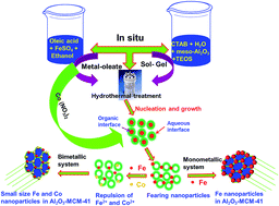
In situ loading of mono and bimetallic nanoparticles in the framework of mesoporous Al2O3–MCM-41 and its effect on the photo-Fenton degradation of dyes and mixed dyes has been reported in the present study. The nanocomposites are synthesized by in situ sol–gel cum hydrothermal method where oleic acid has been used as capping agent for mono and bimetallic nanoparticles. Materials were characterized by high and low angle XRD, N2 sorption, and HRTEM to evaluate mesoporosity, morphology and textural properties. The photoluminescence (PL) study and band gap energy measurement reveals suppression of e and h+ recombination and semiconductor behaviour of bimetallic/Al2O3–MCM-41 in visible region. Both the processes of photo-Fenton and photocatalysis takes place over mesoporous Co–Fe/Al2O3–MCM-41 nanocomposite, which is found to be an efficient material with 100% efficiency for the degradation of dyes and mixed dyes (100 mg L−1) at pH 10 in just 60 minutes. Framework mesoporosity, nanoparticle morphology of the nanocomposite, semiconductor behavior, lowering of the electron–hole recombination and the formation of a large number of ˙OH radicals are the crucial factors for swift degradation of dyes and mixed dyes by mesoporous Co–Fe/Al2O3–MCM-41 nanocomposite.


1. Introduction

Mesoporous materials with regular geometry have been recently paid much attention owing to their great potential in practical applications such as catalysis, adsorption, separation, sensing, medical usage, ecology and nanotechnology. These applications are tailored because of its unique three dimensional structures, high surface area, tuneable and uniform pore channel and wide volumes.1–6 Among the mesoporous material, mesoporous silica (MCM-41) and metals/metal oxides modified MCM-41 have been treated as an efficient support and catalyst. The MCM-41 has been engineered by isomorphous substitution of metals such as Al, Fe, Ti, Cu, Zn, V, Co (ref. 7–9) and extra-framework modification by mesoporous Al2O3 and mesoporous MnO2NPs.10,11 The substitution and extra-framework modification of metals and metal oxides leads to MCM-41 as an excellent catalyst. The catalytic activity depends upon the morphology of the incorporated metals and metal oxides. The best option is zero dimensional nanoparticles which has large surface-to-volume ratio and high surface atomic activity.12 Fabrication of iron based nanoparticles is an important task. Nanoparticles/nanostructured Fe2O3 have been synthesized by hydrothermal method, precipitation method etc.13–15 But fabrication of Fe2O3/Fe nanoparticles by sol–gel method is difficult. Single metal nanoparticles have small surface area that cause poor photocatalytic performances due to insufficient contact with the reactants.16 In order to avoid this problem, now researchers are interested to fabricate metal nanoparticles (MNPs) within the mesoporous support network. But it is also difficult to fabricate MNPs within the mesoporous framework in one pot synthesis. The combination of sol–gel and hydrothermal process in a one pot method by using suitable template/structure-directing agent may be helpful to achieve nanoparticles within mesoporous support strategy. CTAB has been used as a structure directing agent for synthesis of mesoporous Al2O3–MCM-41 because creation longer micelle by the formation of NH4OH during synthesis in aqueous medium.17 Oleic acid has been treated as a capping agent for the synthesis of metal nanoparticles due to good ability and higher affinity to bind with the surfaces of the metal precursor forming metal–oleates that in turn regulate the nucleation and growth of the nanoparticles in organic medium.18 In the combination of aqueous and organic medium, the growth of metal particles in organic medium may slower and restrict to form nanoparticles. This phenomenon will be an innovative approach for synthesis metal nanoparticles into the mesoporous framework. Fabrication of Fe nanoparticle (in situ) into the mesoporous Al2O3–MCM-41 by using oleic acid as capping agent, cetyltrimethylammonium bromide (CTAB) as a surfactant and NH3 as a soft template could be a novel approach. Not merely Fe nanoparticles, the other monometallic (Co and Mn) and bimetallic (Co–Fe, Mn–Co and Fe–Mn) nanoparticles could be formed by the same method.

It has been noted that Fe/iron oxide nanoparticles have been used as a heterogeneous photo-Fenton and photocatalyst for the organic dyes degradation.14,15 The heterogeneous photo-Fenton activity of Fe/iron oxide nanoparticles can be enhanced by incorporating it with another mesoporous support material. For an example, mesoporous Fe3O4@SiO2 composite and Fe3O4@rGO@TiO2 have been treated as a photo-Fenton catalyst for methylene blue degradation.19,20 The photo-Fenton degradation of rhodamine B has been achieved by Fe supported bentonite and graphene oxide–amorphous FePO4.21,22 Fe–MIL-101 has been treated as a heterogeneous photo-Fenton catalyst for reactive dyes.23 Shao et al. synthesized α-Fe2O3–TiO2 for an efficient photo-Fenton degradation of organic pollutant.24 Iron modified Al2O3 nanoparticles pillared montmorillonite nanocomposite has been treated as a brilliant photo-Fenton catalyst for degradation phenolic compounds.25 Lam et al. investigated the catalytic photo-Fenton oxidation of organic II dyes on Fe/MCM-41.26 Mesoporous Fe/Al2O3–MCM-41 has been considered as an efficient photo-Fenton catalyst for degradation of dyes.11 The catalytic activity could be increased by modification of Fe nanoparticles into the mesoporous Al2O3–MCM-41. Firstly, increase in surface active sites by the presence of Fe nanoparticles. The presence of Fe nanoparticles in the framework of Al2O3–MCM-41 and H2O2 in the reaction media will facilitate the photo-Fenton activity efficiently. Secondly, the band gap energy of FeO is 2.4 eV,27,28 which can function as semiconducting oxides under visible light. By incorporation of Fe nanoparticles into the Al2O3–MCM-41, the band gap of Fe/Al2O3–MCM-41 nanocomposite may be reduced. So Fe/Al2O3–MCM-41 composite may be treated as an efficient semiconducting photocatalyst for any photocatalytic applications under visible light. Thirdly, Al2O3–MCM-41 has high surface area, it could act as an efficient catalytic support material. The combination of Al2O3 and MCM-41 is chemically favourable because the ionic radii of Al3+ (53.5 pm) and Si4+ (40 pm).29 It has been reported that in Al–MCM-41/Al2O3–MCM-41 the Al3+ ion either substitutes or coordinates with the Si4+ ion in MCM-41 through oxygen ions.11 These points are necessary and important for making Fe nanoparticles modified mesoporous Al2O3–MCM-41 as a heterogeneous photo-Fenton catalyst/photocatalyst for the degradation of dyes such as methylene blue (MB), Congo red (CR) and mixed dyes (methylene blue and Congo red). These organic dyes have adverse effect even at trace amount.30,31 The heterogeneous photo-Fenton/photocatalytic process is a very quick, low cost and environment friendly process for the degradation of organic dyes and mixed dyes.32,33 The degradation of dyes (MB, CR) and mixed dyes (MB + CR) on mesoporous Fe/Al2O3–MCM-41 nanocomposite by heterogeneous photo-Fenton/photocatalytic process will be more advantage. Hence not merely Fe, but Fe-like metals such as monometallic (Mn, Fe, Co) and bimetallic (Co–Fe, Mn–Co, Fe–Mn) nanoparticles modified Al2O3–MCM-41 nanocomposite can be considered as an effective heterogeneous photo-Fenton catalyst/photocatalyst for degradation of dyes and mixed dyes.

Herein, we report the in situ growth of mono and bimetallic nanoparticles on mesoporous Al2O3–MCM-41 by novel sol–gel cum hydrothermal method. The control concentration of oleic acid is responsible for formation of mono and bimetallic nanoparticles. A possible formation mechanism has been proposed based on the systematic investigation of the morphological evolution and growth processes of Fe nanoparticles into the mesoporous Al2O3–MCM-41 framework. Fabrication and semiconductor behavior of Mn, Fe, and Co/Al2O3–MCM-41 and Co–Fe, Mn–Co, Fe–Mn/Al2O3–MCM-41 nanocomposites are also highlighted in the present study. Furthermore, to demonstrate their potential application for dyes degradation, the as-synthesized materials are used as heterogeneous photo-Fenton catalyst and photocatalyst for degradation of dyes and mixed dyes at pH = 10 under visible light.

2. Materials and methods

2.1 Synthesis of MCM-41

MCM-41 is prepared by sol–gel cum hydrothermal method. 2.4 g of CTAB was dissolved in 120 mL of deionized water under ambient conditions. After complete dissolution, 8 mL of 32% aqueous NH3 was added. Stoichiometry amount of tetraethyl orthosilicate (TEOS, C8H20O4 Si, Aldrich, 99%) was added to the solution under vigorous stirring for 1 h. The precipitate was transferred into stainless steel autoclave and placed in a furnace for 20 h at 120 °C. The final product was filtered and dried at 70 °C for 12 h. The surfactant was removed from the product by calcining at 550 °C in air for 5 h.

2.2 In situ fabrication of mesoporous Al2O3–MCM-41

Mesoporous Al2O3–MCM-41 composite was synthesized by incorporating powder mesoporous Al2O3 into MCM-41 during the synthesis of MCM-41 by in situ, just before the addition of NH3. The sample is prepared at Si/Al ratio of 10. The precipitate was transferred into stainless steel autoclave and placed in a furnace for 20 h at 120 °C. The final product was filtered and dried at 70 °C for 12 h. The composite material is calcined at 550 °C for 5 h. The fabricated powder material is denoted as mesoporous Al2O3–MCM-41 composite.

2.3 In situ sol–gel cum hydrothermal fabrication of mono and bimetallic/Al2O3–MCM-41 nanocomposite

In a typical synthesis, 2.5 g of CTAB was added to 120 mL of H2O and stirred for 1 h and then 0.377 g of mesoporous Al2O3 was added to it. The mixture was continuously stirred by adding 10 mL aqueous NH3 after an hour. Then 8 mL of TEOS as silica source was added to the mixture and stirred for 2 h. Then 1.0 mmol of FeSO4·7H2O dissolved in ethanol and oleic acid was added. The total mixture was transferred into a stainless steel autoclave and put into a furnace for 20 h at 120 °C. The gel was washed with distilled water and ethanol, and further dried in an oven at 70 °C for 12 h. The powder was calcined at 500 °C for 5 h in air. This material is denoted as mesoporous Fe/Al2O3–MCM-41 nanocomposite. Similar procedure was adopted to synthesize Co/Al2O3–MCM-41 and Mn/Al2O3–MCM-41 samples by taking 1.0 mmol of cobalt(II) nitrate hexahydrate (Co(NO3)2·6H2O) and 1.0 mmol of manganese acetate tetrahydrate (C4H6MnO4·4H2O), respectively.

For synthesis of bimetallic/Al2O3–MCM-41, 0.5 mmol of Co (NO3)·6H2O and 0.5 mmol of FeSO4·7H2O are mixed in ethanol and oleic acid. The above synthesis procedure was followed to obtain Co–Fe/Al2O3–MCM-4, Fe–Mn/Al2O3–MCM-41 and Mn–Co/Al2O3–MCM-41 samples.

2.4 Characterization of materials

Transmission electron microscopy (TEM) images were obtained from Philips CM 200 transmission electron microscope with a LaB6 filament and equipped with an ultrathin objective lens and CCD camera. HRTEM images were recorded by using JEOL 3010 machine. Diffuse reflectance UV-visible (DRUV-Vis) spectra of the materials were performed by JASCO V-660 spectrophotometer equipped with 60 mm integrating sphere. The measurements were carried out at a band width of 5 nm and in the wavelength range of 200–800 nm at a scanning speed of 100 nm min−1. The powder X-ray diffraction patterns (PXRD) of the synthesized materials were obtained employing Bruker AXS D8 Advance diffractometer and Cu Kα (λ = 0.15408 nm) radiation. A scan rate of 2° min−1 was used to record higher angle reflections from 10 to 80°, and 0.01° s−1 scan rate for low angle reflections from 0.5 to 10°. The specific surface area, pore size and pore volume were measured by N2 sorption method at liquid nitrogen temperature (−196 °C) using Micromeritics ASAP 2020. The specific surface area and pore size distribution were estimated based on Brunauer–Emmett–Teller (BET) and Barrett–Joyner–Halenda (BJH) methods, respectively. X-ray photoelectron spectroscopy (XPS) measurements were performed using ESCA Probe TPD of Omicron Nanotechnology with polychromatic Mg Kα as the X-ray source ( = 1253.6 eV). All the binding energies were calibrated by using the adventitious carbon (C 1s at 285 eV) as a reference. The photoluminescence spectra were measured on a JASCO FP-6300 fluorescence spectrometer with an excitation at 350 nm light. The FTIR spectra of the samples were recorded with JASCO FTIR-4100 spectrophotometer in the range of 400–4000 cm−1 at room temperature using KBr as reference. The formation of ˙OH radical was studied by replacing phenol with 5 × 10⁻⁴ M terephthalic acid (TPA) and 2 × 10⁻³ M NaOH with the same amount of catalyst.

2.5 Photo-Fenton degradation process

Methylene blue (MB), Congo red (CR) and mixed dyes (MB + CR) used in this study were from Merck, India. A stock solution of 100 mg L−1 was prepared and suitably diluted to the required initial concentration. The photo-Fenton degradation experiment has been carried out by taking 20 mL of 100 mg L−1 dyes solution. The reaction was carried out in 0.02 g catalyst in 20 mL dye solution (1 g L−1) in the presence of visible light (40 W) in 60 minutes with addition 1.0 × 10⁻⁵ mole of H2O2 at pH = 10. The pH of the solution was monitored by Systronics μ pH system 361 with proper addition of 0.01 M HNO3 and/or NH4OH. The solution was centrifuged and analysed by JASCO V-660 UV-visible spectrophotometer. The maximum absorbance of methylene blue (MB) and Congo red (CR) occurs at 664 nm and 498 nm, respectively. The absorbance of mixed dyes (MB + CR) follows both at 664 and 498 nm. The photocatalytic mixed dyes degradation occurred in a similar way as photo-Fenton process, without H2O2. TOC (total organic carbon) analysis has been carried out by TOC analyzer (ANATOC).

3. Results and discussion

3.1 Electron microscopy

The representative HRTEM micrographs of MCM-41 and Al2O3–MCM-41 are shown in Fig. 1. The mesoporous silica exhibited highly ordered hexagonal array of pore structure with amorphous nature of typical MCM-41 materials,34 which is clearly seen in Fig. 1(a)–(c). The HRTEM micrographs of Al2O3–MCM-41 also show similar well-ordered hexagonal pores with amorphous nature, which are shown in Fig. 1(d)–(f). The in situ incorporation of mesoporous Al2O3 into the MCM-41 matrix, did not affect the orders of hexagonal pores. The sustainability of amorphous nature in Al2O3–MCM-41 is due to the presence of very less amount of crystalline Al2O3 in the highly amorphous MCM-41 matrix. The TEM and HRTEM images of Fe/Al2O3–MCM-41, Co/Al2O3–MCM-41 and Co–Fe/Al2O3–MCM-41 nanocomposites are shown in Fig. 2. The well-order Fe nanoparticles (particle size 8 nm) have been observed in TEM and HRTEM image of Fe/Al2O3–MCM-41 nanocomposite (Fig. 2(a) and (b)). The fringes with lattice spacing of 0.280 nm can be indexed to the (200) planes of FeO with the cubic structure. The selected area electron diffraction (SAED) pattern shown in Fig. 2(d) exhibits the diffraction rings (111), (200) and (220) of a cubic structure of FeO in Fe/Al2O3–MCM-41 nanocomposite.35,36 The Fig. 2(e) and (f) shows Co nanoparticles having particle size 9 nm in Al2O3–MCM-41 nanocomposite. In Fig. 2(g), the HRTEM lattice fringes show the d-spacing of 0.250 nm from the (111) plane of cubic CoO phase.37,38 The SAED pattern (Fig. 2(h)) of three diffraction peaks can be assigned to (111), (200), and (220) lattice planes, which are in good agreement with those of the corresponding standard pure rock salt CoO phase (JCPDS card 48-1719). The Fig. 2(i) and (j) shows the TEM and HRTEM micrographs of Co–Fe/Al2O3–MCM-41 nanocomposite. The Fe and Co nanoparticles (4 nm) are shown in Fig. 2(j). The lattice spacing of 0.255 nm between adjacent lattice planes in the image corresponds to the distance between two (111) crystal planes of CoO, while the lattice spacing of 0.285 nm should be assigned to the (200) plane of FeO present in the Co–Fe/Al2O3–MCM-41 nanocomposite (Fig. 2(k)). The SAED pattern (Fig. 2(l)) also revealed the presence of both FeO and CoO in Co–Fe/Al2O3–MCM-41 nanocomposite. The TEM, HRTEM and SAED pattern of MnO in Mn/Al2O3–MCM-41 nanocomposite is shown in the Fig. S1(a)–(c). This result indicates that the presence of Mn nanoparticles of size 12 nm in the network of Al2O3–MCM-41. The observed (111) and (200) planes correspond to the cubic MnO structure.39 Fig. S1(d)–(f) shows TEM, HRTEM and SAED image of Fe–Mn/Al2O3–MCM-41 nanocomposite, respectively. The Fe and Mn nanoparticles of size 6 nm have been observed. The SAED pattern also supports the presence of FeO and MnO nanoparticles in Fe–Mn/Al2O3–MCM-41 nanocomposite. The Mn and Co nanoparticles (particle size 7 nm) have been observed from HRTEM image and the planes (111), (200), (220) and (311) correspond to MnO and CoO in Mn–Co/Al2O3–MCM-41 nanocomposite, shown in Fig. S1(g)–(i). The formation of mono and bimetallic nanoparticles in the framework of Al2O3–MCM-41 has been well established.
image file: c6ra19923b-f1.tif
Fig. 1 The HRTEM images of MCM-41 (a) honeycomb structure, (b) well-ordered, and (c) SAED pattern. HRTEM images of Al2O3–MCM-41 (d) honeycomb structure, (e) hexagonal ordered and (f) SAED pattern of Al2O3–MCM-41.

image file: c6ra19923b-f2.tif
Fig. 2 Characteristics micrographs of Fe/Al2O3–MCM-41 nanocomposite (a) TEM image, (b, c) HRTEM images and (d) SAED pattern. The characteristics images of Co/Al2O3–MCM-41 material (e) TEM image, (f, g) HRTEM images and (h) SAED pattern. The representatives micrographs of Co–Fe/Al2O3–MCM-41 (i) TEM image, (j–k) HRTEM images and (l) SAED pattern.

3.2 Optical analysis

Fig. 3 delineates the UV-Vis diffuse reflectance spectra Fe, Co and Mn/Al2O3–MCM-41 and the Co–Fe, Fe–Mn and Mn–Co/Al2O3–MCM-41 nanocomposite. All the materials showed strong absorption band at 260 nm due to the ligand to metal charge transfer (LMCT) between surface oxygen and isolated metal ions such as Co2+, Mn2+ and Fe2+.40 Moreover, a strong absorption in the visible range is due to the red shift in the band gap transition of the samples. This ascribes the electronic interaction of Al2O3–MCM-41 with metallic center to the influence of charge delocalization. Thus, mesoporous Al2O3–MCM-41 with monometallic and bimetallic system is more responsive for the visible light. The red shifting is shifted from monometallic to bimetallic/Al2O3–MCM-41 is due to the high electronic interaction of more charged metallic center. The band gap energy of a semiconductor can be calculated by using the following equation.41
αhν = A(Eg)n
where α, ν, A, and Eg are the absorption coefficient, light frequency, proportionality constant, and band gap, respectively. The band transition depends upon the value of n, it is ½ for direct and 2 for indirect transition, respectively. The method to evaluate the value of n is given somewhere else.42 It has been found that all the material shows direct allowed transitions. The band gap energies of the all samples can be estimated from the plots of (αhν)2 versus photon energy (). The intercept of the tangent to the X axis would give a good approximation of the band gap energies for the synthesized products, as shown in Fig. 4. It is observed that the band gap energy of the Co/Al2O3–MCM-41 and Mn/Al2O3–MCM-41 nanocomposite is 3.2 eV and 3 eV, respectively (Fig. 4(a) and (b)). It has been also observed that the band gap energy of neat CoO and MnO is 5 eV and 4 eV, respectively.29,43 The band gap of Co/Al2O3–MCM-41 decreases to 3.2 eV while Co nanoparticles incorporate to the framework of Al2O3–MCM-41. This might be due to the formation of a localized state by mixing of Co 2p, Al 2p and Si 2p. Similarly, the band gap energy of Mn/Al2O3–MCM-41 nanocomposite decreases to 3 eV, which might be due to the formation of a localized state by mixing of Mn 2p, Al 2p and Si 2p. The band gap energy of Fe/Al2O3–MCM-41 nanocomposite is 2.05 (Fig. 4(d)) which is narrower than the band gap energy of pure FeO (2.4 eV) due the mixing of Fe 2p, Al 2p and Si 2p.27 That means the formation and combination of localized metallic orbitals has great impact to narrow the band gap energy. Tang et al. observed that the band gap of TiO2 decreases by incorporating ZnFe2O4–TiO2 nanoparticles within mesoporous MCM-41.44 But the bimetallic/Al2O3–MCM-41 nanocomposite system, the band gap energy decreases drastically as compared to monometallic system which resulting a red shift. The most interesting thing is that the band gap energy of bimetallic Mn–Co/Al2O3–MCM-41 nanocomposite (Fig. 4(c)) is 2.70 eV which is in visible region. This band gap narrowing (BGN) of 2.70 eV as compared to individual monometallic system Mn/Al2O3–MCM-41 (3 eV) and Co/Al2O3–MCM-41 (3.2 eV) is due to the mixing and overlapping of the impurity Mn and Co, which resulting an impurity states in between Mn and Co energy band.45 The presence of the charge impurities (Mn2+ and Co2+) leads to the formation of the localized state in the energy band gap. The Mn 2p and Co 2p localized states are formed close to the conduction band of Mn–Co/Al2O3–MCM-41 nanocomposite. The high concentration of Mn 2p and Co 2p states causes the band structures to perturb, resulting in the formation of BGN in Mn–Co/Al2O3–MCM-41 nanocomposite. In the Fig. 4(e) and (f), band gap narrowing of Fe–Mn/Al2O3–MCM-41 (1.93 eV) and Co–Fe/Al2O3–MCM-41 (1.89 eV) as compared to individual monometallic system is due to the similar reason as in Mn–Co/Al2O3–MCM-41 nanocomposite. Hence, the formation of semiconductor mono and bimetallic/Al2O3–MCM-41 nanocomposite has been established.

image file: c6ra19923b-f3.tif
Fig. 3 Diffuse reflectance UV-visible absorption spectra of (a) Co/Al2O3–MCM-41, (b) Mn/Al2O3–MCM-41, (c) Mn–Co/Al2O3–MCM-41, (d) Fe/Al2O3–MCM-41, (e) Fe–Mn/Al2O3–MCM-41, (f) Co–Fe/Al2O3–MCM-41 nanocomposites.

image file: c6ra19923b-f4.tif
Fig. 4 Plots of (αhν)2 vs. photon energy () for the band gap energy of (a) Co/Al2O3–MCM-41, (b) Mn/Al2O3–MCM-41, (c) Mn–Co/Al2O3–MCM-41, (d) Fe/Al2O3–MCM-41, (e) Fe–Mn/Al2O3–MCM-41, (f) Co–Fe/Al2O3–MCM-41 nanocomposites.

3.3 X-ray diffraction studies

The representative low angle XRD pattern for MCM-41, Al2O3–MCM-41 and Co–Fe/Al2O3–MCM-41 samples are shown in the Fig. 5. The three samples exhibited high intense d100 diffraction peak at low angle indicating the mesoporous nature of the materials.46 It is observed that the peak intensity (d100) slightly decreases from MCM-41 to Al2O3–MCM-41 and extensively decreases from Al2O3–MCM-41 to Co–Fe/Al2O3–MCM-41 nanocomposite. This is due to the incorporation of the mesoporous Al2O3 into the extra-framework of the MCM-41 and incorporation of the (Co & Fe) onto the surface of the Al2O3–MCM-41. The other three reflections indexed as d110, d200 and d210 are comparatively less intense. This suggests the presence of a periodic hexagonal arrangement of the channel.47 MCM-41 shows all above said reflections, indicating well-ordered hexagonal mesoporous channel. The (100) and (110) diffraction peaks related to Al2O3–MCM-41 are intact, indicating an addition of mesoporous Al2O3 powder into the MCM-41 which does not affect the mesoporosity and periodic hexagonal arrangement of the channel. This phenomenon is due to (i) the isomorphous of substitution of Si4+ by Al3+ in MCM-41 without affecting its mesoporosity and periodicity.48 and (ii) the coordination of Al3+ ions with Si4+ through oxygen leading to extra-framework modification of MCM-41.11 Incorporation of Fe and Co onto the mesoporous Al2O3–MCM-41 nanocomposite leads to the degeneration of structural order as shown by weak d100 plane and disappearance of reflections (110), (200) and (210) in Fig. 5(c). This is because some Co and Fe particles tend to block the pores of Al2O3–MCM-41 network. Similar behaviour can be predicted in all monometallic and bimetallic/Al2O3–MCM-41 nanocomposites.
image file: c6ra19923b-f5.tif
Fig. 5 Low angle X-ray diffractograms of (a) MCM-41, (b) Al2O3–MCM-41 and (c) Co–Fe/Al2O3–MCM-41 materials.

Fig. 6 shows the high angle XRD pattern of mesoporous Al2O3, mono and bimetallic/Al2O3–MCM-41 samples. Mesoporous γ-Al2O3 is crystalline in nature showing diffraction peaks at (440), (400) and (311).49 However, the diffraction peaks of γ-Al2O3 phase are very feeble in monometallic and bimetallic/Al2O3–MCM-41 systems. The metal incorporated Al2O3–MCM-41 samples are substantially amorphous in nature and do not show any X-ray reflections of the respective metal oxide phases. The broad band centred at 2θ = 22° can be assigned to the characteristic refection from amorphous SiO2 (JCPDS 29-0085). The XRD results indicate that Al2O3–MCM-41 matrix is highly amorphous in which cluster-like Fe, Co, Mn, Co–Fe, Mn–Co, Fe–Mn oxide species are embedded.


image file: c6ra19923b-f6.tif
Fig. 6 High angle X-ray diffractograms of (a) Al2O3, (b) MCM-41, (c) Co/Al2O3–MCM-41, (d) Mn/Al2O3–MCM-41, (e) Fe/Al2O3–MCM-41, (f) Mn–Co/Al2O3–MCM-41, (g) Fe–Mn/Al2O3–MCM-41, (h) Co–Fe/Al2O3–MCM-41 nanocomposites.

3.4 N2 sorption studies

The N2 sorption isotherms of monometallic (Fe, Co, Mn) and bimetallic (Co–Fe, Fe–Mn, Mn–Co)/Al2O3–MCM-41 nanocomposite systems are presented in Fig. 7. The entire materials show type-IV isotherm with H1 hysteresis. This isotherm represents the mesoporous behaviour of these materials.50 The adsorption step of all the materials centred in the relative pressure (P/P0) region from 0.1 to 0.5. This phenomenon indicates the presence of framework-confined mesopores or framework mesoporosity/intra-particle mesoporosity.51,52 The pore size distribution curves shown in Fig. 8 are mono modal and they occur in the narrow mesoporous range between 2–3 nm. This indicates the presence of framework mesoporosity in mesoporous mono and bimetallic/Al2O3–MCM-41 nanocomposite.
image file: c6ra19923b-f7.tif
Fig. 7 N2 sorption isotherms of Mn/Al2O3–MCM-41, Co–Fe/Al2O3–MCM-41, Fe– Mn/Al2O3–MCM-41, Fe/Al2O3–MCM-41, Mn–Co/Al2O3–MCM-41 and Co/Al2O3–MCM-41 nanocomposites.

image file: c6ra19923b-f8.tif
Fig. 8 Pore size distributions of Fe–Mn/Al2O3–MCM-41, Co–Fe/Al2O3–MCM-41 Fe/Al2O3–MCM-41, Co/Al2O3–MCM-41, Mn/Al2O3–MCM-41 and Mn–Co/Al2O3–MCM-41 nanocomposites.

The surface area, pore diameter, pore volume of samples are summarized in Table 1. These values are obtained from N2 sorption isotherms. The high specific surface area of Al2O3–MCM-41 as compared to MCM-41 indicates that the meso-Al2O3 is incorporated into the extra-framework of the MCM-41. In other words, the high texture mesoporous Al2O3 has been incorporated into the extra-framework of MCM-41 by coordination of Al3+ to Si4+. After incorporation of monometallic system such as Fe, Co and Mn nanoparticles into the surface of mesoporous Al2O3–MCM-41, the surface area decreases as compared to pure Al2O3–MCM-41. This is because some metals nanoparticles are blocking the mesoporous network. The bimetallic system, Co–Fe, Fe–Mn and Mn–Co nanoparticles incorporated Al2O3–MCM-41 nanocomposite show much less surface area as compared to monometallic/Al2O3–MCM-41. The pore blocking is more effective in the case of bimetallic materials compared to monometallic materials.

Table 1 Textural property of Al2O3, MCM-41, Al2O3–MCM-41, monometallic/Al2O3–MCM-41 nanocomposite and bimetallic/Al2O3–MCM-41 materials measured by BET method
Samples Surface area (m2 g−1) Pore volume (cm3 g−1) Pore diameter (nm) Average particle size (nm)
Al2O3 268 0.67 3.6
MCM-41 800 1.10 2.2
Al2O3–MCM-41 870 1.78 2.1
Fe/Al2O3–MCM-41 379 0.96 2.2 8.0
Co/Al2O3–MCM-41 321 0.95 2.2 9.0
Mn/Al2O3–MCM-41 308 0.94 2.2 12
Co–Fe/Al2O3–MCM-41 300 0.92 2.3 4.0
Mn–Co/Al2O3–MCM-41 275 0.84 2.2 7
Fe–Mn/Al2O3–MCM-41 265 0.79 2.3 6


3.5 XPS studies

XPS is an excellent technique to understand the oxidation state of the transition metal ion and the relative composition of the synthesized material. Fig. 9(a)–(e) shows the core level XPS spectra of Fe 2p, Co 2p, Si 2p, Al 2p and O 1s in mesoporous Co–Fe/Al2O3–MCM-41 nanocomposite. The electronic states of samples were investigated by XPS study. The binding energy of Al 2p in pure Al2O3 and Si 2p in pristine SiO2 is 75 and 103.5 eV, respectively.53,54 Incorporation of meso-Al2O3 into the framework of MCM-41 (mesoporous Al2O3–MCM-41) and the incorporation of Fe and Co into the mesoporous support Al2O3–MCM-41 is due to the higher shifting of Si 2p binding energy from 103.5 eV to 104.45 eV. The BEs of Si 2p shift towards higher value by nearly 1 eV might be due to the strong interaction between Al3+ and Si4+ through oxygen atom. The higher shift of BEs for Si 2p and lower shift for Al 2p (74.30 eV) might be due to the transfer of electrons from Si4+ to Al3+ and also suggesting some Si4+ ions is replaced by Al3+. It is concluded that mesoporous support Al2O3–MCM-41 consists of Si–O–Al network. The present study depicts that the binding energies (BEs) of Fe 2p3/2 and Al 2p3/2 at 709.41 eV and 74.18 eV, suggesting the presence of Fe in 2+ oxidation state and Al in 3+ oxidation state. It has been reported that the binding energy of Fe 2+ is 709 eV.55,56 The BEs of Fe 2p3/2 shifted towards higher value and the Al 2p3/2 lower values indicates that a strong metal–support interaction. The strong interaction might be due to transfer of electron from Fe2+ to Al3+. Moreover, the binding energy of CoO is 780.7 eV.37 But the binding energy of CoO is 781.8 eV in mesoporous Co–Fe/Al2O3–MCM-41 nanocomposite. The higher shifting of binding energy is due to strong interaction between Co2+ and Al3+. The electronic interaction leads to transfer of electron from Co2+ to Al3+. Conclusively, the mesoporous nanocomposite Co–Fe/Al2O3–MCM-41 nanocomposite consists of Fe–Co–Al–O–Si linkage. The BEs of O 1s was 532.63 eV which might be due close interaction of oxygen atom with Si compared to Al atoms.
image file: c6ra19923b-f9.tif
Fig. 9 XPS of (a) Fe 2p core-level spectrum, (b) Co 2p core level spectrum, (c) Si 2p spectrum, (d) representative Al 2p spectrum and (e) O 1s core-level spectrum of mesoporous Co–Fe/Al2O3–MCM-41 nanocomposite.

3.6 Growth mechanism of mesoporous Fe/Al2O3–MCM-41 and Co–Fe/Al2O3–MCM-41 nanocomposite

Size control of metallic nanoparticles depends on the growth mechanism. Ostwald ripening, phase transfer phenomena, hydrothermal, and orientation attachment are the vital processes that have been highlighted.57,58 These process require suitable capping agent, reaction environment and temperature for nucleation and growth of the nanoparticles. In that context, mesoporous Fe/Al2O3–MCM-41 and Co–Fe/Al2O3–MCM-41 nanocomposite have been synthesized via in situ sol–gel cum hydrothermal process. The role of oleic acid as capping agent, CTAB and optimum temperature are key factors for the formation of Fe and Co–Fe nanoparticles in the framework of mesoporous Al2O3–MCM-41. The entire growth mechanism is shown in Scheme 1. At the initial step, mixing of CTAB with H2O formed a micelle which is susceptible for allowing powder Al2O3 and TEOS, resulting in aggregation of the micelle with Al and Si. On the other hand Fe–oleate formed by mixing of oleic acid and ethanol. The oleic acid has hydrophilic –COOH functional group with long hydrophobic alkyl moiety. Nucleation and growth takes place in both cases of Fe-oleate and –Al–O–Si– (sol form) placed in a stainless steel autoclave. The carboxylate groups of oleic acid may form stable complexes with Fe2+, which prevent the fast growth of Fe particle.36 The replacement of sulphate to oleate may be the key step in slowing down particle growth. The high decomposition temperature Fe–oleate also stabilizes the primary growth of nanoparticles and prevents the Fe nanoparticles growth.59 This is because the oleate molecules with abundant hydroxyl groups are favorably adsorbed on the surface of the generated primary nanoparticles, forming large steric barriers and slowing down the growth rate of primary nanoparticles.60 Moreover, Fe–oleate and sol –Al–O–Si– may generate both organic interface and aqueous interface, respectively. The Fe particle in organic interface may fear the aqueous interface which enable the slow growth Fe particle and prevent the agglomeration. This may be called as “fearing nanoparticles” mechanism. These phenomenon aids to prevent the agglomeration of Fe nanoparticles as well as sol–gel –Al–O–Si–. The Fe nanoparticles are formed in the framework mesoporous Al2O3–MCM-41 by removing oleate molecule and CTAB through calcination. Likewise Co–Fe nanoparticles/Al2O3–MCM-41 is formed by mixing of both precursors (Scheme 1). It has been investigated that the particle size of bimetallic Co–Fe is smaller as compared to monometallic Fe (Table 1). This may be due to the repulsion of similar charge Fe2+ and Co2+ which helps to restrict the growth of nanoparticles. This phenomenon may be called as “similar charge bimetallic repulsion”. Thus, other mono and bimetallic/Al2O3–MCM-41 nanocomposite formed by the similar procedure.
image file: c6ra19923b-s1.tif
Scheme 1 Growth mechanism of mesoporous Fe/Al2O3–MCM-41 and Co–Fe/Al2O3–MCM-41 nanocomposites.

3.7 Photo-Fenton achievement of dyes and mixed dyes

The photo-Fenton degradation of methylene blue (MB), Congo red (CR) and mixed (MB + CR) has been investigated by using mesoporous mono and bimetallic/Al2O3–MCM-41 nanocomposite. The reaction is carried out in the presence of visible light (400 W Xeon source), pH 10 and 1.0 × 10⁻⁶ mole of H2O2. The concentration of methylene blue (MB), Congo red (CR) and mixed dyes (MB + CR) are 100 mg L−1 and catalyst amount is 1.0 g L−1. The photo-Fenton degradation of methylene blue is proceeded very quickly and achieving 100% methylene blue degradation within 60 minute. The methylene blue degradation with different catalysts is shown in Fig. 10. The photo-Fenton degradation order is Co–Fe/Al2O3–MCM-41 > Fe–Mn/Al2O3–MCM-41 > Mn–Co/Al2O3–MCM-41 > Fe/Al2O3–MCM-41 > Co/Al2O3–MCM-41 > Mn/Al2O3–MCM-41 > Al2O3–MCM-41> MCM-41. The high degradation of methylene blue (MB) indicates the decrease in absorbance intensity. The similar trend appeared in the degradation of Congo red (CR) and mixed dyes (MB + CR), which is shown in the Fig. 11 and 12, respectively. The percentage of photo-Fenton degradation of methylene blue, Congo red and mixed methylene blue and Congo is described in Table 2.
image file: c6ra19923b-f10.tif
Fig. 10 UV-Vis spectra of the solutions recorded after photo-Fenton degradation methylene blue (MB). The experiment is carried out on different catalyst in visible light. The concentration of the standard methylene blue solution used is 100 mg L−1. The degradation process is carried out by using 1.0 g L−1 of catalyst, 1.0 × 10⁻⁶ mole of H2O2 for 60 minutes.

image file: c6ra19923b-f11.tif
Fig. 11 UV-Vis spectra of the solutions recorded after photo-Fenton degradation of Congo red (CR). The degradation process is carried out by using different 1.0 g L−1 of catalyst in a 100 mg L−1 Congo red solution. The reaction is accelerated by adding 1.0 × 10⁻⁶ mole of H2O2 in visible light for 60 minute.

image file: c6ra19923b-f12.tif
Fig. 12 UV-Vis spectra of the solutions recorded after photo-Fenton degradation of mixed methylene blue and Congo red (MB + CR). The degradation process is carried out by using different 1.0 g L−1 of catalyst in a 100 mg L−1 (MB + CR) solution. The reaction is accelerated by adding 1.0 × 10⁻⁶ mole of H2O2 in visible light for 60 minutes.
Table 2 Photo-Fenton degradation (%) of methylene blue (MB), Congo red (CR), mixed dyes (MB + CR) and photocatalytic degradation of mixed dyes. The (%) of TOC removal in photo-Fenton mixed dyes (MB + CR) process has also entrenched
Catalysts Photo-Fenton methylene blue (MB) degradation (%) Photo-Fenton Congo red (CR) degradation (%) Photocatalytic mixed dyes (MB + CR) degradation (%) Photo-Fenton mixed dyes (MB + CR) degradation (%) TOC removal (%) in photo-Fenton mixed dyes (MB + CR)
MCM-41 40 30 40 44 15
Al2O3–MCM-41 50 40 53 55 25
Mn/Al2O3–MCM-41 85 66 75 88 70
Co/Al2O3–MCM-41 87 75 79 90 78
Fe/Al2O3–MCM-41 89 78 81 93 82
Mn–Co/Al2O3–MCM-41 95 85 85 97 87
Fe–Mn/Al2O3–MCM-41 97 90 88 99 88
Co–Fe/Al2O3–MCM-41 100 95 90 100 92


The photocatalytic degradation of mixed dyes has been studied (Fig. 13) in order to compare with the photo-Fenton activity. The photocatalytic process is progressed without H2O2 whereas the photo-Fenton process with H2O2. It has been observed from Fig. 13 and Table 2, the photocatalytic mixed dyes degradation is low as compared to photo-Fenton process. Hence, the photocatalytic process progressed solely whereas the photo-Fenton process occurring via photocatalysis.


image file: c6ra19923b-f13.tif
Fig. 13 UV-Vis spectra of the solutions recorded after photocatalytic degradation of mixed methylene blue and Congo red (MB + CR). The degradation process is carried out by using different 1.0 g L−1 of catalyst in a 100 mg L−1 (MB + CR) solution. The reaction is accelerated by adding 1.0 × 10⁻⁶ mole of H2O2 in visible light for 60 minutes.

The TOC removal of mixed dyes (MB + CR) in the case of photo-Fenton degradation is shown in the Table 2. It is observed that 92% of TOC removal occurred in Co–Fe/Al2O3–MCM-41 nanocomposite, indicating that the reaction intermediates are converted into CO2 and H2O. The highest mineralization is due to the presence of nanoparticles Co and Fe in mesoporous Al2O3–MCM-41 support, H2O2 and visible light. This trends decreases from bimetallic/Al2O3–MCM-41 to monometallic/Al2O3–MCM-41 which is due to the unavailability of reacting surface. The very low mineralization occurred in Al2O3 and Al2O3–MCM-41 which showing TOC removal 15% and 25%, respectively. This is because of the absence of metallic surface, the photo-Fenton process will not operate. In absence of metallic surface only adsorption process may occur in presence of H2O2 and visible light. This percentage may be small if it is pure adsorption process in absence of H2O2 and visible light. The presence of H2O2 and visible light, less decomposition of mixed dyes happened due to least decomposition H2O2 by visible light.

3.7.1 Dual mechanism for degradation of dyes and mixed dyes. The degradation of dyes (MB, CR) and mixed dyes (MB + CR) are proceeding by both photo-Fenton and photocatalysis process (dual process). This is because the catalysts exhibits both metallic center as well as semiconductor behavior. The dual mechanism process is initiated by the action of hydroxyl radicals formed during the reaction, which is schematically shown in Scheme 2. In the photo-Fenton system, the degradation of dyes and mixed dyes is progressed in the presence mesoporous Co–Fe/Al2O3–MCM-41 nanocomposite, H2O2 and visible light. The oxidation and reduction reaction is occurred from Co2+–Fe2+/Al2O3–MCM-41 to Co3+–Fe3+/Al2O3–MCM-41 nanocomposite which helps in generating ˙OH radicals. The ˙OH radicals are reacted with the dyes molecule and produced degradation product. It has been observed that the catalyst possesses semiconductor property. The electron and hole are generated when catalytic surface is exposed to visible light. The active species ˙OH radicals are formed when hole (h+) reacts with H2O and photogenerated electron reacts with dissolved O2. The generated ˙OH radicals reacts with dyes and mixed dyes produced degraded product. Hence, the dual mechanism is operating in the dyes and mixed dyes degradation process. Moreover, photocatalytic process is progressed in photo-Fenton system.
image file: c6ra19923b-s2.tif
Scheme 2 Mechanistic pathways of dyes and mixed dyes degradation by mesoporous Co–Fe/Al2O3–MCM-41 nanocomposite in visible light.
3.7.2 Factor affecting the photo-Fenton activity. The degradation of dyes such as methylene blue (MB), Congo red (CR) and mixed dyes (MB + CR) has been achieved by (a) the role of particle size and surface morphology, (b) formation of a high amount of hydroxyl radicals, and (c) lowering the electro-hole recombination.
3.7.2.1 Role of particle size and surface morphology. Particle size has a great deal of importance in the field of catalysis. The small particle (nanoparticle) will act as an efficient catalyst because of high surface to volume ratio. Generally, the surface area increases with decreasing particle size, which generate more active site. This phenomenon aids to increase the catalytic activity. Another thing is that if the nanoparticles are in supported with highly porous network, then the surface area increases as compared to neat nanoparticles. These properties of material help to increase the greater active site for the accommodation of a substrate molecule. In the present study, the photo-Fenton degradation of dyes and mixed dyes strongly depends on the particle size of mono and bimetallic nanoparticles in the framework of mesoporous Al2O3–MCM-41. The particle size of the mono and bimetallic nanoparticles has been calculated from a HRTEM image by ImageJ software is shown in Table 1. It is observed that the particle size of the Co and Fe is very less in mesoporous Co–Fe/Al2O3–MCM-41 nanocomposite which lead to increase the surface area (Table 1). Hence, the dyes degradation activity is more (100%) as compared to other bimetallic/Al2O3–MCM-41 nanocomposite. Moreover, this may be due to the small particle size of Co and Fe (4 nm) of Co–Fe/Al2O3–MCM-41 nanocomposite. This leads to formation of more reactive surface and acceleration of the mass transfer which resulting in more catalytic activity.61 The migrating time of photoinduced charge carriers from inner to surfaces takes much less time which helps to increase the catalytic activity. The monometallic/Al2O3–MCM-41 nanocomposite has high surface area than the bimetallic system, but the photo-Fenton and photocatalytic activity is less as compared to bimetallic/Al2O3–MCM-41 nanocomposite. This is due to the large particle size monometallic system as compared to bimetallic system. It has been observed that the mesoporous Al2O3, MCM-41 and Al2O3–MCM-41 have high surface as compared to mono and bimetallic/Al2O3–MCM-41 nanocomposite. But due to lack of metallic active site, the dyes degradation activity is very less. Mesoporosity which leads to increase the surface area and metallic nanoparticles which leads to increase the catalytic active sites are vital factors for an efficient dyes degradation.
3.7.2.2 Generation of the hydroxyl radical. The hydroxyl radical has an important role for the decomposition of the dyes, and it is necessary to investigate the amount of hydroxyl radicals produced by each catalyst. This analysis is performed by using terephthalic acid (TA) as a probe molecule.62 In this method, TA was attacked by a ˙OH radical, forming 2-hydroxyterephthalic acid (TAOH), which gives a fluorescence signal at 426 nm.63,64 Fig. 14 depicts the fluorescent signal of bimetallic/Al2O3–MCM-41 nanocomposite after reacting with TA solution. It has been observed that generation of the hydroxyl radical is proportional to the PL intensity.65 Hence, higher the generation of hydroxyl radicals indicates the higher yield of TAOH and resulting more intense fluorescence peak. It has been observed that mesoporous Co–Fe/Al2O3–MCM-41 nanocomposite has highest intensity which confirm the high production of ˙OH radical compared to other bimetallic system. The dyes degradation performance of a mesoporous nanocomposite follows the order: Co–Fe/Al2O3–MCM-41 > Fe–Mn/Al2O3–MCM-41 > Mn–Co/Al2O3–MCM-41, which is strongly consistent with fluorescence intensity.
image file: c6ra19923b-f14.tif
Fig. 14 Photoluminescence responses of mesoporous bimetallic/Al2O3–MCM-41 nanocomposite under visible light irradiation for 60 min in 5 × 10⁻⁵ M basic solution of terephthalic acid.

3.7.2.3 Lowering of electron–hole recombination. It has been investigated that the mono and bimetallic/Al2O3–MCM-41 nanocomposite have semiconductor property. In this study, it is understood that photocatalytic reaction is progressed in photo-Fenton system. Hence, efficient photocatalytic reaction leads to high photo-Fenton process. The photoluminescence emission results from the radiative recombination of excited electrons and holes. Hence, it is an essential requirement of a good photocatalyst to have minimum electron–hole recombination. To learn the recombination of charge carriers, PL studies of synthesized materials have been undertaken. PL emission intensity is directly related to recombination of excited electrons and holes.66 The photoluminescence spectra of synthesized photocatalyst are shown in Fig. 15. The result shows that bimetallic system has less PL intensity as compared to monometallic, which is due to the less electron–hole recombination in bimetallic system. This is because of small size and more metallic charged species are available in bimetallic system which leads to accumulate extra electrons onto it and forget to recombine with the hole. In bimetallic system, mesoporous Co–Fe/Al2O3–MCM-41 nanocomposite has the lowest PL intensity. The lowest intensity is due to the least electron–hole recombination. The least electron–hole recombination is because of some generated electrons are engaged to reduce the highly charge species Co2+ and Fe2+ in Co–Fe/Al2O3–MCM-41. Hence, the maximum number of holes can be take part in the photocatalytic system which leads to high production of ˙OH radicals for photocatalytic dyes and mixed dyes degradation. Furthermore, the lowest PL intensity of mesoporous Fe–Co/Al2O3–MCM-41 nanocomposite is due to the small size of the Fe and Co within Al2O3–MCM-41 as compared to other bimetallic system.
image file: c6ra19923b-f15.tif
Fig. 15 Photoluminescence spectra of mesoporous mono and bimetallic/Al2O3–MCM-41 nanocomposite, MCM-41 and Al2O3–MCM-41.

3.7.2.4 Evidence and stability of photo-Fenton degradation of methylene blue. The FTIR spectra of pure methylene blue, Co–Fe/Al2O3–MCM-41 (after MB degradation) and neat Co–Fe/Al2O3–MCM-41 are shown in the Fig. 16(a), (b) and (c), respectively. The two peaks appear at 2816, 2720 cm−1 which represent the stretching vibration of –CH– aromatic and –CH3 methyl groups of pure methylene blue. The spectra range from 1591 to 1363 cm−1, are assigned to the aromatic ring structures in methylene blue.67 The peak at 1170 cm−1 is related to the C[double bond, length as m-dash]C skeleton of the aromatic rings. The absence of methylene blue signature peaks in Fig. 16(b), indicating the complete mineralization of methylene blue on the surface of mesoporous Co–Fe/Al2O3–MCM-41 nanocomposite. The IR spectrum of pure mesoporous Co–Fe/Al2O3–MCM-41 nanocomposite is shown Fig. 16(c) for comparison. The IR spectrum of Co–Fe/Al2O3–MCM-41 after MB degradation (Fig. 16(b)) is exactly similar to neat Co–Fe/Al2O3–MCM-41 (Fig. 16(c)), indicating the stability of the Co–Fe/Al2O3–MCM-41 nanocomposite catalyst after methylene blue degradation. The Fig. 16(d) shows the 100% degradation methylene blue (MB), Congo red (CR) and mixed dyes (MB + CR) on mesoporous Co–Fe@Al2O3–MCM-41 nanocomposite leading to colourless solution at pH = 10.
image file: c6ra19923b-f16.tif
Fig. 16 FTIR spectra of (a) pure methylene blue, (b) methyl blue degraded on Co–Fe/Al2O3–MCM-41 in comparison with (c) Co–Fe@Al2O3–MCM-41 material; (d) colour change after degradation of 100 mg L−1 methylene blue dye using 0.02 g of Co–Fe@Al2O3–MCM-41 at pH = 10.

4. Conclusions

The novelty of the present investigation is the formation of Fe, Co, Mn, Co–Fe, Mn–Co and Fe–Mn nanoparticles in the framework of mesoporousAl2O3–MCM-41 by using oleic acid and CTAB as size controlling (capping agent) and morphology controlling (surfactant) agents, respectively. Both photo-Fenton and photocatalysis (dual) process is operating for the efficient degradation of dyes and mixed dyes over all the mono and bimetallic nanocomposite system. Mesoporous Co–Fe/Al2O3–MCM-41 nanocomposite showed 100% dyes and mixed dyes degradation in 60 min. The high activity of mesoporous Co–Fe/Al2O3–MCM-41 nanocomposite is ascribed to the presence of highly active mono and bimetallic nanoparticles and semiconductor behavior and mesoporosity in mono and bimetallic/Al2O3–MCM-41 nanocomposite. Moreover, (a) nanostructure morphology of catalysts, (b) high surface area due to intraparticle mesoporosity, (c) lowering of electron–hole recombination and (d) swift generation of a large amount of hydroxyl radicals are the key factors which make mesoporous Co–Fe/Al2O3–MCM-41 nanocomposite an efficient versatile photo-Fenton catalyst. It is concluded from the study that Co–Fe/Al2O3–MCM-41, a multifunctional nanocomposites material can be explored for treatment of wastewater containing dyes, phenolic compounds and other organic pollutants by photo-Fenton process.

Acknowledgements

Amaresh Chandra Pradhan thanks IIT Madras for Postdoctoral Fellowship. The instrumental facilities established under the FIST Scheme of SERC division of DST, Ministry of Science and Technology, New Delhi, have been very helpful to carry out this work. Mr A. Narayanan and Mrs S. Srividya carried out BET and XRD measurements.

References

  1. S. Biswas, B. Dutta, K. Mullick, C.-H. Kuo, A. S. Poyraz and S. L. Suib, ACS Catal., 2015, 5, 4394–4403 CrossRef CAS.
  2. C. Gunathilake, M. S. Kadanapitiye, O. Dudarko, S. D. Huang and M. Jaroniec, ACS Appl. Mater. Interfaces, 2015, 7, 23144–23152 CAS.
  3. D. Stoeckel, D. Wallacher, G. A. Zickler, M. Thommes and B. M. Smarsly, Langmuir, 2015, 31, 6332–6342 CrossRef CAS PubMed.
  4. S. Alberti, G. J. A. A. Soler-Illia and O. Azzaroni, Chem. Commun., 2015, 51, 6050–6075 RSC.
  5. F. Razzaghi, C. Debiemme-Chouvy, F. Pillier, H. Perrot and O. Sel, Phys. Chem. Chem. Phys., 2015, 17, 14773–14787 RSC.
  6. C. W. Kim, M. J. Choi, S. Lee, H. Park, B. Moon, Y. S. Kang and Y. S. Kang, J. Phys. Chem. C, 2015, 119, 24902–24909 CAS.
  7. J. González-Rivera, J. Tovar-Rodríguez, E. Bramanti, C. Duce, I. Longo, E. Fratini, I. R. Galindo-Esquivel and C. Ferrari, J. Mater. Chem. A, 2014, 2, 7020–7033 Search PubMed.
  8. B. Li, N. Wu, K. Wu, J. Liu, C. Han and X. Li, RSC Adv., 2015, 5, 16598–16603 RSC.
  9. X. Collard, P. Louette, S. Fiorilli and C. Aprile, Phys. Chem. Chem. Phys., 2015, 17, 26756–26765 RSC.
  10. A. C. Pradhan, B. Nanda, K. M. Parida and G. R. Rao, J. Phys. Chem. C, 2015, 119, 4145–14159 Search PubMed.
  11. A. C. Pradhan and K. M. Parida, J. Mater. Chem., 2012, 22, 7567–7579 RSC.
  12. R. Narayanan and M. A. El-Sayed, J. Am. Chem. Soc., 2003, 125, 8340–8347 CrossRef CAS PubMed.
  13. Z. Xu, C. Huang, L. Wang, X. Pan, L. Qin, X. Guo and G. Zhang, Ind. Eng. Chem. Res., 2015, 54, 4593–4602 CrossRef CAS.
  14. Y. Zhao, F. Pan, H. Li, T. Niu, G. Xu and W. Chen, J. Mater. Chem. A, 2013, 1, 7242–7246 CAS.
  15. G. K. Pradhan, N. Sahu and K. M. Parida, RSC Adv., 2013, 3, 7912–7920 RSC.
  16. R. Jusoh, A. A. Jalil, S. Triwahyono and N. H. N. Kamarudin, RSC Adv., 2015, 5, 9727–9736 RSC.
  17. Q. Cai, Z.-S. Luo, W.-Q. Pang, Y.-W. Fan, X.-H. Chen and F.-Z. Cui, Chem. Mater., 2001, 13, 258–263 CrossRef CAS.
  18. C. Moya, X. Batlle and A. Labarta, Phys. Chem. Chem. Phys., 2015, 17, 27373–27379 RSC.
  19. X. Tan, L. Lu, L. Wang and J. Zhang, Eur. J. Inorg. Chem., 2015, 2015, 2928–2933 CrossRef CAS.
  20. X. Yang, W. Chen, J. Huang, Y. Zhou, Y. Zhu and C. Li, Sci. Rep., 2015, 5, 10632 CrossRef PubMed.
  21. Y. Gao, Y. Wang and H. Zhang, Appl. Catal., B, 2015, 178, 29–36 CrossRef CAS.
  22. S. Guo, G. Zhang and J. C. Yu, J. Colloid Interface Sci., 2015, 448, 460–466 CrossRef CAS PubMed.
  23. T. A. Vu, G. H. Le, C. D. Dao, L. Q. Dang, K. T. Nguyen, P. T. Dang, H. T. K. Tran, Q. T. Duong, T. V. N. Gun and D. Lee, RSC Adv., 2014, 4, 41185–41194 RSC.
  24. P. Shao, J. Tian, B. Liu, W. Shi, S. Gao, Y. Song, M. Ling and F. Cui, Nanoscale, 2015, 7, 14254–14263 RSC.
  25. A. C. Pradhan, G. B. Varadwaj and K. M. Parida, Dalton Trans., 2013, 42, 15139–15149 RSC.
  26. F. L. Y. Lam and X. Hu, Catal. Commun., 2007, 8, 2125–2129 CrossRef CAS.
  27. F. Tran and P. Blaha, Phys. Rev. Lett., 2009, 102, 226401–226404 CrossRef PubMed.
  28. R. Gillen and J. Robertson, J. Phys.: Condens. Matter, 2013, 25, 165502 CrossRef PubMed.
  29. N. N. Greenwood and A. Earnshaw, Chemistry of the Elements, Reed Educational and Professional Publishing Ltd, U.K., 1998 Search PubMed.
  30. A. C. Pradhan, B. Nanda, K. M. Parida and M. Das, Dalton Trans., 2011, 40, 7348–7356 RSC.
  31. J. Luan, B. Pan, Y. Paz, Y. Li, X. Wu and Z. Zou, Phys. Chem. Chem. Phys., 2009, 11, 6289–6298 RSC.
  32. Y. Wang, R. Priambodo, H. Zhang and Y.-H. Huang, RSC Adv., 2015, 5, 45276–45283 RSC.
  33. L. Zhou, Y. Shao, J. Liu, Z. Ye, H. Zhang, J. Ma, Y. Jia, W. Gao and Y. Li, ACS Appl. Mater. Interfaces, 2014, 6, 7275–7285 CAS.
  34. I. Miletto, E. Bottinelli, G. Caputo, S. Coluccia and E. Gianotti, Phys. Chem. Chem. Phys., 2012, 14, 10015–10021 RSC.
  35. A. Lak, M. Kraken, F. Ludwig, A. Kornowski, D. Eberbeck, S. Sievers, F. J. Litterst, H. Weller and M. Schilling, Nanoscale, 2013, 5, 12286–12295 RSC.
  36. Z. Li, Y. Ma and L. Qi, CrystEngComm, 2014, 16, 600–608 RSC.
  37. X. He, X. Song, W. Qiao, Z. Li, X. Zhang, S. Yan, W. Zhong and Y. Du, J. Phys. Chem. C, 2015, 119, 9550–9559 CAS.
  38. B. Liu, L. Ma, L. C. Ning, C. J. Zhang, G. P. Han, C. J. Pei, H. Zhao, S. Z. Liu and H. Q. Yang, ACS Appl. Mater. Interfaces, 2015, 7, 6109–6117 CAS.
  39. C.-H. Kuo, I. M. Mosa, S. Thanneeru, V. Sharma, L. Zhang, S. Biswas, M. Aindow, S. P. Alpay, J. F. Rusling, S. L. Suib and J. He, Chem. Commun., 2015, 51, 5951–5954 RSC.
  40. H. Praliaud, S. Mikhailenko, Z. Chajar and M. Primet, Appl. Catal., B, 1998, 16, 359–374 CrossRef CAS.
  41. J. Cao, B. Xu, H. Lin, B. Luo and S. Chen, Dalton Trans., 2012, 41, 11482–11490 RSC.
  42. J. Luan, S. Zheng, X. Hao, G. Luan, X. Wu and Z. Zou, J. Braz. Chem. Soc., 2006, 17, 1368–1376 CrossRef CAS.
  43. D. K. Kanan and E. A. Carter, J. Phys. Chem. C, 2012, 116, 9876–9887 CAS.
  44. A. Tang, Y. Deng, J. Jin and H. Yang, Sci. World J., 2012, 2012, 480527 Search PubMed.
  45. R. Camacho-Aguilera, Z. Han, Y. Cai, L. C. Kimerling and J. Michel, Appl. Phys. Lett., 2013, 102, 152106 CrossRef.
  46. C. P. Guthrie and E. J. Reardon, J. Phys. Chem. A, 2008, 112, 3386–3390 CrossRef CAS PubMed.
  47. T. A. Konovalova, Y. G. R. Schad and L. D. Kispert, J. Phys. Chem. B, 2001, 105, 7459–7464 CrossRef CAS.
  48. S. Li, Q. Xu, J. Chen and Y. Guo, Ind. Eng. Chem. Res., 2008, 47, 8211–8217 CrossRef CAS.
  49. K. M. Parida, A. C. Pradhan, J. Das and N. Sahu, Mater. Chem. Phys., 2009, 113, 244–248 CrossRef CAS.
  50. S. Brunaure, L. S. Deming, W. E. Deming and E. Teller, J. Am. Chem. Soc., 1940, 62, 723–1732 Search PubMed.
  51. P. T. Tanev and T. J. Pinnavaia, Chem. Mater., 1996, 8, 2068–2079 CrossRef CAS.
  52. K. W. Goyne, A. R. Zimmerman, B. L. Newalkar, S. Komarneni, S. L. Brantley and J. Chorover, J. Porous Mater., 2009, 9, 243–256 CrossRef.
  53. W. E. Morgan and J. R. V. Wazer, J. Phys. Chem., 1973, 77, 964–969 CrossRef CAS.
  54. K. Djebaili, Z. Mekhalif, A. Boumaza and A. Djelloul, J. Spectrosc., 2015, 2015, 1–16 CrossRef.
  55. C. R. Brundle, T. J. Chuang and K. Wandeelt, Surf. Sci., 1977, 68, 459–468 CrossRef CAS.
  56. N. S. McIntyre and D. G. Zetaruk, Anal. Chem., 1977, 49, 1521–1529 CrossRef CAS.
  57. D. G. X. Zhang and W. Gao, ACS Appl. Mater. Interfaces, 2013, 5, 9732–9739 Search PubMed.
  58. M. Raju, A. C. van Duin and K. A. Fichthorn, Nano Lett., 2014, 14, 1836–1842 CrossRef CAS PubMed.
  59. P. Shao, J. Tian, B. Liu, W. Shi, S. Gao, Y. Song, M. Ling and F. Cui, Nanoscale, 2015, 7, 14254–14263 RSC.
  60. S. R. Walsh, I. Rusakova and K. H. Whitmire, CrystEngComm, 2013, 15, 775–784 RSC.
  61. X. Zhong, L. Xiang, S. Royer, S. Valange, J. Barrault and H. Zhang, J. Chem. Technol. Biotechnol., 2011, 86, 970–977 CrossRef CAS.
  62. J. Zhou, Y. Zhang, X. S. Zhao and A. K. Ray, Ind. Eng. Chem. Res., 2006, 45, 3503–3511 CrossRef CAS.
  63. Y. Zhou, B. Xiao, S.-Q. Liu, Z. Meng, Z.-G. Chen, C.-Y. Zou, C.-B. Liu, F. Chen and X. Zhou, Chem. Eng. J., 2016, 283, 266–275 CrossRef CAS.
  64. D. Lu, Y. Zhang, S. Lin, L. Wang and C. Wang, J. Alloys Compd., 2013, 579, 336–342 CrossRef CAS.
  65. J. Zhang, J. Xi and Z. Ji, J. Mater. Chem., 2012, 22, 17700 RSC.
  66. G. Liu, P. Niu, L. Yin and H.-M. Cheng, J. Am. Chem. Soc., 2012, 134, 9070–9073 CrossRef CAS PubMed.
  67. Y.-Y. Lau, Y.-S. Wong, T.-T. Teng, N. Morad, M. Rafatullah and S.-A. Ong, RSC Adv., 2015, 5, 34206–34215 RSC.

Footnote

Electronic supplementary information (ESI) available. See DOI: 10.1039/c6ra19923b

This journal is © The Royal Society of Chemistry 2016
Click here to see how this site uses Cookies. View our privacy policy here.