Preparation of honokiol with biodegradable nanoparticles for treatment of osteosarcoma

Yongzhong Cheng a, Songping Zhenga, Yan Tenga, Yagmur Muftuoglub, Chanjuan Zhaoc, Song Chenb, Xiang Gao*a and Chao You*a
aDepartment of Neurosurgery, State Key Laboratory of Biotherapy, West China Hospital, Sichuan University, Chengdu 610041, PR China. E-mail: xiangxianggao2008@163.com; chaoyouyouchao@163.com; Fax: +86 28 85164060; Tel: +86 28 85164063
bDepartment of Pharmacology, Yale School of Medicine, Yale University, New Haven, CT 06520, USA
cWest China Second University Hospital, Sichuan University, China

Received 26th August 2016 , Accepted 26th September 2016

First published on 28th September 2016


Abstract

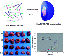
In previous studies, honokiol (Hon) has been demonstrated to have anti-tumorigenic effects. However, its poor water solubility and low bioavailability limit its potential as an orally-administered anti-cancer compound. This study presents work toward improving the water solubility and antitumor effects of honokiol through the use of self-assembling, biodegradable micelles of monomethoxy poly(ethylene glycol)–poly(ε-caprolactone) copolymer (MPEG–PCL). These honokiol-containing polymeric micelles (Hon/MPEG–PCL) release the compound slowly over an extended period of time. In addition, treatment with Hon/MPEG–PCL inhibits the growth of osteosarcoma cells and induces apoptosis of these cells more effectively than free honokiol in vitro. Further, in a nude mouse model of osteosarcoma, Hon/MPEG–PCL inhibits the growth of tumors, induces apoptosis, and inhibits angiogenesis more effectively than the same dose of free honokiol. This shows that Hon/MPEG–PCL enhances the antitumor effects of honokiol in vivo, and, as a result, shows great promise as a clinical treatment for osteosarcoma.


1 Introduction

Among children and adolescents around the world, osteosarcoma is the most common form of primary malignant bone cancer. Osteosarcoma ranks second in causing cancer-related deaths among individuals in these age groups, who usually succumb to metastases occurring in the lung.1–4 At present, treatment for osteosarcoma involves surgical resection of the tumor along with multi-agent chemotherapy; the use of adjuvant chemotherapy is shown to increase the five-year survival rate by approximately 60–70%, but survival rates among patients who relapse or who suffer from metastases remain low.5 Thus, there is a need for novel, more effective therapies to treat osteosarcoma.

Honokiol is a compound extracted from the magnolia tree that has been used for centuries in traditional Chinese and Japanese medicine in a variety of ways: to assuage anxiety, to treat thrombotic stroke, and to mitigate gastrointestinal discomfort,6 for example. Deeper analysis of the effects of this compound have also revealed that it carries cardioprotective,7,8 antimicrobial,9,10 anti-inflammatory,11,12 and neuroprotective13,14 properties. Additionally, researchers have found that honokiol demonstrates anti-neoplastic properties in vitro and in vivo against a number of different cancers.15–20 Following these studies and as a result of growing interest in the pharmacological properties of this compound, honokiol has been both synthesized and modified. Researchers have explored a number of delivery mechanisms – including oral, intravenous, and transdermal preparations – only to discover low bioavailability of honokiol using any modality.6 Hence, it is urgent to find a route to improve the absorption and effectiveness of honokiol.21,22

Advances in nanotechnology can provide breakthroughs for improving aqueous formulations of hydrophobic drugs.23–25 MPEG/PCL used in this study is one of the most common nano-matrices, and it is composed of poly(ε-caprolactone) (PCL) and polyethylene glycol (PEG). PEG is typically used in conjugation to PCL to prevent synthesis of PCL copolymers, and it is neither toxic nor immunogenic. The simple molecular architecture of MPEG–PCL allows for control of the size, thus preventing aggregation and promoting stability; these factors make MPEG–PCL an effective and appropriate candidate for the formation of nano-micelles in drug delivery.26–29 In addition, the high hydration capacity of MPEG/PCL allows it to regulate, or effectively modulate, the hydrophilicity of drugs like honokiol. In fact, a number of systems for drug delivery have already employed MPEG/PCL copolymer nanoparticles,30–34 and previous research demonstrates the advantage of using MPEG/PCL nanoparticles in the treatment of a number of different cancers, owing to its ability to deliver therapeutics that promote apoptosis, suppress angiogenesis, etc.35–39 Furthermore, Samyang Co., based in Korea, has commercialized the formulation of paclitaxel using MPEG–PCL.40

In this work, nano-micelles of MPEG–PCL containing honokiol (Hon/MPEG–PCL) were generated and characterized (Fig. 1), and the in vitro and in vivo anti-tumor properties of this formulation were investigated. Studies were conducted to assess the release of drug, the levels of cellular uptake, and the resulting apoptosis in vitro. In addition, the effects of this formulation on cell proliferation, angiogenesis, and apoptosis in vivo were also studied in detail. Our findings indicate that, in comparison to free honokiol, the formulation of Hon/MPEG–PCL boasts improved anti-tumor activity both in vitro and in vivo, making it a promising drug delivery system for treatment of osteosarcoma.


image file: c6ra21479g-f1.tif
Fig. 1 Preparation of the Hon/MPEG–PCL micelles. Hon/MPEG–PCL were prepared by self-assembly, as demonstrated in this schematic.

2 Materials and methods

2.1 Materials

Reagents were purchased from standard sources as available: honokiol and 3-(4,5-dimethylthiazol-2-yl)-2,5-diphenyl tetrazolium bromide (MTT) from Sigma (USA); Dulbecco's Modified Eagle's Medium (DMEM) and fetal bovine serum (FBS) from Gibco BRL (USA); methanol and acetic acid (HPLC grade) from Fisher Scientific (UK); dimethyl sulfoxide (DMSO) and acetone from KeLong Chemicals (China). Antibodies purchased include: rat anti-mouse CD31 polyclonal antibody (BD Pharmingen™, USA); rabbit anti-human ki67 antibody (Abcam, USA); rhodamine-conjugated secondary antibody (Abcam, USA).

To prepare MPEG(2000)–PCL(2000) diblock copolymer with a molecular weight of 4000, ring-opening of ε-caprolactone (ε-CL) was initiated with MPEG by combining MPEG and ε-CL in a dry, glass ampoule under nitrogen and by adding Sn(Oct)2 into the reaction vessel with mild agitation at 130 °C for 6 hours. For another 45 min, the reaction vessel was degassed under vacuum and cooled to room temperature under nitrogen. Both 1H nuclear magnetic resonance spectroscopy (1H-NMR, Varian 400 spectrometer, Varian, USA) and gel permeation chromatography (GPC, Agilent 110 HPLC, USA) (data not shown) were used to analyze and characterize the resulting MPEG–PCL copolymer. Its molecular weight was found to be 4010 (data not shown), and it was stored inside a desiccator before use. MPEG (molecular weight of 2000) (Sigma-Aldrich) was dried under vacuum in a one-necked flask and stirred for 90 min at 105 °C before use.

UMR106 cells (American Type Culture Collection) were maintained using standard procedures: cultured in DMEM with 10% heat-inactivated FBS and 100 μg mL−1 amikacin, unless specified otherwise, and stored in humidified incubator (37 °C and 5% CO2).

To conduct the in vivo testing and pharmacokinetic analyses, 6–8 week-old female BALB/c mice and female nude BALB/c mice were purchased (Laboratory Animal Center, Sichuan University). This institution houses male and female mice separately in a temperature-controlled environment (20–22 °C) with 12 hour light/dark cycles and a relative humidity of 50–60% with standard laboratory chow and tap water as needed. Animal experiments were performed according to the guidelines of the Animal Care and Use Committee of Sichuan University (Chengdu, Sichuan, China) and approved by the Animal Care and Use Committee of Sichuan University.

2.2 Preparation of MPEG–PCL and Hon/MPEG–PCL nano-micelles

Nanoparticles loaded with honokiol were generated using the method of self-assembly. Honokiol (20 mg) and MPEG–PCL (80 mg) were dissolved together in acetone (2 mL), and this mixture was transferred into water (4 mL), thus loading the MPEG–PCL nanoparticles with honokiol. Removal of acetone was accomplished by subjecting the solution to negative pressure at 55 °C, and the resulting Hon/MPEG–PCL nano-micelles were lyophilized and stored at 4 °C before use. Empty nano-micelles were prepared in parallel.

2.3 Characterization of the Hon/MPEG–PCL nano-micelles

Lyophilized Hon/MPEG–PCL (10 mg) was dissolved in acetonitrile (0.1 mL) to determine values for drug loading (DL, eqn (1)) and encapsulation efficiency (EE, eqn (2)), as described below. The amount of solubilized honokiol was determined using standard HPLC procedures (Shimadzu LC-20AD, Japan).
 
image file: c6ra21479g-t1.tif(1)
 
image file: c6ra21479g-t2.tif(2)

Average particle size and average zeta potential were determined from three independent experiments using dynamic light scattering (Malvern Nano-ZS 90), making sure to maintain 25 °C throughout the analysis. To assess morphology, nano-micelles were observed under a transmission electron microscope (H-6009IV, Hitachi, Japan); samples were diluted in distilled water, placed on a nitrocellulose-covered copper grid, negatively stained using phosphotungstic acid, and allowed to dry at room temperature.

2.4 Calculation of the kinetics of honokiol release in vitro and pharmacokinetics study

To calculate the efficiency of release of honokiol from Hon/MPEG–PCL, Hon/MPEG–PCL nano-micelles (0.5 mL) were placed in a dialysis bag (MWCO of 3.5 kDa), which was then incubated in fresh PBS (pH 7.4) with 0.5% w/v Tween-80 (30 mL). Release of honokiol into the incubation medium was quantified using HPLC (294 nm), repeated in three independent experiments to report the mean value ± SD.

The mice were fasted overnight prior to drug administration and divided into 2 groups: a free Hon treatment group and a Hon/MPEG–PCL micelle treatment group. Free Hon was dissolved in Tween-80 and dehydrated alcohol (1[thin space (1/6-em)]:[thin space (1/6-em)]1, v/v), whereas Hon/MPEG–PCL micelles were dissolved in normal saline. Following intravenous administration of 25 mg kg−1 of free Hon or Hon/MPEG–PCL micelles, blood was collected through removal of the eyeball at different time points (5 mice at each time point). The plasma was separated and extracted with ethyl acetate, and the supernatant fluid was collected and evaporated to dryness. The dry residues were dissolved in methanol for HPLC analysis.

2.5 Cell viability assay

UMR106 cells (5 × 10³ per well) were plated in 96-well plates and, after 24 hours, were washed once with complete DMEM and treated with various concentrations of free honokiol (F-H) or Hon/MPEG–PCL (H–M) in DMEM to determine cell viability after 24 hours or 48 hours using the MTT method with intensity measurements taken at 570 nm (OPTImax, Molecular Dynamics). Values are presented as a percentage compared to untreated cells at 100% survival; mean values from at least three independent experiments are reported as mean ± SD.

2.6 Cellular uptake assay

UMR106 cells (2.5 × 10⁵ cells per well) were plated into six-well plates and, after 24 hours, were incubated for 3–6 hours with fresh DMEM containing free coumarin (50 ng mL−1), drug-encapsulated micelles (100 ng mL−1), or one of two controls: normal saline or empty MPEG–PCL micelles. Cells were then washed thrice with PBS. A confocal laser scanning microscope (LSM 710, Zeiss, Germany) equipped with a multi-argon laser allowed for analysis of coumarin distribution due to the intrinsic green fluorescence of coumarin, the fluorescence intensity of which was confirmed using flow cytometry (BD, USA). From these experiments, we extrapolated to understand the distribution of free honokiol versus Hon/MPEG–PCL.

2.7 Cellular apoptosis assay

UMR106 cells, cultured as described above using 100 units per mL penicillin/streptomycin, were seeded in six-well plates (2.5 × 10⁵ cells per well) and, after 24 hours, were incubated with DMEM containing F-H or H–M. Apoptosis was quantified 48 hours later using the Hoechst method with a fluorescence microscope (400× magnification).

2.8 In vivo mouse model of tumor growth

Six-to-eight week-old female nude BALB/c mice, housed in groups of five in top-filtered cages, were maintained with standard procedures: a regular diet and acidified water lacking antibiotics. Subcutaneous injections into the right flank of a suspension of UMR106 cells (100 μL, 5 × 10⁶ cells) were performed. Tumors were allowed to grow to a mean tumor diameter of about 6 mm. Tumor-bearing mice were then randomly assigned to four different groups to receive daily intravenous tail vein injections for 10 days: normal saline (NS), empty MPEG–PCL micelles (EM), free honokiol (50 mg kg−1), or Hon/MPEG–PCL (50 mg kg−1). When mice in the control NS group could no longer carry the tumor burden, all mice were sacrificed via dislocation of cervical vertebrae; tumors were harvested immediately for further analysis.

2.9 CD31 and ki67 assays

Expression of CD31 and ki67 in tumor tissue was detected by immunofluorescence. Tumor tissue was cut into 5 μm slices; these sections were fixed in acetone, washed with PBS, blocked with goat serum, and incubated separately with rat anti-mouse CD31 polyclonal antibody (1[thin space (1/6-em)]:[thin space (1/6-em)]50) or rabbit anti-human ki67 antibody (1[thin space (1/6-em)]:[thin space (1/6-em)]100) overnight at 4 °C. Sections were washed thrice with PBS and incubated with a rhodamine-conjugated secondary antibody. Microvessel density and expression of ki67 were detected using a microscope to determine the number of microvessels and the index of cell proliferation.

2.10 Tunnel assay

Tumor tissues were fixed over 24 hours in PBS containing 4% paraformaldehyde, washed with water for 4 hours, soaked overnight in 70% ethanol, and dehydrated using graded ethanol, before being embedded in paraffin to cut 3 to 5 μm slices. To quantify the presence of apoptotic UMR106 cells in the tumor samples, a commercially-available kit was used to label deoxynucleotidyl transferase-mediated dUTP nick ends (TUNEL, Promega, Madison, Wisconsin, United States). Analyses were carried out according to specifications of the manufacturer, and samples were confirmed using fluorescence microscopy (400× magnification).

2.11 Statistical analyses

SPSS15.0 (SPSS Inc., Chicago, IL, USA) was used for ANOVA calculations on multiple group comparisons and for the Student's t-test on comparisons between two groups, with p < 0.05 as the cut-off for statistical significance.

3 Results and discussion

In this study, through the application of a new nano-delivery system, an aqueous formulation Hon/MPEG–PCL was obtained and the anticancer activity and mechanism of this formation on osteosarcoma cancer was explored in vitro and in vivo.

3.1 Preparation and characterization of the Hon/MPEG–PCL micelles

We generated a highly soluble formation of honokiol using MPEG–PCL through single-step nano-precipitation, as described here and as reported previously.36 Different ratios of honokiol-to-MPEG–PCL were tested, and nano-micelles with lower DL values were found to have greater stability and greater EE values (Table 1).
Table 1 Characterization of honokiol-containing micelles
Sample HK[thin space (1/6-em)]:[thin space (1/6-em)]MPEG–PCL DL (%) EE (%) Size (nm) PDI Stability (h)
S1 0[thin space (1/6-em)]:[thin space (1/6-em)]100 0 0 21.9 ± 0.3 0.079 ± 0.003 >240
S2 1[thin space (1/6-em)]:[thin space (1/6-em)]99 0.98 ± 0.05 99.03 ± 0.39 23.7 ± 0.5 0.109 ± 0.007 >240
S3 5[thin space (1/6-em)]:[thin space (1/6-em)]95 4.97 ± 0.13 99.05 ± 0.35 25.7 ± 0.3 0.151 ± 0.012 96
S4 10[thin space (1/6-em)]:[thin space (1/6-em)]90 9.90 ± 0.17 99.03 ± 0.29 29.4 ± 2.7 0.152 ± 0.023 72
S5 15[thin space (1/6-em)]:[thin space (1/6-em)]85 14.82 ± 0.47 98.66 ± 1.87 32.7 ± 3.3 0.168 ± 0.03 48
S6 20[thin space (1/6-em)]:[thin space (1/6-em)]80 19.22 ± 0.32 96.1 ± 5.18 35.7 ± 2.7 0.273 ± 0.032 24
S7 25[thin space (1/6-em)]:[thin space (1/6-em)]75 0.1


The developed Hon/MPEG–PCL micelles were further characterized with respect to their particle size or polydispersity index (PDI), their zeta potential, and their overall morphology. The spectrum of size distribution shows that the Hon/MPEG–PCL micelles are monodisperse and have a narrow distribution for particle size (PDI = 0.12 ± 0.03), and their average diameter is approximately 35.7 ± 2.7 nm in aqueous phase (Fig. 2A). The average zeta potential is −3.3 ± 0.7 mV (Fig. 2B). Furthermore, using transmission electron microscopy (TEM), spherical Hon/MPEG–PCL micelles were measured to be about 21.2 ± 0.9 nm in drying phase (Fig. 2C). Since the structure of these amphiphilic particles is usually loose in solution, the size measured by dynamic light scattering is always a little larger than that measured by TEM.


image file: c6ra21479g-f2.tif
Fig. 2 Characterization of Hon/MPEG–PCL. (A) Size distribution of Hon/MPEG–PCL; (B) zeta potential of Hon/MPEG–PCL; (C) TEM image of Hon/MPEG–PCL; (D) photos of PBS solution, a mixture of Hon and MPEG–PCL in PBS solution and Hon/MPEG–PCL in PBS solution (from left to right).

Free honokiol and Hon/MPEG–PCL micelles showed different dissolution behaviors in normal saline solution (Fig. 2D). Compared with free honokiol, which forms a cloudy orange suspension, the solution of Hon/MPEG–PCL micelles was transparent and clear, indicative of complete dispersion in aqueous solution. Finally, from the drug release experiments, we found that honokiol nano-micelles release the drug over a longer timeframe than free honokiol (Fig. 3A).


image file: c6ra21479g-f3.tif
Fig. 3 In vitro release study and pharmacokinetics study. (A) The in vitro release profile of free honokiol (F-H) and Hon/MPEG–PCL (H–M) were examined using dialysis. H–M releases honokiol over a longer timeframe than free honokiol. (B) The concentration–time curve of honokiol in plasma both free honokiol and Hon/MPEG–PCL.

To determine whether Hon/MPEG–PCL nano-micelles improved the pharmacokinetics of Hon in vivo, the pharmacokinetics of free Hon and Hon/MPEG–PCL micelles were studied in mice. BALB/c mice were intravenously administered free Hon or Hon/MPEG–PCL micelles (25 mg kg−1 Hon), and blood was collected after different time intervals. The results of the pharmacokinetics are shown in Fig. 3B. For the MPEG–PCL micelle-encapsulated Hon, the Tmax, T1/2, and Cmax were 5 min, 7.9 h, and 227.738 mg L−1, respectively. For the free Hon, the Tmax, T1/2, and Cmax were 5 min, 0.47 h, and 179.751 mg L−1, respectively. These results suggest that encapsulation of Hon in MPEG–PCL micelles improved the T1/2 and Cmax of Hon in vivo.

3.2 Antitumor activity of honokiol micelles in vitro

We examined the anti-tumor effects of free honokiol versus Hon/MPEG–PCL using the MTT method (Fig. 4). As expected, we found that, as the concentration of honokiol increases, the metabolic activity of the cells gradually decreases. And the half maximal inhibitory concentration (IC50) of free honokiol and Hon/MPEG–PCL were 14.5 μg mL−1 and 10 μg mL−1 at 48 h. Additionally, we found that, under the same conditions of drug concentration and incubation time, the activity of UMR106 cells is reduced more quickly in response to Hon/MPEG–PCL than in response to free honokiol.
image file: c6ra21479g-f4.tif
Fig. 4 Cytotoxicity of Hon/MPEG–PCL on UMR106 cells. Cytotoxicity studies of free honokiol (F-H) and Hon/MPEG–PCL (H–M) in UMR106 cells after treatment for (A) 24 hours or (B) 48 hours using the MTT method. F-H and H–M efficiently decreases the viability of UMR106 cancer cells, and H–M enhances this cytotoxicity.

To investigate how nano-micelles enhance the antitumor activity of free honokiol, we characterized uptake of drug by osteosarcoma cells. In these experiments, for the purpose of visual understanding, we used coumarin as an experimental drug, since it carries intrinsic green fluorescence. After incubation with free coumarin or coumarin-containing nanoparticles, the fluorescence intensity of coumarin contained in the cells was observed under a fluorescence microscope (Fig. 5). We found that, at the same drug concentration, the fluorescence intensity of cells treated with coumarin-containing nanoparticles was higher than that of cells treated with free coumarin. This indicates that use of nano-micelles enhanced drug update. We confirmed these observations using flow cytometry (Fig. 6). These experiments show that the fluorescence intensity of UMR106 cells treated with coumarin-containing nanoparticles exceeds that of cells treated with free coumarin.


image file: c6ra21479g-f5.tif
Fig. 5 Enhanced coumarin uptake into UMR106 cells. Drug uptake of (A) normal saline, (B) empty MPEG–PCL micelles, (C) free coumarin, and (D) coumarin-containing MPEG–PCL by UMR106 cells after treatment for 6 hours, as observed with fluorescence microscopy.

image file: c6ra21479g-f6.tif
Fig. 6 Enhanced coumarin uptake into UMR106 cells. Drug uptake of free coumarin (F-C) and coumarin-containing MPEG–PCL (C–M) in UMR106 cells after treatment for (A) 3 hours and (B) 6 hours, as detected by flow cytometry.

To further explore the effects of free honokiol versus Hon/MPEG–PCL on cellular apoptosis, we utilized the Hoechst method, in which bright spots represent apoptotic cells (Fig. 7). These experiments show that treatment with honokiol enhances the apoptosis of UMR106 cells compared to treatment with normal saline or empty MPEG–PCL micelles. Furthermore, Hon/MPEG–PCL induces a greater level of cellular apoptosis than free honokiol.


image file: c6ra21479g-f7.tif
Fig. 7 The effects of Hon/MPEG–PCL on cell apoptosis in UMR106 cells. UMR106 cells were incubated with (A) normal saline, (B) empty MPEG–PCL micelles, (C) free honokiol, and (D) Hon/MPEG–PCL for 48 hours, with cellular apoptosis determined using the Hoechst method.

In summary, honokiol inhibits the proliferation of UMR106 cells by promoting cellular apoptosis, and honokiol-loaded nano-micelles strengthens this anti-tumor effect compared to free honokiol.

3.3 Antitumor activity of honokiol micelles in an in vivo mouse model

Using a nude mouse model, we evaluated the ability of Hon/MPEG–PCL micelles to hinder osteosarcoma in vivo compared to treatment with normal saline, MPEG–PCL micelles, and free honokiol. Growth curves of tumors from each treatment group are reported (Fig. 8A), showing that treatment with Hon/MPEG–PCL nano-micelles promotes a smaller tumor volume compared to the three other treatments. These findings were statistically significant: p < 0.01 vs. control, p < 0.01 vs. MPEG–PCL, and p < 0.05 vs. free honokiol. This observation is further supported by analysis of tumor mass among the four treatment groups (Fig. 8B), with representative examples from each treatment group shown (Fig. 8C). It is also important to note that treatment with free honokiol or with Hon/MPEG–PCL was well tolerated by the mice in this study, as indicated by steady body weight of mice in these two experimental groups, as well as that of mice in the two control groups (Fig. 8D).
image file: c6ra21479g-f8.tif
Fig. 8 Antitumor effect of Hon/MPEG–PCL in vivo. Female nude mice were injected with UMR106 cells and randomly assigned to four treatment groups to intravenously receive: normal saline (NS), empty MPEG–PCL micelles (EM), free honokiol (F-H) and Hon/MPEG–PCL (H–M). (A) Graph tracking tumor development. (B) Graph tracking mass of tumors. (C) Representative photos of tumors from each treatment group on day 27. (D) Body weight of mice from each treatment group.

3.4 Mechanisms of anti-tumor activity

To better understand the mechanisms of action employed by free honokiol and honokiol nanoparticles, we also studied cell proliferation, apoptosis, and tumor angiogenesis in response to treatment.

The proliferation of osteosarcoma cells was analyzed through the expression of ki67, a marker of proliferation. In these experiments, we found that tumor cell proliferation was significantly inhibited after treatment with free honokiol or with Hon/MPEG–PCL, with the latter more effectively suppressing proliferation than free honokiol (Fig. 9).


image file: c6ra21479g-f9.tif
Fig. 9 Ki67 immunohistochemical analysis. Tumor sections from the groups treated intravenously with (A) normal saline, (B) empty MPEG–PCL micelles, (C) free honokiol, and (D) Hon/MPEG–PCL were subjected to immunohistochemistry to detect ki67. The Hon/MPEG–PCL micelles inhibited osteosarcoma cancer growth more effectively than free honokiol.

The TUNEL Apoptosis Detection Kit allowed for quantification of cellular apoptosis in the four treatment groups (Fig. 10): normal saline (3 ± 2.1%), empty micelles (4 ± 1.4%), free honokiol (26 ± 5.5%) and Hon/MPEG–PCL (43 ± 6.3%). Thus, apoptosis is significantly increased by treatment with free honokiol or with Hon/MPEG–PCL as compared to the control groups, and Hon/MPEG–PCL induces a higher percentage of apoptosis than free honokiol.


image file: c6ra21479g-f10.tif
Fig. 10 TUNEL assay. Tumor sections from the groups treated intravenously with (A) normal saline, (B) empty MPEG–PCL micelles, (C) free honokiol, and (D) Hon/MPEG–PCL were subjected to the TUNEL assay to assess apoptosis. The group treated with the Hon/MPEG–PCL showed the greatest level of apoptosis among cells, indicating that drug delivery using MPEG–PCL improves the anti-osteosarcoma properties of honokiol.

Expression of CD31 in tumor samples from mice in each treatment group allowed for analysis of resulting microvessel density (MVD) (Fig. 11). The Hon/MPEG–PCL treatment resulted in a striking inhibition of angiogenesis in the tumors. MVD after treatment with Hon/MPEG–PCL was found to be 34.7 ± 11.3, significantly lower than that after treatment with free honokiol (116.3 ± 13.7, p < 0.01), empty micelles (237.2 ± 32.7, p < 0.01), or normal saline (241.6 ± 24.3, p < 0.01). These results indicate that honokiol-containing nano-micelles also function to inhibit angiogenesis, thus hindering osteosarcoma in vivo.


image file: c6ra21479g-f11.tif
Fig. 11 CD31 immunohistochemical analysis. Tumor sections from the groups treated intravenously with (A) normal saline, (B) empty MPEG–PCL micelles, (C) free honokiol, and (D) Hon/MPEG–PCL were subjected to immunohistochemistry to detect CD31. Treatment with Hon/MPEG–PCL results in a more striking inhibition of angiogenesis in the osteosarcoma tumors compared to treatment with free honokiol.

Taken together, Hon/MPEG–PCL suppresses tumor growth in vivo by inhibiting cell proliferation, facilitating cell apoptosis, and hindering angiogenesis in tumors.

Honokiol, a compound emerging from ancient Chinese and Japanese medicine, has recently been shown to have anti-tumor properties.41 In particular, honokiol has been shown to exert anti-metastatic properties in studies of osteosarcoma, causing rapid cell death in tumors.21 However, its poor water solubility and low bioavailability dampen its future as a clinical treatment for cancer. Development of an aqueous formulation for honokiol can help overcome these considerations, and the study presented here features a formulation using MPEG–PCL to improve the solubility and uptake of this drug.

Upon delivery with MPEG–PCL nano-micelles, honokiol inhibits the growth of osteosarcoma more effectively than free honokiol both in vitro and in vivo. Hon/MPEG–PCL more severely inhibits the growth of UMR106 cells and induces a greater level of apoptosis in vitro, due to improved delivery and uptake of the compound. Moreover, in vivo, Hon/MPEG–PCL inhibits the growth of osteosarcoma tumors effectively than free honokiol. This study also identifies additional mechanisms of action employed by Hon/MPEG–PCL: suppression of cell proliferation in vivo. Taken together, these results suggest that clinical application of Hon/MPEG–PCL offers great potential in treating osteosarcoma. Further, MPEG–PCL provides a convenient platform to improve the solubility and bioavailability of anti-cancer compounds like honokiol, especially given how well this biodegradable formulation is tolerated in in vivo studies.

4 Conclusions

Biodegradable honokiol-containing nano-micelles were prepared, and their therapeutic potential against UMR106 osteosarcoma was assessed in vitro and in vivo. Loading honokiol into polymeric micelles increases the water solubility and cellular uptake of this compound compared to free honokiol. In addition, Hon/MPEG–PCL releases honokiol over a sustained timeframe in vitro. Furthermore, in comparison to free honokiol, Hon/MPEG–PCL more effectively suppresses growth of tumors in vitro using cell-based experiments and in vivo using a mouse model of osteosarcoma. Mechanisms employed by honokiol and Hon/MPEG–PCL include enhancing apoptosis and inhibiting cell proliferation and angiogenesis.

Acknowledgements

The National Natural Sciences Foundation of China (81502165) supported this study.

References

  1. X. W. Liu, Y. Zi, L. B. Xiang and T. Y. Han, Int. J. Clin. Exp. Med., 2015, 8, 37–44 Search PubMed.
  2. M. Kansara, M. W. Teng, M. J. Smyth and D. M. Thomas, Nat. Rev. Cancer, 2014, 14, 722–735 CrossRef CAS PubMed.
  3. J. C. Clark, C. R. Dass and P. F. Choong, J. Cancer Res. Clin. Oncol., 2008, 134, 281–297 CrossRef CAS PubMed.
  4. N. Jaffe, Cancer Treat. Res., 2009, 152, 239–262 Search PubMed.
  5. M. L. Broadhead, J. C. Clark, D. E. Myers, C. R. Dass and P. F. Choong, Sarcoma, 2011, 2011, 959248 CrossRef PubMed.
  6. A. Woodbury, S. P. Yu, L. Wei and P. Garcia, Front. Neurol., 2013, 4, 130 Search PubMed.
  7. V. B. Pillai, S. Samant, N. R. Sundaresan, H. Raghuraman, G. Kim, M. Y. Bonner, J. L. Arbiser, D. I. Walker, D. P. Jones, D. Gius and M. P. Gupta, Nat. Commun., 2015, 6, 6656 CrossRef CAS PubMed.
  8. V. B. Pillai, N. R. Sundaresan, K. Kim, S. Samant, J. Arbiser and M. P. Gupta, Circulation, 2012, 126, A16685 Search PubMed.
  9. K. Y. Ho, C. C. Tsai, C. P. Chen, J. S. Huang and C. C. Lin, Phytother. Res., 2001, 15, 139–141 CrossRef CAS PubMed.
  10. E. J. Choi, H. I. Kim, J. A. Kim, S. H. Jun, S. H. Kang, D. J. Park, S. J. Son, Y. Kim and O. S. Shin, Appl. Microbiol. Biotechnol., 2015, 99, 4387–4396 CrossRef CAS PubMed.
  11. B. H. Kim and J. Y. Cho, Acta Pharmacol. Sin., 2008, 29, 113–122 CrossRef CAS PubMed.
  12. C. K. Chiang, M. L. Sheu, K. Y. Hung, K. D. Wu and S. H. Liu, Kidney Int., 2006, 70, 682–689 CrossRef CAS PubMed.
  13. S. Chakravarty, S. Maitra, R. G. Reddy, T. Das, P. Jhelum, S. Kootar, W. D. Rajan, A. Samanta, R. Samineni, S. Pabbaraja, S. G. Kernie, G. Mehta and A. Kumar, Sci. Rep., 2015, 5, 14134–14145 CrossRef CAS PubMed.
  14. T. Singh, N. A. Gupta, S. Xu, R. Prasad, S. E. Velu and S. K. Katiyar, Oncotarget, 2015, 6, 21268–21282 CrossRef PubMed.
  15. S. Cheng, M. Welty, I. Eliaz, V. Castillo and D. Sliva, Cancer Res., 2014, 74, 3203 Search PubMed.
  16. C. W. Chi and H. T. Hsu, Cancer Res., 2014, 74, 5494 Search PubMed.
  17. D. B. Avtanski, A. Nagalingam, P. Kupusamy, N. K. Saxena and D. Sharma, Cancer Res., 2013, 73, 299 Search PubMed.
  18. Z. Yuan, H. Liu, F. Yan, Y. Wang, M. L. Gou, C. L. Nie, Z. Ding, S. Lai, Y. Zhao, X. Y. Zhao, J. Li, H. X. Deng, Y. Mao, L. Chen, Y. Q. Wei and X. Zhao, Cancer Sci., 2009, 100, 1757–1766 CrossRef CAS PubMed.
  19. D. Subramaniam, S. Ponnurangam, D. Kwatra, G. Kaushik, P. Ramamoorthy, S. Ramalingam, O. Tawfik, S. J. Weir, S. Padhye, D. A. Dixon, S. Umar, R. A. Jensen and S. Anant, Cancer Res., 2015, 75, 1893 Search PubMed.
  20. P. Steinmann, D. K. Walters, M. J. Arlt, I. J. Banke, U. Ziegler, B. Langsam, J. Arbiser, R. Muff, W. Born and B. Fuchs, Cancer, 2012, 118, 2117–2127 CrossRef CAS PubMed.
  21. Y. Xie, Q. D. Long, Q. J. Wu, S. Shi, M. Dai, Y. W. Liu, L. Liu, C. Y. Gong, Z. Y. Qian, Y. Q. Wei and X. Zhao, RSC Adv., 2012, 2, 7759–7771 RSC.
  22. A. Schroeder, D. A. Heller, M. M. Winslow, J. E. Dahlman, G. W. Pratt, R. Langer, T. Jacks and D. G. Anderson, Nat. Rev. Cancer, 2012, 12, 39–50 CrossRef CAS PubMed.
  23. K. Bourzac, Nature, 2012, 491, S58–S60 CrossRef PubMed.
  24. M. Hamidi, A. Azadi, P. Rafiei and H. Ashrafi, Crit. Rev. Ther. Drug Carrier Syst., 2013, 30, 435–467 CrossRef CAS PubMed.
  25. J. Woodcock, J. Griffin, R. Behrman, B. Cherney, T. Crescenzi, B. Fraser, D. Hixon, C. Joneckis, S. Kozlowski, A. Rosenberg, L. Schrager, E. Shacter, R. Temple, K. Webber and H. Winkle, Nat. Rev. Drug Discovery, 2007, 6, 437–442 CrossRef PubMed.
  26. X. Gao, S. M. Wang, B. L. Wang, S. Y. Deng, X. X. Liu, X. Zhang, L. Luo, R. R. Fan, M. L. Xiang, C. You, Y. Q. Wei, Z. Y. Qian and G. Guo, Biomaterials, 2015, 50, 646–658 CrossRef PubMed.
  27. J. Bidone, A. P. Melo, G. C. Bazzo, F. Carmignan, M. S. Soldi, A. N. Pires and E. L. Senn, Mater. Sci. Eng., C, 2009, 29, 588–593 CrossRef CAS.
  28. L. Yang, Z. Zhao, J. Wei, A. E. Ghzaoui and S. Li, J. Colloid Interface Sci., 2007, 314, 470–477 CrossRef CAS PubMed.
  29. O. Benny, O. Fainaru, A. Adini, F. Cassiola, L. Bazinet, I. Adini, E. Pravda, Y. N. S. Koirala, G. Corfas, R. J. DAmato and J. Folkman, Nat. Biotechnol., 2008, 26, 799–807 CrossRef CAS PubMed.
  30. S. Mura, J. Nicolas and P. Couvreur, Nat. Mater., 2013, 12, 991–1003 CrossRef CAS PubMed.
  31. P. Gao, X. Nie, M. Zou, Y. Shi and G. Cheng, J. Antibiot., 2011, 64, 625–634 CrossRef CAS PubMed.
  32. C. Y. Zhang, Y. Q. Yang, T. X. Huang, B. Zhao, X. D. Guo, J. F. Wang and L. J. Zhang, Biomaterials, 2012, 33, 6273–6283 CrossRef CAS PubMed.
  33. Y. Dong and S. S. Feng, Biomaterials, 2007, 28, 4154–4160 CrossRef CAS PubMed.
  34. C. Y. Gong, S. Y. Deng, Q. J. Wu, M. L. Xiang, X. W. Wei, L. Li, X. Gao, B. L. Wang, L. Sun, Y. Chen, Y. Li, L. Liu, Z. Y. Qian and Y. Q. Wei, Biomaterials, 2013, 34, 1413–1432 CrossRef CAS PubMed.
  35. X. Gao, F. J. Zheng, G. Guo, X. X Liu, R. R. Fan, Z. Y. Qian, N. Huang and Y. Q. Wei, J. Mater. Chem. B, 2013, 1, 5778–5790 RSC.
  36. X. Gao, B. L. Wang, Q. J. Wu, X. W. Wei, F. J. Zheng, K. Men, H. S. Shi, N. Huang, Y. Q. Wei and C. Y. Gong, J. Biomed. Nanotechnol., 2015, 11, 578–589 CrossRef CAS PubMed.
  37. X. Gao, B. L. Wang, X. W. Wei, K. Men, F. J. Zheng, Y. F. Zhou, Y. Zheng, M. L. Gou, M. J. Huang, G. Guo, N. Huang, Z. Y. Qian and Y. Q. Wei, Nanoscale, 2012, 4, 7021–7030 RSC.
  38. X. M. Duan, P. Wang, K. Men, X. Gao, M. J. Huang, M. L. Gou, L. J. Chen, Y. Q. Wei and Z. Y. Qian, Nanoscale, 2012, 4, 2400–2407 RSC.
  39. L. Mu and S. S. Feng, J. Controlled Release, 2001, 76, 239–254 CrossRef CAS PubMed.
  40. X. L. Zheng, B. Kan, M. L. Gou, S. Z. Fu, J. Zhang, K. Men, L. J. Chen, F. Luo, Y. L. Zhao, X. Zhao, Y. Q. Wei and Z. Y. Qian, Int. J. Pharm., 2010, 386, 262–267 CrossRef CAS PubMed.
  41. L. E. Fried and J. L. Arbiser, Antioxid. Redox Signaling, 2009, 11, 1139–1148 CrossRef CAS PubMed.

Footnote

These authors are considered equal first authors.

This journal is © The Royal Society of Chemistry 2016
Click here to see how this site uses Cookies. View our privacy policy here.