Dual-functionalisation of gelatine nanoparticles with an anticancer platinum(II)–bisphosphonate complex and mineral-binding alendronate

Kambiz Farbod a, Alessandra Curci b, Mani Diba a, Tatiana Zinkevich c, Arno P. M. Kentgens c, Michele Iafisco d, Nicola Margiotta b and Sander C. G. Leeuwenburgh *a
aDepartment of Biomaterials, Radboud Institute for Molecular Life Sciences, Radboud University Medical Center, Philips van Leydenlaan 25, 6525 EX Nijmegen, The Netherlands. E-mail: sander.leeuwenburgh@radboudumc.nl
bDipartimento di Chimica, Università degli Studi di Bari Aldo Moro, Via E. Orabona 4, 70125 Bari, Italy
cDepartment of Solid State NMR, Institute for Molecules and Materials, Radboud University, Heyendaalseweg 135, 6525 AJ Nijmegen, The Netherlands
dInstitute of Science and Technology for Ceramics (ISTEC), National Research Council (CNR), Via Granarolo 64, 48018 Faenza, Italy

Received 6th August 2016 , Accepted 15th November 2016

First published on 22nd November 2016


Abstract

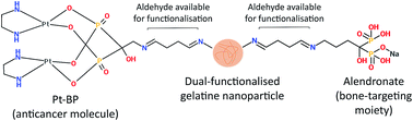
In order to improve the efficacy of therapeutic systems to treat bone tumours, novel drug delivery vehicles should be developed that have strong and specific affinity to mineralised tissue and at the same time are able to release anticancer molecules locally in a controlled and sustained manner. Recently, we developed mineral-binding gelatine nanoparticles with enhanced affinity to calcium phosphate by conjugating alendronate (ALN) molecules onto their surface. Herein, we have enhanced the functionality of these nanoparticles by rendering them potentially therapeutically active via covalent linking of an anticancer platinum–bisphosphonate (Pt–BP) complex. Different functionalisation schemes and molar ratios between reactants were screened and the effective functionalisation of gelatine nanoparticles with Pt–BP (or with both Pt–BP and ALN) was assessed. Our results revealed that the degree of functionalisation could be tailored by varying the molar ratio of Pt–BP and ALN relative to glutaraldehyde used as crosslinker. A sustained and tunable release of platinum as a function of the initial Pt–BP/ALN/glutaraldehyde molar ratio was achieved for both Pt–BP- and dual-functionalised gelatine nanoparticles. Moreover, dual-functionalised gelatine nanoparticles also displayed a high affinity to hydroxyapatite-coated surfaces thanks to the presence of ALN. Summarising, it was demonstrated that mineral-binding gelatine nanoparticles can be loaded with tailored amounts of anticancer molecules, which may benefit the development of bone-seeking carriers for targeted delivery of drugs to treat bone tumours.


Introduction

Bone is a frequent site for metastases, which develop in up to 70% of patients with prostate cancer and breast cancer,1 and in up to 30% of those with cancers of the lung, bladder and thyroid.2 Bone metastases are a health and economic burden as they are associated with numerous skeletal-related issues including pathological fractures, spinal cord compression, pain, need for radiation therapy or surgery to bone.3 Besides the devastating effect of cancer on bone tissue, current treatment modalities not specific to cancer cells result in damaging normal cells and healthy tissues.4 In fact, a major disadvantage of anticancer drugs is their lack of selectivity for tumour tissue, which causes severe side effects and results in low cure rates.5 For instance, therapy with platinum-based chemotherapeutic molecules (e.g. cisplatin (cis-diamminedichloridoplatinum(II))), which are a group of key drugs used in the treatment of several types of cancers, are associated with various secondary effects including anemia, nausea, vomiting, neurotoxicity and nephrotoxicity. These side effects impair the patient's quality of life and can even be life-threatening.6–8

To overcome these side effects, the design of a new generation of molecules to target traditional anticancer platinum-based drugs to bone tissue is strongly required.8 To this end, platinum–bisphosphonate complexes (i.e. bis-{ethylenediamineplatinum(II)}-2-amino-1-hydroxyethane-1,1-diyl-bisphosphonate (Pt–BP) and bis-{ethylenediamineplatinum(II)}-medronate) were recently developed.9–11 Bisphosphonate-functionalised platinum-based molecules not only target bone as a result of the bone-seeking properties of bisphosphonate moieties,12–14 but also specifically select bone tumours15 due to their higher metabolic activity as compared to healthy cells.16

Traditional systemic delivery of chemotherapeutics, however, entails high systemic drug elimination through renal and urinary excretion and limited circulation time.17 On the contrary, chemotherapeutic drugs would be delivered more efficiently to bone tumours upon bone-specific and sustained delivery from suitable carriers,18,19 thereby reducing toxic side effects in unaffected tissues. To this end, novel and more effective drug delivery vehicles should be developed that have strong and specific affinity to mineralised tissue and are able to release anticancer drugs locally in a controlled and sustained manner.5,20

The intrinsic biocompatibility, biodegradability and ease of functionalisation of natural polymers are of great potential value for applications in tissue engineering, regenerative medicine, pharmaceutics and drug delivery.21,22 Gelatine is a particularly attractive candidate material for drug delivery, since it is a low-cost proteinaceous biopolymer that has been extensively used for biomedical purposes thanks to its biocompatible, biodegradable and non-toxic properties.18,22–25 Gelatine can be tuned with respect to its drug loading efficiency,26 degradation rate and release kinetics,25,27 which renders gelatine a remarkably versatile drug carrier for a wide variety of biomedical applications.27,28 For applications as a drug delivery vehicle, gelatine has been processed into different shapes and forms, such as hydrogels,29,30 sponges,31–34 coatings or films,35–37 microparticles38–42 and nanoparticles.6,43–47 Gelatine microparticles have been used as carriers of anticancer drugs such as cisplatin.48,49 Although cisplatin was only adsorbed onto gelatine microparticles, the anticancer effect was enhanced and prolonged as compared to carrier-free delivery of cisplatin alone.48 Microparticles, however, lack the mobility and size required for targeted delivery and are often entrapped in non-target organs.38 Sub-micron or nanoparticles, on the contrary, have gained increased research interest,50 due to their significantly smaller size and thus increased specific surface area, which enhances the drug payload and facilitates targeted delivery to specific tissues.51

Gelatine nanoparticles have been used for the delivery of both hydrophilic and hydrophobic anticancer drugs.18,52 Drug-loaded gelatine nanoparticles have also demonstrated rapid uptake and long-term retention in the tumour after administration.18,19 The superior efficacy of anticancer drug-loaded gelatine nanoparticles compared to free drug has been validated both in vitro (in cancer cell lines) and in vivo (in tumour-bearing animal models).18,19,52–55 Cytotoxicity analysis of anticancer drug-loaded gelatine nanoparticles against different cancer cell lines has shown higher anticancer activity as compared to the free drug. This has been attributed to greater endocytotic uptake of gelatine nanoparticles in cancer cells.52,56,57 Gelatine nanoparticles, however, have not yet been developed for bone-specific drug delivery. Recently, we developed mineral-binding gelatine nanoparticles with enhanced affinity to calcium phosphate (CaP).12 Herein, we attempt to link—for the first time—novel bone-specific gelatine nanoparticles to a chemotherapeutic Pt–BP drug in order to render them potentially chemotherapeutically active. Pt–BP loading was done using two different methods (i.e. via covalent attachment or physical adsorption), while the effect of the drug loading was analysed in detail with respect to the physicochemical properties of the carriers including their mineral-binding capacity as well as the release kinetics of the drug. The functionalisation reactions were carried out using different reaction schemes and molar ratios between the reactants and were characterised using 13C and 31P Solid-State Nuclear Magnetic Resonance (SS-NMR) spectroscopy, Energy-Dispersive X-ray (EDX) spectroscopy and Inductively Coupled Plasma-Optical Emission Spectrometry (ICP-OES). The efficacy of conjugation as well as the retention and release properties of the Pt–BP-loaded gelatine nanoparticles were investigated using ICP-OES. Finally, the influence of dual functionalisation with Pt–BP and ALN on the affinity of gelatine nanoparticles to CaP was assessed using fluorescent microscopy.

Experimental section

Materials

Gelatine type B (from bovine skin, 225 Bloom, average molecular mass: ∼50 kDa, pI ∼ 5, ∼33 millimoles of free amino groups58 and 100–115 millimoles of carboxyl groups per 100 g gelatine) was purchased from Sigma-Aldrich. Glutaraldehyde (GA, 25 wt% solution in water) was purchased from Acros Organics. Alendronic acid (ALN, (4-amino-1-hydroxy-butylidene)diphosphonic acid) was purchased from AK Scientific, Inc. (AKSci, Union City, CA, USA). Acetone was purchased from Boom BV (Meppel, the Netherlands). 2-Ammonium-1-hydroxyethane-1,1-diyl-bisphosphonic acid (AHBP-H5) and [Pt(OSO3)(OH2)(en)] (en = ethylenediamine) were prepared following procedures reported previously.9 Milli-Q water was used to dissolve the platinum compounds. All other reagents were purchased from Sigma-Aldrich.

Preparation and characterisation of Pt–BP complex

[{Pt(en)}2(μ-AHBP-H2)](HSO4) (bis-{ethylenediamineplatinum(II)}-2-amino-1-hydroxyethane-1,1-diyl-bisphosphonate), Pt–BP (Fig. 1), was prepared according to already reported methods.16,59 Briefly, 2-ammonium-1-hydroxyethane-1,1-diyl-bisphosphonic acid (AHBP-H5) (0.055 g, 0.246 mmol) was dissolved in Milli-Q water (18 ml) and the resulting solution, maintained at 40 °C, was first partially neutralised with Ba(OH)2·8H2O (0.082 g, 0.258 mmol) and subsequently treated with a solution of [Pt(OSO3)(OH2)(en)] (0.200 g, 0.541 mmol, in 14 ml of Milli-Q water). The obtained suspension was kept under magnetic stirring at 40 °C overnight and then cooled down in an ice bath. The white precipitate (BaSO4) was removed by filtration through a plug of Celite® and the filtrate was concentrated to a volume of approximately 2 ml. The pH of the concentrated solution was adjusted to ∼1.0 by addition of H2SO4 (1 M) and the acidic solution was treated with methanol, which induced precipitation of the desired product. The precipitate was left standing at 4 °C for 3 h and then isolated by filtration, washed with methanol and diethyl ether and dried under vacuum. Obtained 0.183 g (85% yield). Anal. calc. for [{Pt(en)}2(μ-AHBP-H2)](HSO4)·3H2O (C6H29N5O14P2Pt2S, Mw = 879.4 g mol−1): C, 8.19%; H, 3.32%; N, 7.96%. Found: C, 8.49%; H, 3.19%; N, 7.64%. ESI-MS: calc. for [{Pt(en)}2(μ-AHBP-H2)]+ ([C6H22N5O7P2Pt2]+) = 728.03; found: m/z (% relative to the base peak) = 727.8 (100) [M]+.
image file: c6ra19915a-f1.tif
Fig. 1 Chemical structure of platinum–bisphosphonate (Pt–BP).

The spectroscopic characterisation of Pt–BP was consistent with literature data (1H and 13P NMR spectroscopy and Fourier Transform Infrared Spectroscopy (FTIR)). Electrospray Ionisation-Mass Spectrometry (ESI-MS) was carried out using an electrospray interface and ion trap mass spectrometer (1100 Series LC/MSD Trap system Agilent, Palo Alto, CA). Elemental analyses were carried out using a Hewlett Packard 185 C, H and N analyser.

The dinuclear Pt–BP complex was found to be stable in neutral aqueous solution even after standing for several months at room temperature, as evidenced by NMR spectroscopy (data not shown).

Preparation of unfunctionalised gelatine nanoparticles

Gelatine nanoparticles (Gel) were prepared using a two-step desolvation method as previously reported.12,60,61 Subsequently, GA was used to crosslink the nanoparticles with a molar ratio of GA relative to the amine groups in gelatine (GA/NH2) of 2. After crosslinking at room temperature for 16 h (although 3–4 h crosslinking was sufficient to obtain stable nanoparticles, the reaction was aged for 16 h to obtain the highest yield), 100 ml of glycine solution (100 mM) was added to the gelatine nanoparticles suspension to block the unreacted aldehyde groups. After three cycles of centrifugation (5000 rpm for 60 min, Universal 32 R Hettich Centrifuge, Tuttlingen, Germany) and resuspension in Milli-Q water by vortexing, the pH of the suspensions was adjusted to 7.0. To investigate the properties of the gelatine nanoparticles in swollen state, small fractions of the suspension were stored at 4 °C until further use, whereas the rest of the nanoparticles were lyophilised (no cryoprotectant applied; Freezone 4.5, Labconco, USA) and stored at 4 °C.

Preparation of alendronate-functionalised gelatine nanoparticles

ALN-functionalised gelatine nanoparticles (Gel·ALN) were prepared according to an already reported method.12 Briefly, using a two-step desolvation method, gelatine nanoparticles were prepared as previously described.12,60,61 Subsequently, GA was used to crosslink gelatine nanoparticles with a GA/NH2 molar ratio of 2. Residual aldehyde groups from the GA crosslinking reaction were used for further conjugation with the amine (NH2) groups of ALN. ALN was added to the gelatine nanoparticles suspension after aging the crosslinking reaction for 16 h (post-functionalisation, no pH manipulation (pH ∼ 4)). The conjugation reaction was carried out at room temperature with molar ratios of ALN relative to GA (ALN/GA) of 0.12, 2.5 and 5. After three cycles of centrifugation (5000 rpm for 60 min) and resuspension in Milli-Q water by vortexing, the pH of the suspension was adjusted to 7.0. To investigate the properties of the functionalised gelatine nanoparticles in swollen state, small fractions of the suspension were stored at 4 °C until further use, whereas the rest of the nanoparticles were lyophilised and stored at 4 °C.

Preparation of Pt–BP-functionalised gelatine nanoparticles

Gelatine nanoparticles were prepared using a two-step desolvation method as previously reported.12,60,61 Subsequently, GA was used to crosslink gelatine nanoparticles with a GA/NH2 molar ratio of 2. Residual aldehyde groups from the GA crosslinking reaction were used for further conjugation with the amine (NH2) groups of Pt–BP. Pt–BP was added to the gelatine nanoparticles suspension after aging the crosslinking reaction for 16 h (post-functionalisation, no pH manipulation (pH ∼ 4)). The conjugation reaction was carried out at room temperature. The efficiency of the conjugation reaction was investigated at Pt–BP/GA molar ratios of 0.06, 0.12, 0.24 and 0.48. After three cycles of centrifugation (5000 rpm for 60 min) and resuspension in Milli-Q water by vortexing, the pH of the suspensions was adjusted to 7.0. To investigate the properties of the functionalised gelatine nanoparticles in swollen state, small fractions of the suspension were stored at 4 °C until further use, whereas the rest of the gelatine nanoparticles were lyophilised and stored at 4 °C.

Preparation of dual-functionalised gelatine nanoparticles

Gelatine nanoparticles were prepared using a two-step desolvation method as reported previously.12,60,61 Subsequently, GA was used to crosslink gelatine nanoparticles with a GA/NH2 molar ratio of 2. Residual aldehyde groups from the GA crosslinking reaction were used for further conjugation with the amine (NH2) groups of both ALN and Pt–BP. To evaluate and optimise the dual conjugation reaction, different functionalisation schemes and molar ratios between the reactants were selected for the addition of ALN and Pt–BP to the suspension of gelatine nanoparticles (see Table 1 for an overview of the abbreviations, compositions and functionalisation parameters):
Table 1 Abbreviations, compositions and functionalisation parameters of unfunctionalised and (dual-)functionalised gelatine nanoparticles
Details Abbreviation Experimental parameters
Functionalisation scheme Pt–BP/GA molar ratio ALN/GA molar ratio
a No ALN- or Pt–BP-functionalisation carried out for Gel. b No ALN- or Pt–BP-functionalisation, but only physical adsorption, carried out for Gel·Pt-Ads and Gel·Pt//ALN-Ads.
Gelatine nanoparticles Gel N/Aa N/Aa N/Aa
Gelatine nanoparticles functionalised with Pt–BP or ALN Gel·ALN ALN only 0.12
2.5
5
Gel·Pt Pt–BP only 0.06
0.12
0.24
0.48
Gelatine nanoparticles dual-functionalised with Pt–BP and ALN Gel·ALN·Pt-Sim Simultaneous: ALN and Pt–BP 0.06 0.12
2.5
5
Gel·ALN//Pt Sequential: ALN first 0.06 0.12
2.5
5
Gel·Pt//ALN Sequential: Pt–BP first 0.06 0.12
2.5
5
0.12 5
0.24
0.48
Gelatine nanoparticles with adsorbed Pt–BP and/or ALN Gel·Pt-Ads (Pt–BP-adsorbed) N/Ab 0.06
0.12
0.24
0.48
Gel·Pt//ALN-Ads (Pt–BP and ALN-adsorbed) 0.06 5
0.12
0.24
0.48


• Gelatine nanoparticles functionalised with ALN only (Gel·ALN);

• Gelatine nanoparticles functionalised with Pt–BP only (Gel·Pt);

• Gelatine nanoparticles simultaneously functionalised with ALN and Pt–BP (Gel·ALN·Pt-Sim);

• Gelatine nanoparticles sequentially functionalised with ALN and Pt–BP (Gel·ALN//Pt);

• Gelatine nanoparticles sequentially functionalised with Pt–BP and ALN (Gel·Pt//ALN).

All the conjugation reactions were carried out at room temperature after aging the GA crosslinking reaction for 16 h. After three cycles of centrifugation (5000 rpm for 60 min) and resuspension in Milli-Q by vortexing, the pH of the suspension was adjusted to 7.0 by using 1 or 0.1 M NaOH solutions. To investigate the properties of the functionalised gelatine nanoparticles in swollen state, small fractions of the suspension were stored at 4 °C until further use, whereas the rest of the nanoparticles were lyophilised and stored at 4 °C.

As a control experiment, similar amounts of Pt–BP and ALN (Table 1) were physically adsorbed onto a suspension of unfunctionalised gelatine nanoparticles (GA/NH2 molar ratio = 2; crosslinked at room temperature for 16 h; blocked with 100 ml of 100 mM glycine solution). After addition of Pt–BP and/or ALN to the suspension of unfunctionalised gelatine nanoparticles, the mixture was left stirring at room temperature for 16 h, after which the gelatine nanoparticles were centrifuged (5000 rpm for 60 min) and resuspended in Milli-Q water by vortexing three times. The pH of the final suspension was adjusted to 7.0 by using 1 or 0.1 M NaOH solutions. To investigate the properties of the nanoparticles in swollen state, small fractions of the suspension were stored at 4 °C until further use, whereas the rest of the nanoparticles were lyophilised and stored at 4 °C.

Characterisation of gelatine nanoparticles

Electron microscopy. The morphology and size of the gelatine nanoparticles were characterised in lyophilised state using Scanning Electron Microscopy (SEM, JEOL 6340F; all samples were mounted on aluminium stubs using carbon tape and sputter-coated with gold–palladium). Digital image analysis software (Image J, NIH) was applied to analyse the diameter of at least 200 nanoparticles using scanning electron micrographs in lyophilised state.
Water content. To study the influence of Pt–BP functionalisation as well as the dual functionalisation on the crosslinking efficiency of the gelatine nanoparticles, their water content was measured according to a method described previously.12 Briefly, ∼10 mg of gelatine nanoparticles were soaked overnight at room temperature in 2 ml of Milli-Q water while agitated on a shaking table at 90 rpm. The swollen nanoparticles were collected after 24 h on filter paper to remove surface water and placed in a vial of known weight (Wvial). The weight of the vial including the swollen nanoparticles was then recorded (Wswollen+vial). After lyophilising for 24 h, the weight of the vial including the dry nanoparticles was recorded (Wdry+vial). Accordingly, the water content (%) of gelatine nanoparticles was calculated as follows:
 
image file: c6ra19915a-t1.tif(1)

This parameter provides an indication of the water uptake (wt%) of the gelatine nanoparticles. Five samples were measured for each experimental group.

Dynamic light scattering and laser doppler velocimetry. Dynamic Light Scattering (DLS) and Laser Doppler Velocimetry (LDV) were performed on a Zetasizer® Nano-S (Malvern Instruments Ltd.) to characterise the hydrodynamic size (dispersed in Milli-Q water, solid content 0.01% w/v) and ζ-potential (dispersed in 5 mM HEPES buffer at pH 7.0) of the gelatine nanoparticles, respectively.

ζ-Potential, size and polydispersity index (PDI) of the nanoparticles were also studied as a function of time at storage condition (4 °C) as well as physiological temperature (37 °C).

Elemental analysis. Inductively Coupled Plasma-Optical Emission Spectrometry (ICP-OES, iCap 6300) was used to quantify the degree of functionalisation with Pt–BP, dual functionalisation (Pt–BP and ALN), as well as the retention of Pt–BP and ALN to the gelatine nanoparticles. To quantify the degree of functionalisation, the nanoparticles were digested in concentrated nitric acid (at 37 °C overnight on a shaking table) and subsequently diluted to 1% nitric acid solutions for ICP-OES analysis of the platinum and phosphorus content.
Solid-state nuclear magnetic resonance (SS-NMR) spectroscopy. The conjugation reaction was investigated using 13C and 31P solid-state NMR spectroscopy. All 31P solid-state NMR experiments were performed on a Varian NMR spectrometer operating at proton frequency of 850 MHz. Spectra were acquired using a 1.6 mm magic angle spinning (MAS) HXY probehead at a spinning frequency of 35 kHz. 31P chemical shifts were referenced to the known resonance of H3PO4 (0 ppm). The temperature was stabilised at 298 K. Single pulse excitation was used for obtaining the 31P spectra; no proton decoupling was applied. The recycle delay was 60 s to make sure that the samples can be interpreted quantitatively. 13C solid-state Cross-Polarisation Magic Angle Spinning (CPMAS) spectra were measured by means of a 600 MHz Varian Spectrometer. Samples were packed into 3.2 mm rotors, employing a MAS rate of 10 kHz. A 90° proton pulse of 2.5 μs excited transverse proton magnetisation, then 2 ms cross-polarisation pulses were simultaneously applied on both – proton and carbon channels – to induce magnetisation transfer. Two-pulse phase-modulated (TPPM) decoupling62 at a radiofrequency-field strength of 100 kHz was used during acquisition. Carbon chemical shifts were referenced with respect to tetramethylsilane (TMS) using the adamantane signals as a secondary reference.

Alendronic acid (as reference) was crushed to increase the surface of ALN crystals. As control experiment, Pt–BP and/or Pt–BP mixed with crushed alendronic acid was mixed with unblocked and glycine-blocked unfunctionalised gelatine nanoparticles.

Energy-dispersive X-ray (EDX) spectroscopy. Energy-dispersive X-ray (EDX) measurements were performed using a Field Emission Scanning Electron Microscope (FE-SEM; Sigma 300, Zeiss, Germany) equipped with an EDX detector (XFlash detector 610M, Brucker Nano GmbH, Germany). Prior to the measurements, the samples were coated with a 10 nm thick chromium layer. Each EDX spectrum was collected for 5 min by scanning over an area of 1200 × 900 spots with the spot size of 0.38 μm. The elemental composition of each sample was determined automatically by the EDX (ESPRIT 2.0) software.
Retention and release of alendronate and Pt–BP to gelatine nanoparticles. The retention of Pt–BP and/or ALN to gelatine nanoparticles was evaluated in Milli-Q water as a function of soaking time (1, 3, 7 and 14 days). Phosphate buffered saline (PBS), cell culture medium or similar phosphorus-containing media could not be used for this experiment, since the high phosphate concentration in the media did not allow for detection and differentiation of phosphorus released from dual- or Pt–BP-functionalised gelatine nanoparticles using ICP-OES.

Briefly, 10 mg of lyophilised functionalised gelatine nanoparticles (GA/NH2 molar ratio = 2; pHfunctionalisation = 4) was added to 10 ml of Milli-Q water at 37 °C on a shaking table (70 rpm). At each time point, the suspensions were centrifuged at 5000 rpm for 60 min. The gelatine pellet was separated from the supernatant and re-suspended in 10 ml of fresh Milli-Q water for further time points. After the last time point, to quantify the retained Pt–BP and/or ALN, the gelatine pellet was digested in concentrated nitric acid (at 37 °C overnight on a shaking table) and subsequently diluted to 1% nitric acid solutions for ICP-OES analysis of the platinum and phosphorus content. To evaluate the in vitro release of platinum and ALN from gelatine nanoparticles, the collected supernatants were also mixed with nitric acid to prepare a 1% nitric acid solution (and kept at 37 °C overnight on a shaking table) for further elemental analysis of the platinum and phosphorus content using ICP-OES. The retention and release of Pt–BP and/or ALN to gelatine nanoparticles with adsorbed Pt–BP (and/or ALN) was evaluated similarly as a control experiment.

Affinity of gelatine nanoparticles to calcium phosphate

Commercially available titanium discs of 12 mm diameter and 1.5 mm thickness were grit-blasted with aluminium oxide particles, cleaned ultrasonically in isopropanol (∼15 min) and air-dried overnight. The surface roughness of the titanium discs (Ra = 2.0 ± 0.2 μm) was measured using a universal surface tester (Innowep GmbH, Würzburg, Germany). Titanium discs were subsequently coated with a thin hydroxyapatite film (1.5 ± 0.1 μm) using a commercially available radiofrequency magnetron sputter deposition system (Edwards High Vacuum ESM 100 system, Crawford, England) as described previously.63,64 The target materials were hydroxyapatite (Ca10(PO4)6(OH)2) granules (diameter: 0.5–1.0 mm; CAM Bioceramics BV, Leiden, the Netherlands). The discs were mounted on a rotating water-cooled substrate holder. The distance between target and implant holder was 80 mm. Before sputtering, the discs were cleaned by etching for ∼10 min with argon ions. During deposition (7.5 h), the argon pressure was kept at 5 × 10⁻³ mbar and the sputter power of 2 times 400 W with a coating thickness of ∼1 μm was used. After deposition, the coated implants were subjected to an additional infrared heat treatment for 30 s at 650 °C (Quad Ellipse Chamber, Model E4-10 P, Research Inc.), as previously described.65 The titanium discs were eventually cleaned ultrasonically in isopropanol for ∼15 min to remove any unbound particles, air-dried and stored at room temperature. The coatings were characterised by thin film XRD (PW 1830, PANalytical) and ATR-FTIR (Spectrum One™, Perkin Elmer).

The affinity of gelatine nanoparticles to CaP was evaluated by immersing the hydroxyapatite-coated titanium discs for 16 h in 2 ml of gelatine nanoparticle suspension (dispersed at 2.5 mg ml−1 in 5 mM HEPES buffer) at 37 °C on a shaking plate. The titanium discs were horizontally placed at the bottom of 24-well cell culture plates without any constraint for horizontal movement within the well. The autofluorescence of gelatine was exploited to quantify the amount of surface-bound gelatine nanoparticles by fluorescence microscopy using an automated axio Imager Z1 microscope (Carl Zeiss Micro Imaging GmbH, Göttingen, Germany) and digital image analysis (Image J, NIH) after thoroughly rinsing the discs with Milli-Q water to remove the unbound gelatine nanoparticles. The affinity of gelatine nanoparticles to the hydroxyapatite surfaces was quantified by dividing the areas covered by gelatine nanoparticles to the entire hydroxyapatite-covered surface area of the titanium discs using the autofluorescence detected from gelatine nanoparticles.

Statistical analysis

All results are depicted as mean ± standard deviation. The statistical analyses were performed using GraphPad Prism (version 5.03) software. Differences among groups were determined by one-way analysis of variance (ANOVA) with a Bonferroni (multiple comparisons) post-hoc test, and a value of p < 0.05 was considered as significantly different.

Results and discussion

Preparation and characterisation of Pt–BP

The dinuclear Pt(II) complex bis-{ethylenediamineplatinum(II)}-2-amino-1-hydroxyethane-1,1-diyl-bisphosphonate (Pt–BP, Fig. 1) was prepared according to a method previously described.16 Briefly, the barium salt of the aminobisphosphonate was first prepared and subsequently treated with two equivalents of [Pt(OSO3)(OH2)(en)] (en = ethylenediamine). The reaction mixture was left under magnetic stirring overnight at higher temperature (40 °C) as compared to the previously reported procedure.9 The elemental analysis and the spectroscopic features were consistent with the data reported in the literature (data not shown).9,10,66

Preparation and characterisation of gelatine nanoparticles

Effect of functionalisation scheme. Gelatine nanoparticles (either functionalised or unfunctionalised) were prepared using an established two-step desolvation method60,61 resulting in the formation of spherical nanoparticles. Average diameters of the nanoparticles in lyophilised and swollen state are provided in Table 2. SEM micrographs revealed that the morphology and size of Pt–BP-functionalised or dual-functionalised gelatine nanoparticles in lyophilised state did not change upon functionalisation, as compared to the unfunctionalised ones (Gel) (Fig. 2). DLS measurements confirmed that the size of functionalised gelatine nanoparticles in swollen state did not change upon functionalisation (Table 2). Similar results were obtained for gelatine nanoparticles that were lyophilised and rehydrated/resuspended in Milli-Q water, 5 mM HEPES buffer or phosphate buffered saline (PBS). ζ-Potential, size and polydispersity index (PDI) measurements at storage condition (4 °C) as well as body temperature (37 °C) up to 14 days showed no significant differences as compared to the results shown in Table 2.
Table 2 Particle size, ζ-potential and water content of unfunctionalised and (dual-)functionalised gelatine nanoparticlesa
  Gel Gel·ALN Gel·Pt Gel·ALN·Pt-Sim Gel·Pt//ALN Gel·ALN//Pt
a The presented results are related to gelatine nanoparticles with Pt–BP/GA molar ratio of 0.06 and/or ALN/GA molar ratio of 5. b Particle sizes of lyophilised gelatine nanoparticles were measured by analysing at least 200 particles using electron microscopy, whilst the diameters of swollen nanoparticles were determined by DLS. aDenotes statistical differences versus the other experimental groups within the same row (p < 0.05).
Particle size (lyophilised, nm)b 179 ± 51 181 ± 48 190 ± 36 187 ± 46 188 ± 32 192 ± 40
Particle size (swollen, nm)b 395 ± 4 401 ± 7 387 ± 11 376 ± 20 394 ± 9 382 ± 15
ζ-Potential (mV) −18.7 ± 0.6a −29.3 ± 0.7 −30.4 ± 2.5 −30.2 ± 4.1 −27.3 ± 3.0 −28.6 ± 1.8
Water content (%) 89.3 ± 2.4 90.2 ± 1.8 89.1 ± 3.1 91.0 ± 2.7 89.9 ± 3.3 91.3 ± 1.6


image file: c6ra19915a-f2.tif
Fig. 2 Scanning electron micrographs of gelatine nanoparticles. (a) unfunctionalised (Gel); (b) ALN-functionalised (Gel·ALN); (c) Pt–BP-functionalised (Gel·Pt); (d) simultaneously functionalised with ALN and Pt–BP (Gel·ALN·Pt-Sim); (e) sequentially functionalised with Pt–BP/then/ALN (Gel·Pt//ALN); (f) sequentially functionalised with ALN/then/Pt–BP (Gel·ALN//Pt) (scale bar = 500 nm).

Water content and, hence, the degree of crosslinking of either Pt–BP- or dual-functionalised gelatine nanoparticles did not change as compared to unfunctionalised gelatine nanoparticles (Table 2), as ALN and/or Pt–BP were added after completion of the crosslinking reaction and functioned mainly as blocking agents for the unreacted aldehyde groups. These results were in agreement with our previous findings for ALN-functionalised gelatine nanoparticles.12

ζ-Potential values shown in Table 2 are related to gelatine nanoparticles (dual-)functionalised at a Pt–BP/GA molar ratio of 0.06 and/or an ALN/GA molar ratio of 5. These data revealed that functionalisation of gelatine nanoparticles with negatively charged ALN10 rendered the surface of the nanoparticles more negatively charged, irrespective of the functionalisation scheme or the Pt–BP/GA molar ratio (data not shown). The Pt–BP complex is positively charged and becomes neutral after conjugation with GA and formation of an imine bond between the pending NH2 and the aldehydes. However, functionalisation of gelatine nanoparticles with Pt–BP complex rendered the surface of the nanoparticles more negatively charged, which was similar to the ζ-potential of ALN-functionalised gelatine nanoparticles. This phenomenon may not be completely explained by the experiments performed in this study. Nevertheless, we speculate that the change in ζ-potential may only be the result of avoiding glycine-blocking the unreacted aldehyde groups on the surface of gelatine nanoparticles. Another possible explanation is the hydrolysis of the Pt–O–P bonds in the conjugated Pt–BP complex, that could occur at neutral pH with entering of one or more water molecules in the coordination sphere of platinum and formation of P–O groups. Water molecules coordinated to positively charged platinum ions are acidic and tend to deprotonate with formation of neutral Pt–hydroxido species.67,68 The overall result would be the presence of P–O groups on the surface of nanoparticles, which could contribute to lowering the ζ-potential of the Pt–BP-functionalised gelatine nanoparticles.

As shown in Fig. 3, ICP-OES measurements confirmed the presence of platinum in Pt–BP-functionalised gelatine nanoparticles. At the lowest Pt–BP/GA molar ratio of 0.06, it was possible to link approximately 5.3 moles of Pt–BP to one mole of gelatine (Gel·Pt[0.06]), whereas for Pt–BP-adsorbed gelatine nanoparticles with the same Pt–BP/GA molar ratio (Gel·Pt-Ads[0.06]) only 0.6 moles of Pt–BP per mole of gelatine was detected. By increasing the Pt–BP/GA molar ratio up to 0.48, the amount of Pt–BP linking increased up to approximately 28 moles of Pt–BP per mole of gelatine. The total amount of Pt–BP present on gelatine nanoparticles with adsorbed Pt–BP, however, was significantly lower as compared to those functionalised with Pt–BP at all Pt–BP/GA molar ratios shown in Fig. 3.


image file: c6ra19915a-f3.tif
Fig. 3 Amount of platinum in gelatine nanoparticles measured by ICP-OES (the left Y-axis represents relative weight percentage of Pt–BP present in gelatine nanoparticles (wt%), whereas the right Y-axis represents the molar ratio of Pt–BP relative to gelatine) (p < 0.05); (Gel·Pt = gelatine nanoparticles with covalently conjugated Pt–BP; Gel·Pt-Ads = gelatine nanoparticles with physically adsorbed Pt–BP).

Fig. 4 shows the weight percentages of platinum and phosphorus for all functionalisation schemes in Pt–BP- and dual-functionalised gelatine nanoparticles (at a fixed Pt–BP/GA molar ratio of 0.06). The ALN content in samples was calculated by subtracting the amount of phosphorus derived from Pt–BP from the total amount of detected phosphorus. Corresponding molar ratios of detected Pt–BP or ALN relative to gelatine can be found in Fig. S1. As shown in Fig. 4 and S1, the efficiency of Pt–BP conjugation reaction decreased regardless of the applied ALN/GA molar ratio by simultaneous addition of ALN and Pt–BP to gelatine nanoparticles after completion of the GA crosslinking reaction. This was attributed to the competition between Pt–BP and ALN to react with available free aldehyde groups on gelatine nanoparticles. Moreover, Pt–BP molecules were less likely to link to the available free aldehyde groups on gelatine nanoparticles, since ALN/GA molar ratios were higher than Pt–BP/GA ratios.


image file: c6ra19915a-f4.tif
Fig. 4 Effect of functionalisation scheme and ALN/GA molar ratio on the efficiency of dual-functionalisation of gelatine nanoparticles measured by ICP-OES (Y axis represents wt% of Pt–BP and ALN present in gelatine nanoparticles; Letters “a” and “b” denote statistically significant differences versus Gel·Pt and Gel·ALN[0.06], respectively) (p < 0.05).

The amount of Pt–BP linked to gelatine nanoparticles decreased considerably for all ALN/GA molar ratios (Fig. 4) when the nanoparticles were first functionalised with ALN. This is obviously due to the fact that the majority of available free aldehyde groups on gelatine nanoparticles were already consumed by the reaction with ALN. The amount of either Pt–BP or ALN linked to gelatine nanoparticles did not decrease when the nanoparticles were first functionalised with Pt–BP prior to the reaction with ALN. Therefore, the latter dual-functionalisation scheme and the highest ALN/GA molar ratio were selected for further experiments.

By increasing the Pt–BP/GA molar ratio up to 0.48, the efficiency of the Pt–BP conjugation reaction for dual-functionalised gelatine nanoparticles increased (Fig. 5; molar ratios of detected Pt–BP or ALN in different samples can be found in Fig. S2), which was similar to our results for Pt–BP-functionalised gelatine nanoparticles shown in Fig. 3. However, no significant change was observed with respect to the efficiency of the ALN conjugation reaction (Fig. 5 and S2), which indicated that the ALN-functionalisation reaction was independent from the preceding reaction with Pt–BP.


image file: c6ra19915a-f5.tif
Fig. 5 Effect of Pt–BP/GA molar ratio on the efficiency of dual-functionalisation of gelatine nanoparticles measured by ICP-OES (Y axis represents wt% of Pt–BP and ALN present in gelatine nanoparticles) (p < 0.05).

Energy-dispersive X-ray (EDX) spectroscopy confirmed the presence of platinum on dual- and Pt–BP-functionalised gelatine nanoparticles, whereas no platinum was detected on unfunctionalised and ALN-functionalised gelatine nanoparticles (Fig. S3). The presence of phosphorus was also confirmed for gelatine nanoparticles functionalised with alendronate and Pt–BP (Fig. S3).

Since the amount of Pt–BP attached to gelatine nanoparticles relative to the gelatine matrix itself was very low, the obtained solid-state NMR spectra showed very low signal intensity from the Pt–BP complex linked to gelatine nanoparticles (Fig. S4). Furthermore, 13C spectra are dominated by the gelatine signals, which overlap all signals from other moieties. As a result, we could not observe free alendronate signals in the 13C spectrum. Further investigations using higher Pt–BP concentrations are required to obtain clear and conclusive solid-state NMR spectra from (dual-)functionalised gelatine nanoparticles.

Retention of Pt–BP and alendronate to gelatine nanoparticles. To evaluate the extent and stability of the covalent linking between Pt–BP and gelatine, the retention of Pt–BP to gelatine nanoparticles was studied as a function of soaking time in Milli-Q water. Retention tests showed that at the lowest Pt–BP/GA molar ratio of 0.06, up to 97 wt% of the Pt–BP initially present on Pt–BP-functionalised gelatine nanoparticles (Gel·Pt[0.06]) was retained after 14 days of soaking (Fig. 6a). On the contrary, less retention of Pt–BP was observed for gelatine nanoparticles containing adsorbed Pt–BP (Gel·Pt-Ads[0.06]) as compared to the functionalised ones, since 85 wt% of the initially bound Pt–BP was retained to Gel·Pt-Ads[0.06] after only 14 days of soaking. Similarly, higher cumulative release of platinum (% M/Mtot) was observed for Pt–BP-functionalised gelatine nanoparticles when compared to the Pt–BP-adsorbed ones at the lowest Pt–BP/GA ratio of 0.06 (Fig. 6b; Gel·Pt[0.06] versus Gel·Pt-Ads[0.06]; Fig. 6c and d show statistical analysis of Fig. 6a and b, respectively, for day 14 only). Up to Pt–BP/GA ratio of 0.24, Pt–BP-functionalised and Pt–BP-adsorbed gelatine nanoparticles showed a similar behaviour. Pt–BP-adsorbed gelatine nanoparticles at highest Pt–BP/GA molar ratio (Gel·Pt-Ads[0.48]), however, displayed considerable differences with respect to cumulative platinum release (% M/Mtot) compared to all the other groups (Fig. 6b and d).
image file: c6ra19915a-f6.tif
Fig. 6 (a) Weight percentage of Pt–BP retained to Pt–BP-functionalised versus Pt–BP-adsorbed gelatine nanoparticles as a function of soaking time and Pt–BP/GA molar ratio; (b) cumulative release of total amount of platinum relative to platinum initially detected on 10 mg of gelatine nanoparticles (% M/Mtot) as a function of soaking time and Pt–BP/GA molar ratio; (c) and (d) statistical analysis of (a) and (b), respectively, for day 14 only (*: p < 0.05; NS: not statistically different).

When the Pt–BP/GA molar ratio was increased up to 0.12, 0.24 and 0.48, gelatine nanoparticles functionalised with Pt–BP did not show more Pt–BP retention as compared to the corresponding gelatine nanoparticles containing adsorbed Pt–BP (Fig. 6a and c). Interestingly, even at the highest Pt–BP/GA ratio of 0.48, more than 80 wt% of the Pt–BP initially present on the gelatine nanoparticles was retained to the nanoparticles after 14 days of soaking. Pt–BP generally showed strong retention (higher than 75 wt%) to both types of gelatine nanoparticles containing Pt–BP (adsorbed or functionalised) after 14 days (Fig. 6a and b). Nevertheless, the extent at which Pt–BP can be loaded onto (Fig. 3) and released from gelatine nanoparticles is limited when Pt–BP is adsorbed. For functionalised gelatine nanoparticles, on the other hand, it is possible to adjust the desired amount of released Pt–BP by varying the Pt–BP/GA molar ratio.

Fig. 7a compares the retention of Pt–BP to dual-functionalised gelatine nanoparticles versus gelatine nanoparticles functionalised with Pt–BP only. The results revealed that dual-functionalisation did not have a considerable influence on the retention efficiency of Pt–BP to gelatine nanoparticles as compared to those functionalised with Pt–BP only. Furthermore, Pt–BP generally showed strong retention (higher than 80 wt%) to dual-functionalised gelatine nanoparticles. Cumulative platinum release (% M/Mtot) from dual-functionalised gelatine nanoparticles increased by increasing the Pt–BP/GA molar ratio, which was similar to gelatine nanoparticles functionalised with Pt–BP only (Fig. 7b–d show statistical analysis of Fig. 7a and b, respectively, for day 14 only).


image file: c6ra19915a-f7.tif
Fig. 7 (a) Weight percentage of Pt–BP retained to dual-functionalised versus Pt–BP-functionalised gelatine nanoparticles as a function of soaking time and Pt–BP/GA molar ratio; (b) cumulative release of total amount of platinum relative to platinum initially detected on 10 mg of gelatine nanoparticles (% M/Mtot) as a function of soaking time and Pt–BP/GA molar ratio; (c) and (d) statistical analysis of (a) and (b), respectively, for day 14 only (*: p < 0.05; NS: not statistically different).

Similar to the results obtained for Pt–BP-adsorbed and Pt–BP-functionalised gelatine nanoparticles (Fig. 6b), dual-functionalised gelatine nanoparticles displayed increased cumulative platinum release (Fig. S5b and d) for all Pt–BP/GA molar ratios, when compared to those with adsorbed Pt–BP and ALN. For dual-functionalised gelatine nanoparticles, however, it was possible to detect differences in Pt–BP retention, when compared to those with adsorbed Pt–BP and ALN (Fig. S5a and c). Nevertheless, it should be emphasised that even gelatine nanoparticles adsorbed with Pt–BP and ALN displayed high Pt–BP retention properties after 14 days (approximately higher than 65% for the group with the highest Pt–BP/GA molar ratio of 0.48 (Gel·Pt·ALN-Ads[0.48])).

The Pt–BP conjugation reaction resulted into significantly improved retention of Pt–BP to dual-functionalised gelatine nanoparticles as compared to nanoparticles which were loaded with Pt–BP and ALN by physical adsorption only. The increased amount of Pt–BP linked to chemically functionalised gelatine nanoparticles (Fig. 3) and the improved retention of Pt–BP on dual-functionalised gelatine nanoparticles (Fig. S5) was attributed to the formation of covalent imine linkages between aldehyde groups at the nanoparticle surface and the pending amine group of the Pt–BP complex. Fig. 8 shows potential schemes for the reactions between ALN (Fig. 8a) and the Pt–BP complex (Fig. 8b) with aldehyde residues as present on gelatine nanoparticles. The hydrolytic stability of this bond was more than two weeks, which would be sufficient for the intended applications. Moreover, in the tumour microenvironment, where protease such as matrix metalloproteinases (collagenase and gelatinase) are highly expressed, the proteolytic degradation of the gelatine nanoparticles could possibly occur at a faster rate than in the blood with the simultaneous increased release of the Pt–BP complex.69,70 Several studies from our group confirmed that release of loaded compounds from gelatine nanoparticles is controlled by proteolytic enzymes when sufficiently strong bonds are formed between the loaded compounds and gelatin carriers.71–73 The results presented in the current study showed that Pt–BP complex displays a considerable retention to gelatine nanoparticles; therefore, it can be assumed that the release of linked Pt–BP from these (dual-)functionalized nanoparticles will also be controlled to a certain extent by proteolytic enzymes. This, however, should be investigated in more details, e.g. by using radioactive 195mPt.


image file: c6ra19915a-f8.tif
Fig. 8 Potential schemes for the reaction between (a) ALN and (b) Pt–BP complex with aldehyde residues on gelatine nanoparticles.

The fast release of ∼15–20 wt% of platinum from (dual-)functionalised gelatine nanoparticles at 0.12, 0.24 and 0.48 Pt–BP/GA molar ratios (Fig. 6, 7 and S5) was attributed to physically entrapped Pt–BP rather than cleavage of imine bonds. This phenomenon was in line with the broadly reported burst release of various biomolecules adsorbed onto or encapsulated inside gelatine nanoparticles without formation of covalent bonds between the biomolecules and the polymeric macromers.12,45,74–76

By comparing the ALN retention of dual-functionalised gelatine nanoparticles with ALN-functionalised gelatine nanoparticles (Fig. S6),12 we observed that dual-functionalisation decreased the retention of ALN to gelatine nanoparticles from approximately 70 wt% down to approximately 50 wt%. Increasing the Pt–BP/GA molar ratio, however, did not affect the retention of ALN of the dual-functionalised gelatine nanoparticles. These findings suggest that the affinity of mineral-binding ALN-functionalised gelatine nanoparticles to calcium phosphate (CaP) could be maintained even after dual-functionalisation with high amounts of Pt–BP.

Affinity of gelatine nanoparticles to calcium phosphate. To investigate the effect of Pt–BP-functionalisation as well as dual-functionalisation on the affinity of unfunctionalised and ALN-functionalised gelatine nanoparticles to CaP surfaces, hydroxyapatite-coated titanium discs (corresponding XRD and ATR-FTIR spectra are reported in Fig. S7a and b, respectively) were immersed in gelatine nanoparticle suspensions. Pure titanium discs, hydroxyapatite-coated titanium discs, ALN and Pt–BP did not generate any autofluorescence. Consequently, the detected fluorescence could be only attributed to the autofluorescence of gelatine nanoparticles. Digital image analysis of fluorescence micrographs of hydroxyapatite-coated titanium discs revealed that Pt–BP-functionalisation (Fig. 9a–c) did not increase the affinity of gelatine nanoparticles to hydroxyapatite surfaces (Fig. 9f). This finding is not surprising, since all the oxygen atoms of the bisphosphonate are bound to platinum. ALN- (Gel·ALN) or dual-functionalisation (Gel·Pt//ALN[0.48]) of gelatine nanoparticles (Fig. 9d and e, respectively), however, resulted into approximately sixfold higher fluorescence intensity on the hydroxyapatite surfaces compared to unfunctionalised or Pt–BP-functionalised gelatine nanoparticles. Gelatine nanoparticles dual-functionalised with lower Pt–BP/GA molar ratios (0.06, 0.12 and 0.24) also showed similar fluorescence intensities on the hydroxyapatite surfaces as shown in Fig. 9e for Pt–BP/GA molar ratio of 0.48 (data not shown). These results demonstrate that mineral-binding gelatine nanoparticles functionalised with Pt–BP molecules did not lose their mineral-binding capacity.
image file: c6ra19915a-f9.tif
Fig. 9 Fluorescence images of hydroxyapatite-coated titanium discs immersed in suspensions of (a) Gel, (b) Gel·Pt[0.06], (c) Gel·Pt[0.48], (d) Gel·ALN and (e) Gel·Pt//ALN[0.48] (scale bar = 1 mm). (f) Quantification of the fluorescence intensity derived from the autofluorescence of surface-bound gelatine nanoparticles (p < 0.05).

Conclusions

This study presents a conjugation strategy for the preparation of gelatine nanoparticles functionalised with a chemotherapeutic platinum–bisphosphonate (Pt–BP) complex or dual-functionalised with Pt–BP and mineral-binding alendronate (ALN). The degree of functionalisation could be tuned by varying the molar ratio of Pt–BP and ALN relative to glutaraldehyde used as crosslinker. Sustained and tuneable release of platinum was observed from both Pt–BP- and dual-functionalised gelatine nanoparticles. Moreover, dual- and ALN-functionalised gelatine nanoparticles displayed a high affinity to calcium phosphate as a result of functionalisation with ALN. Summarising, it can be concluded that mineral-binding gelatine nanoparticles can be functionalised with chemotherapeutic drugs, which may benefit the development of bone-seeking and potentially chemotherapeutically active carriers for treatment of bone tumours.

Acknowledgements

The authors are grateful for the funding support from NIRM, the Netherlands Institute for Regenerative Medicine and AgentschapNL (IOP Self Healing Materials, Project no. SHM012014). Tatiana Zinckevich would like to acknowledge the support from funding TA-COAST programme. The Netherlands Organisation for Scientific Research (NWO) is acknowledged for support of the solid-state NMR facility for advanced materials science. Furthermore, the authors would like to thank Dr Vincent M. Cuijpers and Martijn Martens for their practical assistance.

References

  1. K. W. Hagberg, A. Taylor, R. K. Hernandez and S. Jick, Cancer Epidemiol., 2013, 37, 240–246 CrossRef PubMed.
  2. I. G. Finlay, M. D. Mason and M. Shelley, Lancet Oncol., 2005, 6, 392–400 CrossRef CAS PubMed.
  3. M. R. Smith, F. Saad, R. Coleman, N. Shore, K. Fizazi, B. Tombal, K. Miller, P. Sieber, L. Karsh, R. Damiao, T. L. Tammela, B. Egerdie, H. Van Poppel, J. Chin, J. Morote, F. Gomez-Veiga, T. Borkowski, Z. Ye, A. Kupic, R. Dansey and C. Goessl, Lancet, 2012, 379, 39–46 CrossRef CAS.
  4. J. R. Gralow, J. S. Biermann, A. Farooki, M. N. Fornier, R. F. Gagel, R. N. Kumar, C. L. Shapiro, A. Shields, M. R. Smith, S. Srinivas and C. H. Van Poznak, J. Natl. Compr. Cancer Network, 2009, 7(suppl. 3), S1–S35 Search PubMed.
  5. A. Muvaffak, I. Gurhan, U. Gunduz and N. Hasirci, J. Drug Targeting, 2005, 13, 151–159 CrossRef CAS PubMed.
  6. C. L. Tseng, W. Y. Su, K. C. Yen, K. C. Yang and F. H. Lin, Biomaterials, 2009, 30, 3476–3485 CrossRef CAS PubMed.
  7. N. J. Wheate, S. Walker, G. E. Craig and R. Oun, Dalton Trans., 2010, 39, 8113–8127 RSC.
  8. T. C. Johnstone, K. Suntharalingam and S. J. Lippard, Chem. Rev., 2016, 116, 3436–3486 CrossRef CAS PubMed.
  9. N. Margiotta, R. Ostuni, D. Teoli, M. Morpurgo, N. Realdon, B. Palazzo and G. Natile, Dalton Trans., 2007, 3131–3139 RSC.
  10. B. Palazzo, M. Iafisco, M. Laforgia, N. Margiotta, G. Natile, C. L. Bianchi, D. Walsh, S. Mann and N. Roveri, Adv. Funct. Mater., 2007, 17, 2180–2188 CrossRef CAS.
  11. M. Iafisco, B. Palazzo, M. Marchetti, N. Margiotta, R. Ostuni, G. Natile, M. Morpurgo, V. Gandin, C. Marzano and N. Roveri, J. Mater. Chem., 2009, 19, 8385–8392 RSC.
  12. K. Farbod, M. Diba, T. Zinkevich, S. Schmidt, M. J. Harrington, A. P. M. Kentgens and S. C. G. Leeuwenburgh, Macromol. Biosci., 2016, 16, 717–729 CrossRef CAS PubMed.
  13. K. Farbod, M. R. Nejadnik, J. A. Jansen and S. C. G. Leeuwenburgh, Tissue Eng., Part B, 2014, 20, 173–188 CrossRef CAS PubMed.
  14. S. E. Papapoulos, Bone, 2006, 38, 613–616 CrossRef PubMed.
  15. L. E. Cole, T. Vargo-Gogola and R. K. Roeder, Adv. Drug Delivery Rev., 2015, 12–27 Search PubMed.
  16. K. Farbod, K. Sariibrahimoglu, A. Curci, A. Hayrapetyan, J. N. W. Hakvoort, J. J. J. P. van den Beucken, M. Iafisco, N. Margiotta and S. C. G. Leeuwenburgh, Tissue Eng., Part A, 2016, 22, 788–800 CrossRef CAS PubMed.
  17. K. Park, Quant. Imag. Med. Surg., 2012, 2, 106–113 Search PubMed.
  18. A. O. Elzoghby, J. Controlled Release, 2013, 172, 1075–1091 CrossRef CAS PubMed.
  19. Z. Lu, T. K. Yeh, M. Tsai, J. L. Au and M. G. Wientjes, Clin. Cancer Res., 2004, 10, 7677–7684 CrossRef CAS PubMed.
  20. M. A. Lopez-Heredia, G. J. Kamphuis, P. C. Thüne, F. C. Öner, J. A. Jansen and X. F. Walboomers, Biomaterials, 2011, 32, 5411–5416 CrossRef CAS PubMed.
  21. H. Wang, S. C. G. Leeuwenburgh, Y. Li and J. A. Jansen, Tissue Eng., Part B, 2011, 18, 24–39 CrossRef PubMed.
  22. D. Ding, Z. Zhu, Q. Liu, J. Wang, Y. Hu, X. Jiang and B. Liu, Eur. J. Pharm. Biopharm., 2011, 79, 142–149 CrossRef CAS PubMed.
  23. A. Ethirajan, U. Ziener, A. Chuvilin, U. Kaiser, H. Colfen and K. Landfester, Adv. Funct. Mater., 2008, 18, 2221–2227 CrossRef CAS.
  24. N. Doi, J. I. Jo and Y. Tabata, J. Biomater. Sci., Polym. Ed., 2012, 23, 991–1004 CrossRef CAS PubMed.
  25. M. Santoro, A. M. Tatara and A. G. Mikos, J. Controlled Release, 2014, 190, 210–218 CrossRef CAS PubMed.
  26. Y. Ikada and Y. Tabata, Adv. Drug Delivery Rev., 1998, 31, 287–301 CrossRef PubMed.
  27. S. Young, M. Wong, Y. Tabata and A. G. Mikos, J. Controlled Release, 2005, 109, 256–274 CrossRef CAS PubMed.
  28. H. Wang, Q. Zou, O. C. Boerman, A. W. Nijhuis, J. A. Jansen, Y. Li and S. C. Leeuwenburgh, J. Controlled Release, 2013, 166, 172–181 CrossRef CAS PubMed.
  29. J. P. Chen, Y. L. Leu, C. L. Fang, C. H. Chen and J. Y. Fang, J. Pharm. Sci., 2011, 100, 655–666 CrossRef CAS PubMed.
  30. F. A. Dorkoosh, M. P. Stokkel, D. Blok, G. Borchard, M. Rafiee-Tehrani, J. C. Verhoef and H. E. Junginger, J. Controlled Release, 2004, 99, 199–206 CrossRef CAS PubMed.
  31. J. Liu, D. Meisner, E. Kwong, X. Y. Wu and M. R. Johnston, Biomaterials, 2007, 28, 3236–3244 CrossRef CAS PubMed.
  32. J. Liu, D. Meisner, E. Kwong, X. Y. Wu and M. R. Johnston, Cancer Res., 2009, 69, 1174–1181 CrossRef CAS PubMed.
  33. S. M. Selkirk, S. J. Greenberg, R. J. Plunkett, T. A. Barone, A. Lis and P. O. Spence, Gene Ther., 2002, 9, 432–443 CrossRef CAS PubMed.
  34. D. Y. Sze, J. Vasc. Intervent. Radiol., 2006, 17, 163–167 CrossRef PubMed.
  35. C. Lawlor, M. P. O'Sullivan, N. Sivadas, S. O'Leary, P. J. Gallagher, J. Keane and S. A. Cryan, Mol. Pharm., 2011, 8, 1100–1112 CrossRef CAS PubMed.
  36. A. S. Mundada and B. K. Shrikhande, Drug Dev. Ind. Pharm., 2006, 32, 443–448 CrossRef CAS PubMed.
  37. M. Rajan, V. Raj, A. A. Al-Arfaj and A. M. Murugan, Int. J. Pharm., 2013, 453, 514–522 CrossRef CAS PubMed.
  38. M. Saravanan, K. Bhaskar, G. Maharajan and K. S. Pillai, Int. J. Pharm., 2004, 283, 71–82 CrossRef CAS PubMed.
  39. Y. Fan, W. Shan-Guang, P. Yu-Fang, S. Feng-Lan and L. Tao, Drug Dev. Ind. Pharm., 2009, 35, 630–645 CrossRef PubMed.
  40. C. Goldenstedt, A. Birer, D. Cathignol, S. Chesnais, Z. El Bahri, C. Massard, J. L. Taverdet and C. Lafon, Ultrason. Sonochem., 2008, 15, 808–814 CrossRef CAS PubMed.
  41. M. Saravanan, J. Anbu, G. Maharajan and K. S. Pillai, J. Drug Targeting, 2008, 16, 366–378 CrossRef CAS PubMed.
  42. D. Thakor, I. Spigelman, Y. Tabata and I. Nishimura, Mol. Ther., 2007, 15, 2124–2131 CrossRef CAS PubMed.
  43. C. Bourquin, D. Anz, K. Zwiorek, A. L. Lanz, S. Fuchs, S. Weigel, C. Wurzenberger, P. von der Borch, M. Golic, S. Moder, G. Winter, C. Coester and S. Endres, J. Immunol., 2008, 181, 2990–2998 CrossRef CAS.
  44. M. Brzoska, K. Langer, C. Coester, S. Loitsch, T. O. Wagner and C. Mallinckrodt, Biochem. Biophys. Res. Commun., 2004, 318, 562–570 CrossRef CAS PubMed.
  45. A. Kaur, S. Jain and A. K. Tiwary, Acta Pharm., 2008, 58, 61–74 CrossRef CAS PubMed.
  46. S. J. Lee, J. Y. Yhee, S. H. Kim, I. C. Kwon and K. Kim, J. Controlled Release, 2013, 172, 358–366 CrossRef CAS PubMed.
  47. D. Wu and M. Wan, J. Pharm. Pharm. Sci., 2008, 11, 32–45 CAS.
  48. S. Ohta, N. Nitta, A. Sonoda, A. Seko, T. Tanaka, M. Takahashi, S. Takemura, Y. Tabata and K. Murata, Eur. J. Radiol., 2009, 72, 534–540 CrossRef PubMed.
  49. S. Gunji, K. Obama, M. Matsui, Y. Tabata and Y. Sakai, Surgery, 2013, 154, 991–999 CrossRef PubMed.
  50. A. K. Gupta, M. Gupta, S. J. Yarwood and A. S. Curtis, J. Controlled Release, 2004, 95, 197–207 CrossRef CAS PubMed.
  51. D. A. Heller, Y. Levi, J. M. Pelet, J. C. Doloff, J. Wallas, G. W. Pratt, S. Jiang, G. Sahay, A. Schroeder, J. E. Schroeder, Y. Chyan, C. Zurenko, W. Querbes, M. Manzano, D. S. Kohane, R. Langer and D. G. Anderson, Adv. Mater., 2013, 25, 1449–1454 CrossRef CAS PubMed.
  52. N. Dixit, K. Vaibhav, R. S. Pandey, U. K. Jain, O. P. Katare, A. Katyal and J. Madan, Biomed. Pharmacother., 2015, 69, 1–10 CrossRef CAS PubMed.
  53. Y. W. Won, S. M. Yoon, C. H. Sonn, K. M. Lee and Y. H. Kim, ACS Nano, 2011, 5, 3839–3848 CrossRef CAS PubMed.
  54. E. Leo, R. Arletti, F. Forni and R. Cameroni, Farmaco, 1997, 52, 385–388 CAS.
  55. G. Y. Lee, K. Park, J. H. Nam, S. Y. Kim and Y. Byun, J. Drug Targeting, 2006, 14, 707–716 CrossRef CAS PubMed.
  56. K. Karthikeyan, P. Gunasekaran, N. Ramamurthy and S. Govindasamy, Pharm. Biol., 1999, 37, 285–290 CrossRef.
  57. J. Madan, N. Dhiman, S. Sardana, R. Aneja, R. Chandra and A. Katyal, Anticancer Drugs, 2011, 22, 543–555 CrossRef CAS PubMed.
  58. W. A. Bubnis and C. M. Ofner, Anal. Biochem., 1992, 207, 129–133 CrossRef CAS PubMed.
  59. S. Piccinonna, N. Margiotta, C. Pacifico, A. Lopalco, N. Denora, S. Fedi, M. Corsini and G. Natile, Dalton Trans., 2012, 41, 9689–9699 RSC.
  60. H. Wang, M. B. Hansen, D. W. Lowik, J. C. van Hest, Y. Li, J. A. Jansen and S. C. G. Leeuwenburgh, Adv. Mater., 2011, 23, H119–H124 CrossRef CAS PubMed.
  61. H. Wang, M. Bongio, K. Farbod, A. W. G. Nijhuis, J. J. J. P. van den Beucken, O. C. Boerman, J. C. van Hest, Y. Li, J. A. Jansen and S. C. G. Leeuwenburgh, Acta Biomater., 2014, 10, 508–519 CrossRef CAS PubMed.
  62. A. E. Bennett, C. M. Rienstra, M. Auger, K. V. Lakshmi and R. G. Griffin, J. Chem. Phys., 1995, 103, 6951–6958 CrossRef CAS.
  63. J. G. Wolke, J. P. van der Waerden, H. G. Schaeken and J. A. Jansen, Biomaterials, 2003, 24, 2623–2629 CrossRef CAS PubMed.
  64. H. S. Alghamdi, R. Bosco, J. J. J. P. van den Beucken, X. F. Walboomers and J. A. Jansen, Biomaterials, 2013, 34, 3747–3757 CrossRef CAS PubMed.
  65. M. Yoshinari, T. Hayakawa, J. G. Wolke, K. Nemoto and J. A. Jansen, J. Biomed. Mater. Res., 1997, 37, 60–67 CrossRef CAS PubMed.
  66. N. Margiotta, R. Ostuni, V. Gandin, C. Marzano, S. Piccinonna and G. Natile, Dalton Trans., 2009, 10904–10913 RSC.
  67. M. El-Khateeb, T. G. Appleton, B. G. Charles and L. R. Gahan, J. Pharm. Sci., 1999, 88, 319–326 CrossRef CAS PubMed.
  68. N. Summa, W. Schiessl, R. Puchta, N. van Eikema Hommes and R. van Eldik, Inorg. Chem., 2006, 45, 2948–2959 CrossRef CAS PubMed.
  69. E. I. Deryugina, M. A. Bourdon, R. A. Reisfeld and A. Strongin, Cancer Res., 1998, 58, 3743–3750 CAS.
  70. C. Wong, T. Stylianopoulos, J. Cui, J. Martin, V. P. Chauhan, W. Jiang, Z. Popović, R. K. Jain, M. G. Bawendi and D. Fukumura, Proc. Natl. Acad. Sci. U. S. A., 2011, 108, 2426–2431 CrossRef CAS PubMed.
  71. S. Young, M. Wong, Y. Tabata and A. G. Mikos, J. Controlled Release, 2005, 109, 256–274 CrossRef CAS PubMed.
  72. H. A. Wang, O. C. Boerman, K. Sariibrahimoglu, Y. B. Li, J. A. Jansen and S. C. G. Leeuwenburgh, Biomaterials, 2012, 33, 8695–8703 CrossRef CAS PubMed.
  73. J. K. Song, J. C. E. Odekerken, D. W. P. M. Lowik, P. M. Lopez-Perez, T. J. M. Welting, F. Yang, J. A. Jansen and S. C. G. Leeuwenburgh, Macromol. Biosci., 2015, 15, 901–911 CrossRef CAS PubMed.
  74. A. K. Bajpai and J. Choubey, J. Mater. Sci.: Mater. Med., 2006, 17, 345–358 CrossRef CAS PubMed.
  75. M. G. Cascone, L. Lazzeri, C. Carmignani and Z. Zhu, J. Mater. Sci.: Mater. Med., 2002, 13, 523–526 CrossRef CAS PubMed.
  76. J. K. Li, N. Wang and X. S. Wu, J. Microencapsulation, 1998, 15, 163–172 CrossRef CAS PubMed.

Footnote

Electronic supplementary information (ESI) available: Additional characterisation of functionalised gelatine nanoparticles and characterisation of hydroxyapatite sputter coating on titanium discs. See DOI: 10.1039/c6ra19915a

This journal is © The Royal Society of Chemistry 2016