DOI:
10.1039/C6RA19821J
(Paper)
RSC Adv., 2016,
6, 110571-110582
Design, synthesis and evaluation of diphenyl ether analogues as antitubercular agents†
Received
5th August 2016
, Accepted 29th October 2016
First published on 8th November 2016
Abstract
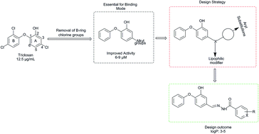
We herein report the investigation of new diphenyl ethers as Mycobacterium tuberculosis enoyl-acyl carrier protein reductase (InhA) inhibitors by structure-based drug design approach. The virtual library of diphenyl ethers was designed and molecules with appreciable physicochemical and ADMET properties were docked. The best ranked molecules based on docking studies were synthesized and characterized by spectral studies. Synthesized compounds were evaluated for in vitro antitubercular activity against Mycobacterium tuberculosis H37Rv strain by Microplate Alamar Blue Assay. Among the tested compounds, DE3 and DE2 exhibited substantial antitubercular potential at 3.125 and 6.25 μg mL−1 concentrations, respectively. The most active compounds were further evaluated for cytotoxicity studies against Vero and HepG2 normal cell lines by microculture tetrazolium assay and ascertained to be safe against normal cell. The molecular dynamic study reveals that the best active compounds show better binding free energy than the reference compounds TCl and JPL at Mtb InhA binding site.
1. Introduction
Mycobacterium tuberculosis (Mtb) infection is responsible for the mortality of over two million people worldwide every year. World Health Organization reports indicated that at the end of 2020 (ref. 1) more than one billion people worldwide may be infected with tuberculosis (TB). Clinical studies indicate that TB is more prevalent in AIDS patients and there has been an increase in TB epidemics over the last five years that may be associated with HIV co-infection. The current status of tuberculosis is magnified due to the increase in multidrug resistance to existing drug therapy.1–3
Currently, the three major challenges that hinder our ability to eradicate TB effectively are drug-resistant strain infection, HIV co-infection and regimen non-compliance. Consequently, there is a need to address the issues of multidrug resistance and persistent TB infection. In view of these facts, the development of drugs with novel modes of action has been the focus of the investigators. The pursuit of inhibitors targeting enzymes that are deemed specific and essential for the replication and persistence of Mtb has been the core of antitubercular research. The target-based screening approach for the discovery of new drugs was rendered possible with the advances in proteomics, genomics and molecular genetics of Mycobacterium. The structure based drug design approach yielded remarkable results in the field of cancer drug discovery and hope that the same would results for TB drug discovery.
The mycobacterial fatty acid synthase I (FAS I) and FAS II contribute to the biosynthesis of mycolic acids, which are components of the mycobacterial cell wall. FAS II has a predominant role in the elongation of fatty acids derived from the end product of FAS I.4 The terminal step of FAS II, trans-enoyl reduction, is catalyzed by InhA and is responsible for this significant role. InhA catalyze the elongation of C16 fatty acid and longer, which is different from the enoyl-ACP reductase (ENR) of other bacterial species. InhA has been validated as a promising target for antitubercular drug discovery. Isoniazid (INH) and ethionamide (ETH), the first line agents and most prescribed drugs to treat tuberculosis (TB), inhibit an NADH-dependent InhA that provides precursors of mycolic acids. They are pro-drugs that need activation to form the inhibitory INH/ETH-NAD adduct by the KatG/EthA encoding enzyme. These adducts are marked as tight binding inhibitors of Mtb InhA.5,6 Mutations of KatG and EthA have been identified and are associated with the development of Mtb resistance to INH and ETH, respectively.7,8 Inhibitors that can directly inhibit InhA without activation by KatG/EthA are able to circumvent the INH/ETH resistance mechanism. This approach is useful in the rational design of potential antitubercular agents against MDR-TB and XDR-TB strains.
The pursuit of small molecules as potential Mtb InhA inhibitors is an effective strategy to eradicate MDR-TB. Clinical studies highlight the importance of direct InhA inhibitors and investigation of new small molecules for the same is ongoing worldwide. The new small molecules like pyrazoles,9 indole-5-amides,10 diphenyl ethers,11,12 pyrrolidine carboxamides,13 arylamides,14 imidazopiperidines15 and 4-hydroxy-2-pyridones16 have been reported as direct InhA inhibitors. These molecules can inhibit InhA without prior activation by KatG and have shown potential activity against MDR/INH-resistant TB strains. Triclosan (TCl), a diphenyl ether derivative, was found to be a potent inhibitor of Mtb InhA with a Ki value of 0.22 mM and an MIC value 12.5 μg mL.−17 However, use of TCl as antitubercular agent is limited due to its poor bioavailability. The biochemistry involved in bacterial inhibition of InhA with TCl is possible through the π–π ring stacking interactions between the aromatic ring of TCl and the pyridine of the NAD+ cofactor. TCl also exhibits strong hydrogen bonding with Tyr158 residue at the catalytic site and the NAD+ cofactor.18
In order to improve the bioavailability, TCl-modified derivatives with improved pharmacokinetic parameters have been synthesized in the last decade, and these new compounds have shown significant activity against both susceptible and resistant Mtb strains. In continuation and exploring the structure–activity relationship (SAR), the alkyl substituted diphenyl ethers were synthesized with an improved affinity towards InhA inhibition. In this regard, 5-octyl-2-phenoxyphenol has shown potential activity (MIC: 6–9 μM) against both drug-sensitive and drug resistant strains of Mtb and, more importantly, the mechanism of inhibition of InhA is KatG independent.4 The SAR highlights that the InhA inhibitory potency also depends on the length of the alkyl chain at the 4th position of the A-ring. The optimal activity was observed with alkyl chain length between 3 and 5 carbons. The removal of the two chlorine atoms on the diphenyl ether B-ring moiety and also replacement of the chlorine function with an ethyl group in the A-ring resulted in a 2-fold increase in the IC50 value in comparison to that for TCl (Fig. 1). In spite of these structural modifications in improving the antitubercular efficiency, the limited bioavailability was the foremost drawback of these alkyl chain-substituted diphenyl ether derivatives and may be attributed to the higher log
P values (>5). Hence, the investigation was focused to develop druggable diphenyl ethers with improved antitubercular activity with appreciable ADMET properties using a structure-based drug design approach. We report herein the molecular docking, synthesis, antimycobacterial evaluation and molecular dynamics study of new diphenyl ether derivatives.
2. Results and discussion
2.1. Strategies for drug design
In view of facts discussed earlier, the present work was aimed to develop diphenyl ethers with optimal lipophilicity (log
P values between 3 and 5) as this is favorable for drug likeness properties. Prior to the drug design strategy, the physicochemical properties of the reported diphenyl ethers were studied. The results highlights that most of the compounds have significantly higher log
P values (>5), while other physicochemical parameters were well within the acceptable range. These compounds exhibit potent in vitro antitubercular activity and their clog
P values ranged between 4 and 7. Despite their promising in vitro activity, some of the compounds exhibit poor in vivo efficacy, which may be attributed to the high clog
P values. The SAR of diphenyl ethers indicates that the higher log
P values can be attributed to the dichloro substitutions on ring-B of TCl. The studies also indicated that the compounds were involved in unfavorable steric interactions with the enzyme and the removal of chlorine functionality from the scaffold would increase the affinity of inhibitors by seven fold towards the enzyme.19 Consequently, in the present investigation, diphenyl ethers were designed with hydrophilic substitutions at the 4th position of ring-A and exclusion of chlorine substitutions on ring-B. The hydrophilic linkers were employed to achieve desired lipophilicity. The substitutions on the aryl moiety with both electron donating and electron withdrawing functions were made in order to experiment and understand the possible interaction that effects the inhibition of the enzyme.20 Overall, modifications of diphenyl ethers were aimed to reduce the inherent lipophilicity of TCl derivatives without compromising their orientations and catalytic interactions at the InhA binding site. The drug design strategy is depicted in Fig. 1.
 |
| Fig. 1 Design strategy for diphenyl ether analogues. | |
2.2. Molecular modelling studies
2.2.1. In silico ADMET studies. The designed compounds were screened for their appreciable ADMET properties using the online tool PreADME.21 All the compounds have appropriate values towards the evaluated in silico parameters. The Lipinski parameters of the molecules towards their biological efficacy were encouraging with zero Lipinski violation. The in silico ADMET results shows that the selected compounds have appreciable oral bioavailability and protein binding efficiency. The predicted oral availability was excellent as the molecules exhibited a calculated percentage of absorption (HIA) values ranging from 92 to 96%. The predicted plasma protein binding (PPB) exhibits parentage binding values ranging from 89 to 100%. The compounds also show moderate Caco2 cell permeability effect, which is in the range of 15–32 nm s−1. The compounds which have blood–brain barrier penetration values <2 indicate poor penetration. The ADMET properties data is provided as ESI.† Compounds that satisfied the ADMET properties and Lipinski's parameters of drug likeness were docked against Mtb InhA.
2.2.2. Molecular docking study. The molecular docking technique was used to explore, predict and understand the protein/enzyme interactions with designed diphenyl ethers at the Mtb InhA binding site. The docking study was performed on Mtb InhA pdb protein (PDB ID: 3FNG) using the SYBYL-X 2.1 molecular modelling software. The molecular docking study was executed initially by the analysis of binding site structural features, followed by docking and interpretation of the results and finally the validation of the docking protocol.
Mtb InhA binding site analysis. The structural features of InhA reveal that it has four monomer units and each consists of 269 amino acid residues. The molecular weight of each monomer unit is ∼29
000 Da. The monomer unit builds with Rossmann fold structure and the core of the binding site contains eight α helices and seven β sheets, representing the dinucleotide binding for the cofactor, NADH. The substrate binding loop consists of 15 amino acid residues (194–208) and is further categorized into three regions, upper, middle and lower substrate binding loops (USL, MSL, and LSL) (Fig. 2). The top USL covers the substrate binding pocket (SBP) by forming inter-loop interactions. The dinucleotide binding site is close to the LSL and forms interactions with NADH.
 |
| Fig. 2 Substrate binding pocket of InhA and structural components (USL: upper substrate binding loop, MSL: middle substrate binding loop and LSL: lower substrate binding loop). | |
The InhA complex of fatty acyl substrate and NAD+ cofactor crystal structure reveals that the fatty acyl substrate binds in a U-shaped conformation. The residues Tyr158 and Lys165 are very much essential for the trans-enoyl reduction. They promote the removal of a proton from the 2′-nicotinamide –OH group. The InhA enzyme plays a crucial role in the reduction of the C2–C3 double bond by transfer of a hydrate ion to the C3 carbon. The Tyr158 hydroxyl group donates a proton to the carbonyl oxygen of C1 fatty acid, which results in an enolate anion. The substrate fatty acyl chain is present in the core of lipophilic residues and most of the lipophilic residues are abreast of the SBL (Met103, Phe149, Tyr158, Lys165, Thr196, Met199, Leu207 and Ile215). The SBL of InhA is larger than the enoyl-ACP reductases of other organisms and facilitates a deeper substrate binding area. The Tyr158 interaction with the fatty acyl substrate is the crucial feature in all enoyl-ACP reductases.
2.2.3. Docking results. Molecular docking results revealed that the basic scaffolds of DE1–10 (5-((substituted imino)methyl)-2-phenoxyphenol) fit in the binding pocket of InhA similar to TCl and 5-(cyclohexylmethyl)-2-(2,4-dichlorophenoxy)phenol (co-crystallized ligand of 3FNG; JPL) (Fig. 3a & b). Furthermore, docking study revealed the formation of hydrogen bonding interactions between the phenolic –OH and the ether oxygen of the designed compounds with Tyr-158 and NAD+ of Mtb InhA, which is vital as per the docking literature. The hydrogen bonding network is consistent in all the designed compounds, and this feature is reported to be a conserved one among all the InhA-inhibitor complexes including TCl. Hence, the core structural features of TCl (diphenyl ether nucleus) and the hydroxyl group in ring-A were kept intact in all the designed analogues. Based on docking scores (>7), interactions and binding with the above mentioned crucial residues, the best ranked molecules were considered for synthesis. The docking score data of the synthesized compounds is presented in Table 1.
 |
| Fig. 3 (a) Binding poses of TCl and JPL at InhA binding site. Green: TYR158; purple: NAD+; yellow lines: hydrogen bonding. (b) Binding pose of compounds DE3 and JPL at InhA binding site. Green: TYR158; purple: NAD+; yellow lines: hydrogen bonding. | |
Table 1 Molecular docking scores of synthesized compounds DE1–10a
Compound |
Docking score |
Total |
Crash |
Polar |
Total score = the total Surflex-Dock score expressed as −log(Kd). Crash = the degree of inappropriate penetration by the ligand into the protein. Polar = contribution of the polar interactions to the total score. |
DE1 |
7.74 |
−2.75 |
2.25 |
DE2 |
8.07 |
−3.15 |
2.11 |
DE3 |
7.06 |
−1.96 |
2.12 |
DE4 |
8.25 |
−1.44 |
1.02 |
DE5 |
8.56 |
−1.11 |
2.18 |
DE6 |
8.07 |
−1.02 |
1.13 |
DE7 |
8.71 |
−2.61 |
2.02 |
DE8 |
8.81 |
−1.58 |
2.21 |
DE9 |
8.04 |
−1.21 |
1.26 |
DE10 |
8.05 |
−2.08 |
1.84 |
JPL |
8.86 |
−0.58 |
2.24 |
TCl |
5.07 |
−0.29 |
2.41 |
2.2.4. Docking validation. The accuracy of the docking protocol detailed under the method was extensively validated by reproducing the ligand–receptor (Mtb InhA) complex (3FNG) deposited in the RCSB PDB. The Root Mean Square Deviation (RMSD) value and poses were used for validation of docking protocol. The co-crystallized ligand (JPL) of the PDB structure was extracted and docked along with the designed ligands. The docking poses of the designed ligands were compared with the binding poses of JPL and TCl (Fig. 3a). The RMSD value was calculated between the co-crystallized and docked ligand. The RMSD value for the co-crystallized and docked ligand was 0.948 Å and is lower than the acceptable limit (<1.5 Å), accordingly indicating that the docking protocol is validated.
2.3. Chemistry
The synthetic protocol for the new series of diphenyl ether scaffolds is outlined in Scheme 1. The starting material, 3-methoxy-4-phenoxybenzaldehyde (1a), was prepared according to Lam-Chan O-arylation with vanillin and phenyl boronic acid. Demethylation of methoxy aldehyde derivative 1 was carried out in the presence of boron tribromide (BBr3) at −78 °C (dry ice and acetone) to afford 3-hydroxy-4-phenoxybenzaldehyde (2a), which was further condensed with various acid hydrazides to afford title compounds DE1–10 (Table 2). The synthesized compounds were characterized by IR, 1H NMR, 13C NMR, mass spectral and elemental analysis data. In the IR spectra, an absorption band around ∼1590 cm−1 was observed in all the synthesized derivatives and may be attributed to –C
N stretching. The characterization by 1H NMR spectra of the derivatives indicates chemical shift signals in the range of δ 6.6–7.9 ppm (Ar-H) as a multiplet for aromatic protons. The NH proton of the amide group resonated as a singlet around δ 11.5 ppm, which was attributed to the amide bond formation. The mass spectra of the compounds exhibit molecular ion peaks (M+) corresponding to their respective molecular weights.
Table 2 Physicochemical properties data of diphenyl ether derivatives
The 1H NMR spectrum of compound 1a exhibited a singlet at δ 9.9 ppm value corresponding to aldehyde functionality and 8 protons in the aromatic region (δ 7.6–7.0 ppm), confirming the formation of a diphenyl ether nucleus. In the 1H NMR spectrum of compound 1b, the singlets at δ 10.1 and δ 9.9 ppm were attributed to the proton of the aldehyde functional group and the phenolic OH of the diphenyl ether, respectively. The 1H NMR spectral data confirms the formation of the diphenyl ether and its demethylation product. The IR spectrum of DE3 exhibited absorption bands at 3435 cm−1 attributed to N–H stretching and 1732 cm−1 accounting for stretching of the C
O functionality. The 1H NMR spectrum of the compound DE3 exhibited singlet protons at δ 11.8 and 9.8 ppm corresponding to the NH of the hydrazide functionality and the phenolic OH group of the diphenyl ether, respectively. The spectral characterization data of all the synthesized compounds are in accordance with the proposed structures as depicted in Scheme 1. The experimental log
P values of all the synthesized compounds were determined by RP-HPLC method to affirm and towards a comparative study with respect to the calculated log
P values. The physicochemical properties data of DE1–10 are listed in Table 2.
 |
| Scheme 1 Synthesis of designed compounds. Reagents and conditions: (i) Cu(OAc)2, C5H5N, CH2Cl2, 25–27 °C, 72 h. (ii) BBr3, CH2Cl2, −78 to 10 °C, 10 h. (iii) EtOH, reflux. | |
2.4. Biological activity
2.4.1. Antitubercular activity. The synthesized compounds were evaluated for in vitro antitubercular activity by Microplate Alamar Blue Assay (MABA) against Mtb H37Rv strain. The antitubercular activities of DE1–10 are expressed as minimum inhibitory concentration (MIC) values using INH and TCl as the reference drugs towards a comparative study. The in vitro antitubercular evaluation of diphenyl ethers indicates that few of the tested compounds exhibit significant activity (Table 3). DE3 was found to be the most potent antitubercular among all tested compounds with an MIC value of 3.125 μg mL−1. The results indicate that the better activity of compound DE3 may be attributed to the electron withdrawing chlorine at the para position and a comparatively higher log
P value (4.8). In support of the above view, compound DE2, the phenyl derivative, exhibited a MIC value of 6.25 μg mL−1, whilst the methyl group substituted derivatives DE8 and DE9, examples of electron donating groups, exhibited trivial antitubercular activity with MIC values >100 μg mL−1. The o-chloro derivative (DE4) exhibited a higher MIC value (12.5 μg mL−1) in comparison to the p-chlorophenyl derivative (DE3; MIC = 3.125 μg mL−1), but was comparatively better than that of the p-nitrophenyl (DE6 MIC = 50 μg mL−1). The better activity of compound DE3 in comparison to DE4 and DE6 may be attributed to its log
P value. Compound DE3's log
P value (4.80) is higher than that of compound DE6 and DE4, which have log
P values of 4.43 and 4.44, respectively. The efficiency of the drug to produce an antitubercular effect depends on the accumulation of the same in the cell and resulting in cell death. The higher lipophilic nature of compound DE3 may be correlated to its potential to cross the phospholipid membrane of Mtb leading to a significant accumulation of it in the cell and resulting in considerable antitubercular activity. The compounds with pyridinyl (DE1 and DE5) substitutions demonstrated poor activity (MIC > 100 μg mL−1). The activity data reveals that the antitubercular activity of the derivatives drastically improved with the replacement of pyridinyl moiety (DE1 and DE5 MIC > 100 μg mL−1) by a phenyl/substituted phenyl moiety (DE2; MIC = 6.25 μg mL−1). Moreover, the low log
P values of DE1 (3.34) and DE5 (2.56) may also be correlated with the lower antitubercular activity of pyridinyl derivatives. The investigation also indicated that the antitubercular activity decreases with increasing carbon chain length between the amide function and the phenyl moiety; compound DE10, a benzyl derivative, exhibited an MIC value of 12.5 μg mL−1 in contrast to DE2 (MIC = 6.25 μg mL−1). The considerable antitubercular activity of DE3 may be attributed to the electronegative functional group substitutions on the phenyl hydrazide moiety and significantly higher log
P (4–5) values.
Table 3 In vitro antitubercular, antibacterial activity (MIC) and cytotoxicity (CC50) of diphenyl ether derivativesa
Compounds |
MICb (μg mL−1) |
CC50c (μg mL−1) |
Mtb H37Rv |
S. aureas |
E. coli |
Vero |
HepG2 |
SId |
nc: not carried out. MIC = minimum drug concentration required to stop the growth of bacteria. CC50 = minimum drug concentration required for 50% death of viable cells. SI (selective index) = CC50/MIC. |
DE1 |
>100 |
6.25 |
12.5 |
nc |
nc |
— |
DE2 |
6.25 |
3.125 |
3.125 |
>300 |
>300 |
>48 |
DE3 |
3.125 |
3.125 |
6.25 |
>300 |
>300 |
>96 |
DE4 |
12.5 |
3.125 |
12.5 |
>300 |
>300 |
>24 |
DE5 |
>100 |
25 |
12.5 |
nc |
nc |
— |
DE6 |
50 |
12.5 |
25 |
nc |
nc |
— |
DE7 |
25 |
12.5 |
50 |
>300 |
>300 |
>12 |
DE8 |
>100 |
25 |
12.5 |
nc |
nc |
— |
DE9 |
>100 |
25 |
100 |
nc |
nc |
— |
DE10 |
12.5 |
12.5 |
25 |
>300 |
262 |
>24 |
INH |
<3.125 |
— |
— |
nc |
nc |
— |
TCl |
12.5 |
— |
— |
nc |
nc |
— |
Ampicillin |
— |
<3.125 |
<3.25 |
nc |
nc |
— |
The investigation towards the antitubercular efficacy of the molecules with respect to the docking results did not show a significant correlation; this can be related towards the better binding free energy of the compounds (Table 4). Compounds with better in vitro antitubercular activity exhibited a substantial binding free energy, which may be attributed to the considerable activity of a few of the compounds.
Table 4 Calculated MMPBSA binding free energies of the compounds DE1–10, TCl and JPL
Cpd code |
Avg. RMSD (Å) |
MM-PB/SA results |
ΔVDW |
ΔEEL |
ΔEPB |
ΔG |
DE1 |
2.05 |
−49.73 |
−20.08 |
49.03 |
−21.25 |
DE2 |
1.73 |
−49.80 |
−11.39 |
39.96 |
−25.46 |
DE3 |
1.84 |
−54.54 |
−35.90 |
65.15 |
−29.56 |
DE4 |
1.86 |
−49.24 |
−9.26 |
36.86 |
−26.04 |
DE5 |
1.88 |
−43.39 |
−7.95 |
34.66 |
−20.73 |
DE6 |
1.85 |
−44.09 |
−27.89 |
43.91 |
−24.29 |
DE7 |
1.95 |
−49.28 |
−15.84 |
41.70 |
−28.01 |
DE8 |
2.01 |
−40.12 |
−16.45 |
45.14 |
−22.62 |
DE9 |
2.26 |
−47.13 |
−5.14 |
36.35 |
−20.49 |
DE10 |
2.29 |
−54.26 |
−22.06 |
51.43 |
−29.34 |
TCl |
1.87 |
−36.41 |
−10.10 |
34.90 |
−14.70 |
JPL |
1.96 |
−49.97 |
−3.36 |
28.69 |
−28.96 |
The compounds exhibiting significant antitubercular activity (DE2, DE3, DE4, DE7 and DE10) with MIC values less than 25 μg mL−1 were further screened for cell viability assay by Microculture Tetrazolium Assay (MTT) against Vero (epithelial cells) and HepG2 (hepatocytes) cell lines to ascertain that their antitubercular activity is not due to cytotoxicity and also to highlight their safety profile on normal cells. The approximate CC50 values and selectivity index (SI) are tabulated in Table 3. These findings indicate that the active derivatives target Mtb to a greater extent compared to macrophage cell lines.
2.4.2. Antibacterial activity. The synthesized compounds were also evaluated for in vitro antibacterial activity by tube dilution method22 against Gram-positive Staphylococcus aureus (ATCC-25923) and Gram-negative Escherichia coli (ATCC-25922). The antibacterial data (Table 3) reveal that few of the diphenyl ether derivatives exhibited significant activity against the tested strains. Among the tested compounds, DE2, DE3 and DE4 showed potential antibacterial activity with an MIC value of 3.125 μg mL−1 against S. aureus, while compound DE3 was also effective against E. coli with an MIC value of 6.25 μg mL−1.
2.5. Molecular dynamic studies
The synthesized compounds were subjected to molecular dynamics (MD) simulation studies in comparison to the well-known inhibitors, TCl and JPL (3FNG co-crystalized ligand). The MD trajectories from the end of the simulation time were used to apply the MM-PB/SA method. The absolute free energy of a system is estimated from a combination of molecular mechanics following the Poisson–Boltzmann protocol. This protocol involves the estimation of electrostatic free energy determined from the exposed surface area and estimation of the entropy of the system derived from normal mode calculations. RMSD plots of the simulation studies are presented in Fig. 4. The RMSD plot shows that all the molecular dynamic simulation systems were stabilized and the results highlight that the designed ligands are stable in the InhA core. The MM-PB/SA results and the average of the RMSD values (0–5 ns) for all the evaluated molecules are presented in Table 4. The studies indicate that a few of the compounds have shown better binding free energy (ΔG binding > −29) than that of the TCl (ΔG binding = −14.70) and JPL (ΔG binding = −28.96). The results highlight that the best active compounds would have a better inhibitory efficacy than TCl and comparable to that of JPL at the Mtb InhA binding site. The video files of the molecular dynamic trajectories and detailed report files of MM-PB/SA are given as ESI.†
 |
| Fig. 4 MD simulations RMSD plots of TCl (green), JPL (blue) and DE3 (red). | |
3. Conclusion
In the present investigation, a library of diphenyl ethers was designed by structure-based drug design approach. The ADMET studies indicate that the designed compounds have drug-like properties. The docking study indicates that the best ranked diphenyl ether molecules exhibit major interactions with the 2-hydroxyl moiety of the nicotinamide ribose and the hydroxyl group of the Tyr158 residue at InhA, which are similar to TCl and JPL. The best ranked diphenyl ether derivatives were synthesized and evaluated for antitubercular and antibacterial activities. The antitubercular activity of the tested compounds was encouraging, wherein compounds DE2 and DE3 were found to be the most potent molecules with MIC values 3.125 and 6.25 μg mL−1, respectively. The cytotoxicity results of the most active compounds indicate that the tested compounds are non-toxic. The selectivity index values were found to be >10. The antibacterial activity of the test compounds was promising and compound DE3 was found to be an effective antibacterial agent against the tested bacterial strains. The molecular dynamics study showed that the best active molecules form a stable protein–ligand complex and exhibit better binding free energy.
4. Materials and methods
4.1. Molecular docking study
The 3D structures of the designed compounds were generated using SYBYL-X 2.1 molecular modelling software (Tripos Associates, St. Louis, MO, USA).23 The molecules were subjected to energy minimization with MMFF94s force field using a distance dependent-dielectric function, an energy gradient of 0.001 kcal mol−1 and electrostatics. The Surflex-Dock tool was used to dock the designed molecules against the InhA binding site to identify the binding mode and structural optimization. Surflex-Dock adopted is an empirical scoring function and a patented searching engine was employed for molecular docking. The crystal structure of Mtb InhA complexed with 5-(cyclohexylmethyl)-2-(2,4-dichlorophenoxy)phenol (PDB ID: 3FNG, 1.97 Å X-ray resolution) was taken from the protein databank (http://www.rcsb.org/pdb).24 The protein structure was prepared for the molecular docking study by removing all water molecules. The missing hydrogen atoms were assigned to the InhA crystal structure. The co-crystalized ligand (JPL) was extracted from the protein and used as reference molecule for the validation study. The protomol was generated by keeping the parameters of the co-crystalized ligand (JPL), and threshold and bloat unchanged from the default values of 0.50 and 0 Å. The mode of interaction of the co-crystalized ligand against 3FNG (InhA crystal structure) was used as a standard docking model. The molecular docking was performed by analyzing 20 poses per ligand without any constraints. The docked complex was assumed to represent the protein–ligand interactions, which was selected based on docking score, the orientation of the ligands at the active site in a similar way to reference ligands and preservation of the two key interactions (H-bonds with Tyr158 and NAD+).
4.2. Synthesis and characterization of designed molecules
All the chemicals and solvents used in this study were procured from Aldrich Chemical Co., Spectrochem Ltd., and Sd Fine Chemicals. All commercially available reagents procured were used without further purification. Column chromatography was carried out on 100–200 mesh silica gel. The progress of the reactions was monitored by TLC using aluminum-backed sheets of silica gel-60 F24 (Merck). Melting points were recorded using laboratory melting point apparatus and are uncorrected. 1H NMR spectra were recorded on an NMR spectrometer (VNMRS400, 400 MHz) using DMSO-d6 as the solvent. Mass spectroscopy was performed using LC-MS with methanol as the solvent. IR spectra were obtained using an FTIR spectrophotometer (Shimadzu, Japan) using KBr pellets.
4.2.1. 3-Methoxy-4-phenoxybenzaldehyde (1a). To a stirred solution of vanillin (1 mmol) in anhydrous dichloromethane (60 mL), activated molecular sieves (4 Å, 2.5 g), phenylboronic acid (1.5 mmol), copper(II) acetate (1.5 mmol) and anhydrous pyridine (2 mmol) were added successively. The resulting suspension was stirred at 25–27 °C for 72 h.25 The progress of the reaction was monitored by TLC, using hexane
:
ethyl acetate (4
:
1) as the mobile phase. After the completion of the reaction (72 h), the reaction mixture was diluted with dichloromethane and filtered under vacuum. The filtrate was washed with dilute aqueous hydrochloric acid solution (2 M), followed by water, then dried over anhydrous MgSO4 and evaporated under vacuum. The crude compound obtained was purified by column chromatography over silica 100–200 with hexane
:
ethyl acetate (4
:
1) as the mobile phase to afford the target compound.
4.2.2. 3-Methoxy-4-phenoxybenzaldehyde (1a). Yield = 75%; mp = 40–42 °C; anal. calcd for C14H12O3: C, 73.67; H, 5.30; found: C, 73.62; H, 5.36%; IR (KBr, νmax, cm−1): 3051 (Ar-H), 2928 (C–H), 1674 (C
O), 1583, 1489, 1413 (Ar-C
C), 1276 (asym. C–O–C), 1134 (sym. C–O–C); 1H NMR (400 MHz, DMSO-d6): 9.94 (s, 1H, CHO), 7.62 (s, 1H, Ar-H), 7.54–7.56 (d, 1H, Ar-H), 7.38–7.42 (t, 2H, Ar-H), 7.15–7.18 (t, 1H, Ar-H), 7.05–7.07 (d, 1H, Ar-H), 7.00–7.01 (d, 2H, Ar-H), 3.87 (s, 3H, CH3); LC-MS (m/z): 229.2 [M+].
4.2.3. 3-Hydroxy-4-phenoxybenzaldehyde (2a). To a solution of compound 1a (3 g, 13.16 mmol) in dichloromethane (50 mL), BBr3 (1 mmol in dichloromethane 26.32 mmol) was added at −78 °C, and the reaction continued for 6 h at 0–10 °C. The progress of the reaction was monitored by TLC, using hexane
:
ethyl acetate (4
:
1) as the mobile phase. The resulting reaction mixture was poured into water and extracted with ethyl acetate (3 × 50 mL). The organic layers were combined and washed with saturated sodium bicarbonate solution, dried over anhydrous magnesium sulfate and evaporated under vacuum. The crude product obtained was purified by column chromatography over silica 100–200 with hexane
:
ethyl acetate (4
:
1) as the mobile phase to afford the target compound.Yield = 55%; mp = 82–84 °C; anal. calcd for C13H10O3: C, 72.89; H, 4.71; found: C, 72.92; H, 4.65%; IR (KBr, νmax, cm−1): 3051 (Ar-H), 2928 (C–H), 1674 (C
O), 1583, 1489, 1413 (Ar-C
C), 1276 (asym. C–O–C), 1134 (sym. C–O–C); 1H NMR (400 MHz, DMSO-d6): 10.08 (s, 1H, CHO), 9.85 (s, 1H, OH), 7.54 (d, 1H, Ar-H), 7.42–7.32 (m, 3H, Ar-H), 7.32–7.20 (m, 1H, Ar-H), 7.20–7.07 (m, 2H, Ar-H), 6.86–6.84 (d, 1H, Ar-H); LC-MS (m/z): 215.1 [M+].
Preparation of acid hydrazides. The acid hydrazides (3a–h) were prepared as per the reported procedure.26
4.2.4. Synthesis of (E)-N′-(3-hydroxy-4-phenoxybenzylidene) isonicotinohydrazide (DE1). To a solution of 3-hydroxy-4-phenoxybenzaldehyde (1 mmol) in absolute ethanol (10 mL), isoniazid (1 mmol) was added and refluxed for 4 h, then the solution was allowed to cool for 12 h. The progress of the reaction was monitored by TLC using hexane
:
ethyl acetate (1
:
1) as the mobile phase. The reaction mixture was cooled, poured into water and the precipitate was filtered then dried. The crude compound obtained was purified by column chromatography over silica 100–200 with hexane
:
ethyl acetate (1
:
1) as the mobile phase to afford the target compound. A similar procedure was adopted to synthesize title compounds DE2–10 using appropriate acid hydrazides.
4.2.5. (E)-N′-(3-Hydroxy-4-phenoxybenzylidene)isonicotinohydrazide (DE1). Yield = 75%; mp = 186–188 °C; anal. calcd for C19H15N3O3: C, 68.46; H, 4.54; N, 12.61; found: C, 68.41; H, 4.60; N, 12.58%; IR (KBr, νmax, cm−1): 3556 (–OH), 3435 (–NH), 3055 (Ar, –CH), 1722 (–C
O), 1592 (C
N), 1221 (C–O–C); 1H NMR (400 MHz, DMSO-d6, δ ppm): 11.98 (s, 1H, CONH), 9.84 (s, 1H, OH), 8.77 (s, 2H, Ar-H), 8.35 (s, 1H, N
CH), 7.80–7.78 (d, 2H, Ar-H), 7.43–7.42 (d, 1H, Ar-H), 7.31–7.29 (dd, 2H, Ar-H), 7.12–6.88 (m, 5H, Ar-H); 13C NMR (400 MHz, DMSO-d6, δ ppm): 161.92, 157.74, 150.77, 149.88, 149.01, 145.49, 140.96, 131.40, 130.17, 122.98, 121.93, 121.73, 120.60, 117.32, 114.81; LC-MS (m/z): 334.14 [M+].
4.2.6. (E)-N′-(3-Hydroxy-4-phenoxybenzylidene)benzohydrazide (DE2). Yield: 68%; mp = 118–120 °C (ethanol); anal. calcd for C20H16N2O3: C, 72.28; H, 4.85; N, 8.43; O; found: C, 72.30; H, 4.90; N, 8.40%; IR (KBr, νmax, cm−1): 3565 (–OH), 3435 (–NH), 3058 (Ar, –CH), 1714 (–C
O), 1590 (C
N), 1228 (C–O–C); 1H NMR (400 MHz, DMSO-d6, δ ppm): 11.77 (s, 1H, CONH), 9.82 (s, 1H, OH), 8.35 (s, 1H, N
CH), 7.90–7.89 (d, 2H, Ar-H), 7.52–7.49 (m, 3H, Ar-H), 7.43–7.42 (d, 1H, Ar-H), 7.34–6.89 (m, 7H, Ar-H); 13C NMR (400 MHz, DMSO-d6, δ ppm): 163.47, 157.83, 149.89, 147.77, 145.26, 133.95, 132.12, 131.77, 130.14, 128.90, 128.02, 122.90, 121.79, 120.33, 117.24, 114.71, 114.71; LC-MS (m/z): 333.15 [M+].
4.2.7. (E)-N′-(3-Hydroxy-4-phenoxybenzylidene)-4-chlorobenzohydrazide (DE3). Yield: 70%; mp = 154–156 °C (ethanol); anal. calcd for C20H15ClN2O3: C, 65.49; H, 4.12; N, 7.64; found: C, 65.55; H, 4.16; N, 7.66%; IR (KBr, νmax, cm−1): 3564 (–OH), 3435 (–NH), 3056 (Ar, –CH), 1732 (–C
O), 1592 (C
N), 1230 (C–O–C); 1H NMR (400 MHz, DMSO-d6, δ ppm): 11.83 (s, 1H, CONH), 9.82 (s, 1H, OH), 8.34 (s, 1H, N
CH), 7.93–7.91 (d, 2H, Ar-H), 7.59–7.58 (d, 2H, Ar-H), 7.42 (s, 1H, Ar-H), 7.34–7.29 (m, 2H, Ar-H), 7.11–6.89 (m, 5H, Ar-H); 13C NMR (400 MHz, DMSO-d6, δ ppm): 162.39, 157.80, 149.88, 148.12, 145.37, 136.98, 132.64, 131.65, 130.38, 130.14, 129.96, 129.01, 122.92, 121.77, 120.40, 118.31, 117.26, 114.75; LC-MS (m/z): 367.11 [M+].
4.2.8. (E)-N′-(3-Hydroxy-4-phenoxybenzylidene)-2-chlorobenzohydrazide (DE4). Yield: 65%; mp = 152–153 °C (ethanol); anal. calcd for C20H15ClN2O3: C, 65.49; H, 4.12; N, 7.64; found: C, 65.6; H, 4.10; N, 7.61; IR (KBr, νmax, cm−1): 3552 (–OH), 3425 (–NH), 3042 (Ar, –CH), 1734 (–C
O), 1594 (C
N), 1225 (C–O–C); 1H NMR (400 MHz, DMSO-d6, δ ppm): 11.87 (s, 1H, CONH), 9.84 (s, 1H, OH), 8.37 (s, 1H, N
CH), 7.94–7.89 (d, 2H, Ar-H), 7.59–7.58 (d, 2H, Ar-H), 7.44 (s, 1H, Ar-H), 7.41 (s, 1H, Ar-H) 7.36–7.34 (d, 1H, Ar-H), 7.13–6.92 (m, 5H, Ar-H); 13C NMR (400 MHz, DMSO-d6, δ ppm): 167.42, 159.78, 152.14, 149.82, 146.31, 139.26, 133.27, 131.93, 129.91, 129.64, 127.76, 126.41, 122.52, 119.73, 119.48, 117.39, 116.76; LC-MS (m/z): 367.11 [M+].
4.2.9. (E)-N′-(3-Hydroxy-4-phenoxybenzylidene)isonicotinamide (DE5). Yield: 54%; mp = 135–136 °C (ethanol); anal. calcd for C19H14N2O3: C, 71.69; H, 4.43; N, 8.80; found: C, 71.70; H, 4.48; N, 8.76%; IR (KBr, νmax, cm−1): 3568 (–OH), 3048 (Ar, –CH), 1715 (–C
O), 1602 (C
N), 1216 (C–O–C); 1H NMR (400 MHz, DMSO-d6, δ ppm): 9.87 (s, 1H, OH), 8.64 (s, 2H, Ar-H), 8.37 (s, 1H, N
CH), 7.81–7.79 (d, 2H, Ar-H), 7.44–7.42 (d, 1H, Ar-H), 7.33–7.32 (dd, 2H, Ar-H), 7.08–6.82 (m, 5H, Ar-H); 13C NMR (400 MHz, DMSO-d6, δ ppm): 167.92, 161.74, 154.77, 147.68, 147.14, 144.42, 141.16, 134.47, 130.87, 124.38, 123.23, 121.73, 121.61, 119.32, 114.81; LC-MS (m/z): 319.15 [M+].
4.2.10. (E)-N′-(3-Hydroxy-4-phenoxybenzylidene)-4-nitrobenzohydrazide (DE6). Yield: 62%; mp = 154–156 °C (ethanol); anal. calcd for C20H15N3O5: C, 63.66; H, 4.01; N, 11.14; found: C, 63.70; H, 4.15; N, 11.20%; IR (KBr, νmax, cm−1): 3560 (–OH), 3441 (–NH), 3065 (–CH), 1708 (–C
O), 1590 (C
N), 1225 (C–O–C); 1H NMR (400 MHz, DMSO-d6, δ ppm): 12.04 (s, 1H, CONH), 9.83 (s, 1H, OH), 8.34–8.32 (m, 3H, N
CH and Ar-H), 8.14–8.12 (d, 2H, Ar-H), 7.45 (s, 1H, Ar-H), 7.34–7.30 (m, 2H, Ar-H), 7.13–6.89 (m, 5H, Ar-H); 13C NMR (400 MHz, DMSO-d6, δ ppm): 162.39, 157.80, 149.88, 148.12, 145.37, 136.98, 132.64, 131.65, 130.38, 130.14, 129.96, 129.01, 122.92, 121.77, 120.40, 118.31, 117.26, 114.75; LC-MS (m/z): 378.5 [M+].
4.2.11. (E)-N′-(3-Hydroxy-4-phenoxybenzylidene)-2-hydroxybenzohydrazide (DE7). Yield: 54%; mp = 154–156 °C (ethanol); anal. calcd for C20H16N2O4: C, 68.96; H, 4.63; N, 8.04; found: C, 68.90; H, 4.68; N, 8.07%; IR (KBr, νmax, cm−1): 3563 (–OH), 3430 (–NH), 3058 (–CH), 1726 (–C
O), 1590 (C
N), 1225 (C–O–C); 1H NMR (400 MHz, DMSO-d6, δ ppm): 11.88 (s, 1H, CONH), 11.79 (s, 1H, OH), 9.85 (s, 1H, OH), 8.37 (s, 1H, N
CH), 7.89–7.87 (d, 1H, Ar-H), 7.45–7.40 (d, 2H, Ar-H), 7.33–7.29 (m, 2H, Ar-H), 7.13–7.02 (m, 2H, Ar-H), 6.98–6.89 (m, 5H, Ar-H); 13C NMR (400 MHz, DMSO-d6, δ ppm): 165.22, 159.59, 157.77, 149.89, 148.76, 145.51, 134.26, 131.50, 130.16, 128.91, 122.95, 121.74, 120.56, 119.38, 117.74, 117.30, 116.26, 114.87; LC-MS (m/z): 349.13 [M+].
4.2.12. (E)-N′-(3-Hydroxy-4-phenoxybenzylidene)-4-methylbenzohydrazide (DE8). Yield: 60%; mp = 150–151 °C (ethanol); anal. calcd for C21H18N2O3: C, 72.82; H, 5.24; N, 8.09; found: C, 72.85; H, 5.19; N, 8.12%; IR (KBr, νmax, cm−1): 3558 (–OH), 3438 (–NH), 3051 (Ar, –CH), 1728 (–C
O), 1585 (C
N), 1238 (C–O–C); 1H NMR (400 MHz, DMSO-d6, δ ppm): 11.62 (s, 1H, CONH), 9.79 (s, 1H, OH), 8.34 (s, 1H, N
CH), 7.89–7.87 (d, 2H, Ar-H), 7.51–7.49 (m, 3H, Ar-H), 7.42 (s, 1H, Ar-H), 7.33–7.27 (m, 6H, Ar-H), 1.17 (s, 3H, CH3); 13C NMR (400 MHz, DMSO-d6, δ ppm): 161.34, 158.11, 148.32, 146.27, 143.26, 131.96, 131.12, 129.77, 129.14, 127.92, 126.22, 121.96, 121.19, 119.73, 116.24, 114.21, 24.71; LC-MS (m/z): 347.2 [M+].
4.2.13. (E)-N′-(3-Hydroxy-4-phenoxybenzylidene)-2-methylbenzohydrazide (DE9). Yield: 60%; mp = 146–148 °C (ethanol); anal. calcd for C21H18N2O3: C, 72.82; H, 5.24; N, 8.09; found: C, 72.80; H, 5.22; N, 8.10%; IR (KBr, νmax, cm−1): 3562 (–OH), 3435 (–NH), 3048 (Ar, –CH), 1736 (–C
O), 1590 (C
N), 1233 (C–O–C); 1H NMR (400 MHz, DMSO-d6, δ ppm): 11.57 (s, 1H, CONH), 9.77 (s, 1H, OH), 8.35 (s, 1H, N
CH), 7.87–7.84 (d, 2H, Ar-H), 7.53–7.51 (m, 3H, Ar-H), 7.41 (s, 1H, Ar-H), 7.29–7.22 (m, 6H, Ar-H), 1.19 (s, 3H, CH3); 13C NMR (400 MHz, DMSO-d6, δ ppm): 163.11, 159.26, 148.12, 146.74, 144.16, 131.27, 130.82, 129.93, 128.74, 126.92, 126.31, 121.56, 119.84, 119.13, 117.44, 113.20, 22.64; LC-MS (m/z): 347.3 [M+].
4.2.14. N′-[(E)-(3-Hydroxy-4-phenoxyphenyl)methylidene]-2-phenylacetohydrazide (DE10). Yield: 65%; mp = 146–148 °C (ethanol); anal. calcd for C21H18N2O3: C, 72.82; H, 5.24; N, 8.09; found: C, 72.90; H, 5.20; N, 8.12%; IR (KBr, νmax, cm−1): 3556 (–OH), 3441 (–NH), 3052 (Ar, –CH), 1726 (–C
O), 1594 (C
N), 1236 (C–O–C); 1H NMR (400 MHz, DMSO-d6, δ ppm): 11.71 (s, 1H, CONH), 9.86 (s, 1H, OH), 8.33 (s, 1H, N
CH), 7.89–7.87 (d, 2H, Ar-H), 7.57–7.54 (m, 3H, Ar-H), 7.47 (s, 1H, Ar-H), 7.41–7.10 (m, 7H, Ar-H), 3.02 (s, 2H,
); 13C NMR (400 MHz, DMSO-d6, δ ppm): 162.64, 159.71, 149.22, 147.43, 144.26, 133.95, 132.12, 131.77, 130.14, 128.90, 128.02, 122.90, 121.79, 120.33, 117.24, 114.71, 114.71, 32.47; LC-MS (m/z): 347.4 [M+].
4.3. Antitubercular activity
The synthesized compounds were screened against M. tuberculosis H37Rv using MABA27,28 and Middlebrook 7H9-S broth was used as the media. A frozen stock culture suspension of M. tuberculosis H37Rv (ATTCC 27294) from Lowenstein–Jensen slants in complete 7H9 broth was vortexed and then adjusted to a turbidity equivalent to that of a 1 McFarland standard (3 × 10⁸ CFU mL−1). It was further diluted to a concentration of 2 × 10⁵ CFU mL−1 and used as the inoculum in the MABA assay. Test samples and standard compounds (isoniazid and TCl) were uniformly dissolved in DMSO and sterilized by filtering through syringe-driven filters (0.22 μm) to prepare stock solutions of concentration 20
000 μg mL−1. The stock solutions (4×) were diluted serially with media in a 96 deep well plate to afford the working solutions. Dehydration of the perimeter wells of the 96 well plates was prevented by filling with sterile deionized water during the incubation period. Twofold serial dilutions of the test compounds were placed directly on the plate by using a multichannel micropipette by adding media (100 μL) and 100 μL of inoculum (2 × 10⁵ CFU mL−1) was added to each well of the 96 well plate to obtain 200 μL and the final drug concentrations tests were 3.125–100 μg mL−1. Isoniazid and TCl were used as standards and DMSO as the blank. The error was minimized by applying a positive control (inoculum) and a negative control (media) in the plate. The plates were then incubated at 37 °C under aeration. On the seventh day of incubation, 20 μL of Alamar blue reagent solution was added to each well of the plate and the plate was then incubated for another 24 h at 37 °C. A change in color from blue to pink was considered as indicating the growth of the Mycobacterium at that concentration of the drug. For better interpretation of the results, the color was compared to the color present in the growth control wells. The MIC was defined as the lowest concentration of drug that inhibited bacterial growth.
4.4. Cytotoxicity screening
The cytotoxicity of the best active compounds was assessed by MTT29 and against Vero and HepG2 cells (NCCS, Pune, India). The stock solutions of the test compounds were diluted in a 96 deep well plate aseptically with MEM (without FBS) to achieve concentrations of 300, 250, 200, 150, 100 and 50 μg mL−1. The 96 well plates containing the cells were taken and kept inverted on filter paper to remove the supernatant media and then washed gently with PBS and decanted. 100 μL of sterile water was added to the outer perimeter wells. Then 100 μL of each test compound dilution was added to the wells. DMSO was used as a control and the plates were incubated in an incubator (5% CO2) at 37 °C 72 h and 24 h for Vero and HepG2 cells, respectively. After the incubation, plates were inverted on filter paper to remove the supernatant media followed by PBS washing. To this, 50 μL of MTT solution was added to each well in a dark place and then incubated for 3 h. After the incubation, the MTT solution was removed from the well by inverting gently on filter paper and 50 μL of DMSO was added to each well and then kept in a dark place for 2 h. Then the optical density readings of the plates were taken using an ELISA reader at 540 nm. The safety profile (CC50) was determined using eqn (1) and (2).
Determination of safety profile (CC50)
|
 | (1) |
|
Percentage cell inhibition = 100 − % cell viability
| (2) |
CC50 was calculated by extrapolating from a graph with % cell inhibition on the Y-axis against the concentration of the test compound on X-axis.
4.5. Determination of log
P
A reverse phase HPLC method was applied to determine the log
P (lipophilicity) of the compounds.30,31 All the chromatographic runs were conducted by HPLC (Shimadzu, Japan) at room temperature using an ODS-4 (Intersil ODS-4, 5 μm, 4.6, 150 mm, GL Science Inc., Japan) column and a PDA detector. Numerical analysis and data processing were done using Lab solution-2013 software. 3-Morpholinopropane-1-sulfonic acid (MOPS, 4.18 g) was added to 900 mL of octanol-saturated MilliQ water, and the volume was made up to 1 L. The pH of the buffer was adjusted to 7.4. A mixture of methanol (0.25% v/v octanol) and buffer at ratios of 60
:
40, 65
:
45 and 70
:
30 was used to elute the test sample. 5 μL of the sample was injected, and the flow rate was kept at 1 mL min−1. The signal was detected at λmax 254 nm. The sample runtime was kept below 20 min. The capacity factor (k0) was calculated for each run by using eqn (3).32where tR is the retention time of the sample and t0 is the retention time of the blank (methanol). A graph was plotted of log
k′ (y-axis) against % methanol (3–4% concentrations) (x-axis). The logarithm of k′ was extrapolated to a 0% concentration of methanol in the graph. log
k′ at 0% methanol was calculated from the regression equation (R2 = 0.99) generated from the graph to determine the log
P of the compounds. The log
P calculation of compound DE-3 is given in the ESI.†
4.6. Molecular dynamics
The molecular dynamic simulations were carried out by AmberTools 15.0 software. The topology and coordinate files were prepared for the selected Mtb InhA crystal structure complexes. The initial structure of the Mtb InhA-ligand complex was taken from the molecular docking study. The AMBER (Assisted Model Building with Energy Refinement) LeaP module33,34 was used to set up FF99SB force field parameters for the protein and the ligand force field parameters were taken from the General Amber Force Field (GAFF) and AM1 RSP atomic partial charges were assigned. The parameters missing for the NADH were taken from the AMBER parameter database from the University of Manchester.35,36 The prepared complexes were solvated with a TIP3P water model by creating an isometric water box, where the distance of the box was set to 10 Å from the periphery of the protein.37 Molecular complexes were neutralized through the AMBER tleaP module by the addition of a necessary amount of counter ions (Na+) to construct the system in electrostatically preferred positions. The whole assembly was then saved as per the requirements for free energy calculation. This involved the preparation of parameter and coordinate files for the complex, protein and the ligand without solvation. Further, the prepared topology and coordinate files of the solvated complexes were used as inputs for the sander module of AMBER.38 The optimization and relaxation of solvent and ions were performed by means of two energy minimization cycles using 1500 and 2000 steps. The initial 1500 steps of each minimization cycle were performed using steepest descent followed by conjugate gradient minimization for the rest of the steps. In the first part of the minimization, the InhA-ligand complex was kept fixed to allow water and ion molecules to move, followed by the minimization of the whole system (water, ions and complex) in the second part. Heating was performed by six steps using an NVT ensemble for 500 ps where the InhA-ligand complex was restrained with a very small force constant of 5 kcal mol−1 Å−1. The temperature was allowed to rise slowly from 0 to 300 K in the first 50 ps. Then the system was further equilibrated under constant pressure at 300 K for a period of 200 ps without restraining the complex. Final simulation (production phase) was performed for 5 ns on the NPT ensemble at 300 K temperature and 1 atm pressure. The step size of 1 fs was kept for the entire simulation study. A Langevin thermostat and barostat were used for temperature and pressure coupling, respectively. The SHAKE algorithm was applied to constrain all bonds containing hydrogen atoms. Trajectory snapshots were taken at each 2 ps of the production phase, which were used for final analysis. The minimization, equilibration and production were performed by the sander module of AmberTools 15. The production run was considered for the analysis, which was carried out using the cpptraj module of AmberTools 15 (ref. 39) and Chimera.40
4.6.1. MM-PB/SA calculations. The MM-PBSA method41 was used to calculate the binding free energy of the protein–ligand complex. The binding free energy was estimated using eqn (4):T is the temperature of the system at 300 K. The binding free energy (ΔG) of the protein–ligand complex was computed as per eqn (5). |
ΔG = Gcomplex − [Gprotein + Gligand]
| (5) |
Detailed calculations of MM-PB/SA are given in the ESI.†
Conflict of interest
Authors declare no conflict of interest.
Acknowledgements
Authors are thankful to The Principal, JSS College of Pharmacy, JSS University, Mysuru, India for providing the necessary facilities. Authors are beholden to DBT for the financial assistance (Ref. No. BT/PR5594/MED/29/540/20l2). Authors express gratitude to The Director, University of Mysore, Central Instrumentation and Research Facility for spectral data.
References
- A. A. Velayati, P. Farnia and M. R. Masjedi, Int. J. Clin. Exp. Med., 2013, 6, 307–309 Search PubMed.
- K. Chung-Delgado, S. Guillen-Bravo, A. Revilla-Montag and A. Bernabe-Ortiz, PLoS One, 2015, 10, e0119332 CrossRef PubMed.
- B. C. E. Lafeuille, N. Veziris, W. Sougakoff, F. Roure, D. Le D
, N. Dournon, E. Caumes, V. Jarlier, A. Aubry and J. Robert, Médecine Mal. Infect., 2016, 46, 52–55 CrossRef PubMed. - T. J. Sullivan, J. J. Truglio, M. E. Boyne, P. Novichenok, X. Zhang, C. F. Stratton, H.-J. Li, T. Kaur, A. Amin, F. Johnson, R. A. Slayden, C. Kisker and P. J. Tonge, ACS Chem. Biol., 2006, 1, 43–53 CrossRef CAS PubMed.
- R. Rawat, A. Whitty and P. J. Tonge, Proc. Natl. Acad. Sci. U. S. A., 2003, 100, 13881–13886 CrossRef CAS PubMed.
- N. Vale, P. Gomes and H. A. Santos, Curr. Drug Metab., 2013, 14, 151–158 CrossRef CAS PubMed.
- M. Seifert, D. Catanzaro, A. Catanzaro and T. C. Rodwell, PLoS One, 2015, 10, e0119628 CrossRef PubMed.
- G. P. Morlock, B. Metchock, D. Sikes, J. T. Crawford and R. C. Cooksey, Antimicrob. Agents Chemother., 2003, 47, 3799–3805 CrossRef CAS PubMed.
- P. Kamsri, A. Punkvang, S. Hannongbua, P. Saparpakorn and P. Pungpo, RSC Adv., 2015, 5, 52926–52937 RSC.
- M. R. Kuo, H. R. Morbidoni, D. Alland, S. F. Sneddon, B. B. Gourlie, M. M. Staveski, M. Leonard, J. S. Gregory, A. D. Janjigian, C. Yee, J. M. Musser, B. Kreiswirth, H. Iwamoto, R. Perozzo, W. R. Jacobs, J. C. Sacchettini and D. A. Fidock, J. Biol. Chem., 2003, 278, 20851–20859 CrossRef CAS PubMed.
- M. Chhibber, G. Kumar, P. Parasuraman, T. N. C. Ramya, N. Surolia and A. Surolia, Bioorg. Med. Chem., 2006, 14, 8086–8098 CrossRef CAS PubMed.
- J. S. Freundlich, F. Wang, C. Vilchèze, G. Gulten, R. Langley, G. A. Schiehser, D. P. Jacobus, W. R. Jacobs and J. C. Sacchettini, ChemMedChem, 2009, 4, 241–248 CrossRef CAS PubMed.
- X. He, A. Alian, R. Stroud and P. R. Ortiz de Montellano, J. Med. Chem., 2006, 49, 6308–6323 CrossRef CAS PubMed.
- X. He, A. Alian and P. R. Ortiz de Montellano, Bioorg. Med. Chem., 2007, 15, 6649–6658 CrossRef CAS PubMed.
- M. D. Wall, M. Oshin, G. A. C. Chung, T. Parkhouse, A. Gore, E. Herreros, B. Cox, K. Duncan, B. Evans, M. Everett and A. Mendoza, Bioorg. Med. Chem. Lett., 2007, 15, 2740–2744 CrossRef PubMed.
- U. H. Manjunatha, S. P. S. Rao, R. R. Kondreddi, C. G. Noble, L. R. Camacho, B. H. Tan, S. H. Ng, P. S. Ng, N. L. Ma, S. B. Lakshminarayana, M. Herve, S. W. Barnes, W. Yu, K. Kuhen, F. Blasco, D. Beer, J. R. Walker, P. J. Tonge, R. Glynne, P. W. Smith and T. T. Diagana, Sci. Transl. Med., 2015, 7, 269ra3 CrossRef PubMed.
- A. B. Dann and A. Hontela, J. Appl. Toxicol., 2011, 31, 285–311 CrossRef CAS PubMed.
- R. Petersen, J. Nat. Sci., 2015, 73, 389–400 Search PubMed.
- S. Sivaraman, T. J. Sullivan, F. Johnson, P. Novichenok, G. Cui, C. Simmerling and P. J. Tonge, J. Med. Chem., 2004, 47, 509–518 CrossRef CAS PubMed.
- H. Y. Jong, K. K. Chang, D. Yong, K. Lee, Y. Chong, M. K. Cheol, M. K. Jeong, S. Ro and M. C. Joong, Antimicrob. Agents Chemother., 2007, 51, 2591–2593 CrossRef PubMed.
- K. T. N. S. K. Lee, I. H. Lee, H. J. Kim, G. S. Chang and J. E. Chung, in EuroQSAR 2002 Designing Drugs and Crop Protectants: processes, problems and solutions, Blackwell Publishing, Massachusetts, USA, 2003, pp. 418–420 Search PubMed.
- M. K. Lalitha, Manual on Antimicrobial Susceptibility Testing, 2004 Search PubMed.
- I. Certara, USA, NJ 08540, USA.
- J. S. Freundlich, F. Wang, C. Vilchèze, G. Gulten, R. Langley, G. A. Schiehser, D. P. Jacobus, W. R. Jacobs and J. C. Sacchettini, ChemMedChem, 2009, 4, 241–248 CrossRef CAS PubMed.
- T. A. Cinu, S. K. Sidhartha, B. Indira, B. G. Varadaraj, P. S. Vishnu and G. G. Shenoy, Arab. J. Chem., 2015 DOI:10.1016/j.arabjc.2015.09.003.
- V. Judge, B. Narasimhan, M. Ahuja, D. Sriram, P. Yogeeswari, E. De Clercq, C. Pannecouque and J. Balzarini, Med. Chem. Res., 2012, 21, 1451–1470 CrossRef CAS.
- B. Leonard, J. Coronel, M. Siedner, L. Grandjean, L. Caviedes, P. Navarro, R. H. Gilman and D. A. J. Moore, J. Clin. Microbiol., 2008, 46, 3526–3529 CrossRef CAS PubMed.
- C. Ryan, B. T. Nguyen and S. J. Sullivan, J. Clin. Microbiol., 1995, 33, 1720–1726 CAS.
- K. Mohan Krishna, B. Inturi, G. V. Pujar, M. N. Purohit and G. S. Vijaykumar, Eur. J. Med. Chem., 2014, 84, 516–529 CrossRef CAS PubMed.
- S. Ong, H. Liu and C. Pidgeon, J. Chromatogr. A, 1996, 728, 113–128 CrossRef CAS PubMed.
- S. K. Poole and C. F. Poole, J. Chromatogr. B: Biomed. Sci. Appl., 2003, 797, 3–19 CrossRef CAS.
- S. Gocan, G. Cimpan and J. Comer, Adv. Chromatogr., 2006, 44, 79–176 CAS.
- V. Hornak, R. Abel, A. Okur, B. Strockbine, A. Roitberg and C. Simmerling, Proteins, 2006, 65, 712–725 CrossRef CAS PubMed.
- V. Kumar and M. E. Sobhia, PLoS One, 2015, 10, e0144635 CrossRef PubMed.
- J. J. Pavelites, J. Gao, P. A. Bash and A. D. Mackerell, J. Comput. Chem., 1997, 18, 221–239 CrossRef CAS.
- R. C. Walker, M. M. de Souza, I. P. Mercer, I. R. Gould and D. R. Klug, J. Phys. Chem. B., 2002, 106, 11658–11665 CrossRef CAS.
- W. L. Jorgensen, J. Chandrasekhar, J. D. Madura, R. W. Impey and M. L. Klein, J. Chem. Phys., 1983, 79, 926 CrossRef CAS.
- D. A. Case, T. E. Cheatham, T. Darden, H. Gohlke, R. Luo, K. M. Merz, A. Onufriev, C. Simmerling, B. Wang and R. J. Woods, J. Comput. Chem., 2005, 26, 1668–1688 CrossRef CAS PubMed.
- T. J. G. D. A. Case, J. T. Berryman, R. M. Betz, D. S. Cerutti, T. E. Cheatham III, T. A. Darden, R. E. Duke, S. L. H. Gohlke, A. W. Goetz, N. Homeyer, S. Izadi, P. Janowski, J. Kaus, A. Kovalenko, T. S. Lee, I. P. Li, T. Luchko, R. Luo, B. Madej, K. M. Merz, G. Monard, P. Needham, H. Nguyen, H. T. Nguyen, J. S. Omelyan, A. Onufriev, D. R. Roe, A. Roitberg, R. Salomon-Ferrer, C. L. Simmerling, W. Smith, D. M. Y., P. A. K. R. C. Walker, J. Wang, R. M. Wolf and X. Wu, in AMBER 2015, University of California, San Francisco, 2015 Search PubMed.
- E. F. Pettersen, T. D. Goddard, C. C. Huang, G. S. Couch, D. M. Greenblatt, E. C. Meng and T. E. Ferrin, J. Comput. Chem., 2004, 25, 1605–1612 CrossRef CAS PubMed.
- I. B. R. Miller, J. T. Dwight McGee, J. M. Swails, N. Homeyer, H. Gohlke and A. E. Roitberg, J. Chem. Theory Comput., 2012, 8, 3314–3321 CrossRef PubMed.
Footnote |
† Electronic supplementary information (ESI) available. See DOI: 10.1039/c6ra19821j |
|
This journal is © The Royal Society of Chemistry 2016 |
Click here to see how this site uses Cookies. View our privacy policy here.