Nanoporous gold–copper oxide based all-solid-state micro-supercapacitors†
Balwant Kr Singh,
Aasiya Shaikh,
Subramanya Badrayyana,
Debananda Mohapatra,
Rajiv O. Dusane and
Smrutiranjan Parida*
Department of Metallurgical Engineering and Materials Science, Indian Institute of Technology-Bombay, Powai, Mumbai – 400076, Maharashtra, India. E-mail: paridasm@iitb.ac.in; Tel: +91-22-2576-7643
First published on 17th October 2016
Abstract
The rapid growth of miniaturized electronic devices has increased the demand for energy storage devices with small dimensions. Micro-supercapacitors have great potential to supplement or replace batteries and electrolytic capacitors for a wide range of applications. Micro-supercapacitor can be fabricated with micro-electronic devices for efficient energy storage unit. However, the lower energy densities of micro-supercapacitors are still a bigger challenge to its application in micro devices. In this paper, we report all-solid-state nanoporous gold (NPG)–copper oxide (CuO) based micro-supercapacitor prepared using a simple fabrication process. In this process, first NPG interdigital patterns were developed by using a simple annealing and dealloying procedure, and then CuO was electrodeposited on NPG interdigital microelectrodes. The nanoporous gold substrate provides good electronic/ionic conductivity with high intrinsic surface area for the electrodeposition of CuO, which forms a novel hybrid electrode. The NPG–CuO micro-supercapacitor exhibits maximum areal capacitance 26 mF cm−2, maximum specific energy 3.6 μW h cm−2 and maximum specific power 646 μW cm−2. NPG–CuO micro-supercapacitors show excellent cyclic stability with 98% capacitance retention after 10![[thin space (1/6-em)]](https://www.rsc.org/images/entities/char_2009.gif) 000 cycles.
000 cycles.
1. Introduction
Miniaturization of portable micro-electronics devices and microelectromechanical systems (MEMS) has increased the demand of small scale energy storage systems that can be integrated on an electronic chip. As the size of individual devices get smaller, the power consumption also decreases to a reasonably low level.1,2 Presently most of microdevices are powered by rechargeable microbatteries, but the microbatteries are limited by their cycle life, low power density and abrupt failure.3 On the other hand supercapacitor is an energy storage device with high power density, fast charge and discharge time, and long service life.4,5 As conventional sandwich supercapacitor design is incompatible with microdevices, new supercapacitor design with interdigital pattern has developed to reduce the size and enhance charge transfer characteristics.6 Miniaturized supercapacitors or micro-supercapacitor can be fabricated with microelectronic devices to work as efficient energy storage units.5,7 Development of microelectronic fabrication technology has enabled us to integrate on-chip interdigital planner micro-supercapacitor for high energy and high power delivery.7The performance of micro-supercapacitor depends on intrinsic property of electrode materials, electrolyte and architectural design of device and fabrication process.7,8 In literature to increase the energy density and the power density of interdigital planner micro-supercapacitor with a graphene,9,10 activated carbon,11,12 onion-like carbon,13 and photoresist derived carbon14,15 have been investigated and are capable of delivering high power density while their energy density (0.1–1.0 mW h cm−3)16 and operation time are not sufficient to meet microdevices requirements. To achieve the high energy density micro-supercapacitor with transition metal oxides (RuO2/carbon nanowalls,17 tubular RuO2,18 MnO2,19 MnOx/Au,20 VN–NiO,21 MWCNT/V2O5,22 CoO/CNT23 etc.) has been also developed, but most of transition metal oxide suffer from poor electrical conductivity except RuO2. Application of RuO2 micro-supercapacitor is limited due to its high cost. Among transition metal oxides, CuO is a promising candidate because of its lower cost, larger abundance, non-toxicity, environmental stability and desirable electrochemical properties. The theoretical capacitance of CuO is nearly 1800 F g−1, which is comparable with theoretical capacitance of most widely used pseudocapacitance material MnO2 (1370 F g−1) and hydrated ruthenium oxide (RuO2·nH2O) (2200 F g−1).24,25 The available literature on CuO as electrode material for supercapacitor suggest that they suffer from lower electrical conductivity due to the method of synthesis followed.19,26–30 Compared to other methods of synthesis of CuO, electrodeposition16,31,32 demonstrate multiple benefits such as mass control, excellent conductivity and precise control of the oxidation.
The electrical conductivity and surface area of the substrate are two important factors affecting the supercapacitor performances. Nanoporous gold (NPG), a three-dimensional gold network with continuous skeleton and interconnected pores, is an attractive and promising material to be used as substrate for loading active materials.31,33 Since the electrical conductivity of NPG is approximately 3–5 orders of magnitude higher than that of traditional carbon substrates, they are expected to enhance the electronic and ionic conductivity for high rate performance in solid/gel electrolytes.31 Due to its high surface area and electrical conductivity, NPG has been employed as an attractive substrate for active electrode materials in fabricating micro-supercapacitor devices. Nanostructured metal oxides deposited on NPG becomes an attractive component for micro-supercapacitor devices with high energy and power densities.
We report the successful fabrication of a micro-pseudocapacitor based on NPG–CuO for the first time, with performance substantially exceeding that of previously reported micro-pseudocapacitors. It is observed that both nanoporous channels with high ion-accessible ability and interconnected skeletons with high conductivity enable the design of pseudocapacitive micro-supercapacitors with high performances. The work leads to a maximum areal capacitance of 26 mF cm−2, energy density of 3.6 μW h cm−2 and power density of 646 μW cm−2.
2. Experimental details
2.1 Chemicals and reagents
All chemical reagents were of reagent grade and were used without any further purification. Copper acetate Cu (CH3COO)2, nitric acid (HNO3, 69%), sulphuric acid (H2SO4 98%), sodium acetate anhydrous (NaCH3COO) were purchased from Sigma Aldrich. Gold, silver and chromium used are 99.99% pure purchased from Kart J. Lasker USA. Polyvinyl alcohol was obtained from Merck Millipore. Double distilled water was used in all experiments at room temperature.2.2 Fabrication of NPG interdigital electrode
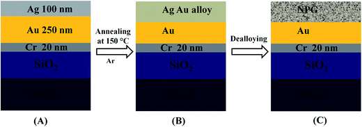
The interdigital patter of the NPG was prepared using a through-mask deposition procedure. Before deposition a mask of brass (0.15 mm) was cut by laser to prepare the required interdigital device. This way multiple numbers of devices with ten interdigits each were prepared. This procedure cuts down multiple steps required in a photolithographic method. Briefly, a silicon wafer of orientation (100) with 50.8 mm diameter was used as substrate. Initially silicon wafers were cleaned in the RCA process to remove organic and inorganic contaminants. After that, a thin film of silicon dioxide (250 nm) was grown by a wet thermal oxidation process. Before the deposition of metals a shadow mask with interdigital patterns was attached on silicon dioxide/silicon substrate to pattern the planner micro-supercapacitor (Fig. S1, ESI†). The chromium, gold and silver thin films were sequentially deposited using a four target e-beam evaporation machines. The deposition was carried out at the pressure of 3.5 × 10⁻⁶ mbar. The distance between graphite crucibles (source) and substrate was 18.5 cm during e-beam evaporation. The thicknesses of chromium, gold and silver layers were 20 nm, 250 nm and 100 nm respectively (Fig. 1). Chromium was used as adhesion promoter layer between silicon dioxide and gold layer. After deposition, the individual micro-supercapacitor devices were separated by cutting with a diamond cutter. The precursor AgAu alloy was then formed by annealing the device at an optimized temperature for a given time in argon (Ar) atmosphere. The optimized condition for annealing was 150 °C for 15 minutes, which lead to formation of Ag76Au24 alloy (Fig. S2, ESI†). After Ag76Au24 alloy formation the NPG interdigital patterns were formed by electroless dealloying in 3.5 M nitric acid (HNO3) for 30 minutes. The interdigit length, inter-digit separation and the channel width of planner micro-supercapacitor are 4.5 mm, 300 μm and 500 μm respectively (Fig. S1, ESI†), giving a footprint area of 44.66 mm2 (including interspaces gap between microelectrodes) excluding the contact pads.2.3 Deposition of copper oxide thin films
After dealloying, copper oxide (CuO) was deposited on NPG interdigital micro-electrodes by electrodeposition process. An acetate bath was used with 1.81 g copper acetate and 0.8 g sodium acetate dissolved in 120 ml water. The electrodeposition was carried out at a constant potential of −0.5 V (vs. MSE reference electrode). In order to vary the thickness of the CuO layer the deposition time was varied as 0.5, 1, 10 and 30 minutes.2.4 Preparation of solid electrolyte and micro-supercapacitor device
The PVA–H2SO4 gel was used as solid electrolyte for NPG–CuO micro-supercapacitors. The solid electrolyte was prepared by adding 3 g of PVA powder to 30 ml deionized water and 3 ml concentrated H2SO4. The mixture was then stirred at rotating speed of 1200 rpm at 85 °C for 2 hours to obtain transparent PVA–H2SO4 gel electrolyte. After formation the gel electrolyte was kept at room temperature for cooling.20,34 The cooled electrolyte was then applied on NPG–CuO interdigital micro-electrodes. Finally NPG–CuO interdigital micro-electrodes with gel electrolyte were kept in vacuum for drying.2.5 Materials characterizations
The microscopic morphology nanoporous gold (NPG) and NPG–CuO interdigital micro-electrodes was characterized using a Hitachi Model S-3400N field-emission gun scanning electron microscope (FEG-SEM). Cross-sectional imaging of all sample was done using dual-beam system with focus ion beam and field-emission gun scanning electron microscope (FIB-FEG-SEM) of Carl Zeiss Auriga Compact-4558. The structure of NPG–CuO samples were characterized by high-resolution transmission electron microscope TecnaiG2, F30 (HR-TEM) with 2.0 angstrom point resolution 1.0 angstrom line resolution and accelerating voltage of 300 kV. X-ray diffraction (XRD) patterns were obtained on a Philips X-Pert PRO X-ray diffractogram with Ni-filtered Cu Kα radiation (λ = 1.5418 Å) in the range 2θ = 10–90°. Surface analysis of electrodeposited CuO were performed by XPS using Kratos Analytical, UK (AXIS Supra), having analysis pressure of <2 × 10⁻⁹ Torr and fitted with a monochromatic Al Kα (1486.6 eV) X-ray source. Spectral analysis was performed using the peak fitting software (XPSPEAK version 4.1).2.6 Electrochemical measurements
The electrochemical performance of NPG–CuO micro-supercapacitor with PVA–H2SO4 solid electrolyte was measured by performing cyclic voltammetry (CV) and galvanostatic charge–discharge measurements using a Biologic potentiostat (SP-300) instrument. The cyclic voltammetry test was performed at different scan rates from 10 to 200 mV s−1 in potential window of −0.5 V to 0.5 V. All current values were normalized using the footprint area (i.e. 44.66 mm2 including interspaces gap between microelectrodes and excluding the contact pads) of the NPG–CuO micro-supercapacitor.3. Results and discussion
3.1 Microstructure studies
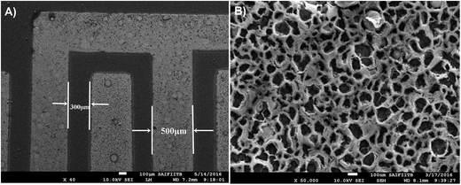
In this work NPG micro-electrodes patterns were prepared by following a sequential deposition procedure using a shadow mask, where individual devices were laser patterned in the mask. This method cuts down several steps used in a standard lithographic procedure. The top view SEM of a single device after dealloying is presented in Fig. 2A, which shows no edge-spreading after the deposition, indicating the formation of precise interdigital patterns. Another simplicity adapted in the work is a simple annealing method to prepare the AgAu alloy of required composition. | ||
| Fig. 2 (A) low magnification FEG-SEM image of NPG interdigital electrodes, (B) high magnification FEG-SEM of nanoporous gold layer showing typical nanoporous morphology. | ||
Due to fast diffusivity of the gold, the intermixing of the Au and Ag lead to formation of Ag76Au24 alloy layer (confirmed by XPS, Fig. S2 ESI†), with composition confirming to optimum percolation path required for the dealloying.35,36 The thickness of the intermixed alloy layer is about the thickness of the initial Ag layer, which is 100 nm. Fig. 2B represents the surface morphology of NPG layer. The crack free nano porous gold layer has a sponge like morphology with interconnected gold ligaments and a pore size distribution. The average pore size in the NPG layer is 77 nm ± 33 nm.
The electrodeposition of the CuO was carried out on the NPG interdigital patterns to form NPG–CuO micro-supercapacitor. The potentiostatic deposition was carried out for different time intervals such as 0.5, 1, 10 and 30 minutes, in order to form CuO layers of different thickness. The cyclic voltammetry after the deposition confirms to the CuO formation with the appearance of the signature redox peaks of the CuO (Fig. S3, ESI†).37
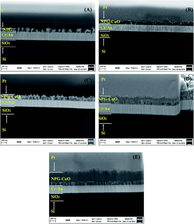
The growth of the CuO from within the NPG layers was studied by taking cross-sectional view in a dual beam FIB-SEM. Fig. 3 depicts cross-sectional view and average thickness of nanoporous gold (NPG) and electrodeposited copper oxide (CuO) on NPG for 0.5, 1, 10 and 30 minutes. In all samples except NPG, first platinum was deposited on top, and then gallium ion beam was used to cut a trench. Finally, cross-sectional imaging was done in FEG-SEM mode. For NPG sample carbon was deposited in place of platinum to get a better contrast image.
From the cross-sectional view of the NPG in Fig. 3A, the porous nature of the NPG layer is clearly visible. The porous layer is ca. 110 nm thick and contains many open pores. These open porous structures are essential for further deposition of active materials such as CuO by electrodeposition. Fig. 3B–E, represents cross-sectional view of the CuO deposited sample for 0.5, 1, 10 and 30 minutes, respectively. In all the samples thickness of silicon dioxide, chromium, gold and NPG are almost equal, but the thickness of copper oxide is increasing with deposition time. After half and one minute of deposition (Fig. 3B and C), it is very difficult to identify the copper oxide layer as the deposition has taken place throughout the pores of the NPG. From the cross-sectional view the imprints of the porosity of the NPG are still discernible. The deposition has filled the most of the pores of the NPG, while some surface pores and highly corrugated rough surface is still visible. From Fig. 3C, thickness of the NPG–CuO layer after 1 min deposition is 130 nm, about 20 nm thicker than pristine NPG layer. In case of 10 and 30 minutes deposited samples (Fig. 3D and E), the copper oxide layers are easily visible. The pores of NPG are completely filled (with some occluded pores) by copper oxide in all deposited samples. The via-filling of the pores indicates that the nucleation has started at the bottom of the pores because of the lower surface energy.34 The CuO layers have grown over the NPG layer for 10 and 30 min deposited samples. The average thickness of NPG–CuO layer after 10 and 30 minutes of deposition are ca. 220 nm and 500 nm, respectively (Fig. 3D and E). The top-view SEM of the NPG–CuO with different deposition times are presented in Fig. S4A–D.† In all deposited samples, CuO covers the whole surface of NPG. For 0.5 min sample, a few surface nanopores are visible (Fig. S4A†). However, the flake-like structure of the deposited CuO with higher surface roughness is clearly visible in all electrodeposited samples.
The high resolution transmission electron microscopy (HRTEM) image of the NPG and the NPG–CuO sample are presented in Fig. 4. Fig. 4A shows the inter planner distance between two gold planes. An interplanar spacing of 0.234 nm corresponds to (111) plane of NPG. From the HRTEM image of NPG–CuO (Fig. 4B) distance between two neighboring lattice fringes of CuO nanostructure is 0.253 nm which corresponds to (−111) plane of CuO.38 The interface between CuO and NPG in HRTEM confirms anchoring of CuO nanostructure on nanoporous gold substrate (Fig. 4B).
 | ||
| Fig. 4 HRTEM image of electrodeposited copper oxide on nanoporous gold micro-electrodes for 1 minute (A) NPG layer after dealloying, (B) NPG–copper oxide layer. | ||
The XRD of samples are presented in Fig. S5.† In the diffractogram of 30 min deposited sample (−111) peak of CuO is visible (JCPDS-050-6601) along with Au and Si peaks. This is in agreement with HRTEM data (Fig. 4B). The lattice spacing calculated from HRTEM matches with XRD. This indicates that there is a preferential (−111) orientation.
XPS analysis was carried out for elemental identification, finding the chemical state of element and relative composition of the constituent in the surface region. Fig. 5 shows representative XPS survey spectra for all NPG–CuO micro-supercapacitors and the inset shows high-resolution scan of Cu 2p core level. In XPS survey spectrum all the indexed peak correspond to Cu and O for all NPG–CuO samples, illustrating the purity of composite obtained by the electrodeposition. Intense lines in the survey spectrum at 930–950 eV, 121 eV and 74 eV corresponds to Cu 2p doublet (Cu 2p1/2 and Cu 2p3/2), Cu 3s and Cu 3p levels respectively. In addition, an Auger Cu LMM triplet, which represents energy levels of the Cu Auger process, was detected between 568 and 720 eV.39 Oxygen O 1s, at 530 eV and carbon C 1s, at 284 eV were present in all the samples. The main copper LMM peak at 568.8 eV corresponds to the copper peak position for the copper in bivalent oxidation state.40 The high-resolution Cu 2p spectrum has been depicted in the inset of Fig. 5. The Cu 2p3/2 peak lies at about 930.9 eV while the peak at 950.8 eV is assigned to Cu 2p1/2 peak. The binding energy gap between these two peaks is 20 eV which is comparable to the values of the standard spectrum of CuO.26
 | ||
| Fig. 5 XPS spectrum of copper oxide electrodeposited on nanoporous gold micro-electrodes for (A) 1 minute (B) 10 minute (C) 30 minutes deposition time. | ||
3.2 Electrochemical performance
Cyclic voltammetry (CV) was performed to investigate capacitive performance of NPG–CuO micro-supercapacitor for all the samples. CV of all samples was done at scan rates 10–200 mV s−1 in potential window of 1 V. Fig. 6A shows the CV plot of all samples at scan rate of 100 mV s−1 in PVA–H2SO4 electrolyte in a potential window of −0.5 V to 0.5 V. The nature of the CV is similar to that reported earlier.41,42 The pair of anodic and cathodic peaks within 0.2 V to −0.2 V can be recognized to faradaic redox reaction of CuO similar to previously reported literature.30,42 In this process Cu2+ converted into Cu+ during charging and reversible process occurs during discharging,27,43 shows pseudocapacitive behavior of NPG–CuO micro-supercapacitor. The charge storage mechanism of a pseudocapacitive material can be expressed by plotting peak currents as a function of scan rates from cyclic voltammetry experiment. For surface redox reaction the peak current varies directly with scan rates and for a semi-infinite bulk diffusion process the peak current varies as square root of scan rates.28,44 Fig. 7B shows the peak current as a function of square root of scan rate for NPG–CuO micro-supercapacitors. The plots show linear relationship between peak currents and square root of scan rate, which suggests semi-infinite bulk diffusion controlled process. Another feature is the one-to-one relation between anodic and cathodic peak current densities, which indicates excellent reversibility of the redox process in the given potential window.The areal capacitance was calculated by using eqn (1)9,45,46
 | (1) |
 is scan rate (V s−1) and A is the footprint area including interspaces gap (cm2) of the micro-supercapacitor. The areal capacitance of NPG–CuO micro-supercapacitors with different deposition times 0.5, 1, 10 and 30 minutes have been calculated using eqn (1). Fig. 6C shows areal capacitances as a function of scan rates (10–200 mV s−1) for all samples. In all the samples areal capacitance decreases with increasing scan rate from 10 mV s−1 to 200 mV s−1. The areal capacitance of micro-supercapacitor increases with decrease in scan rate because as scan rate decreases the electrolyte ions has longer time to enter in the bulk of electrode surface, but at higher scan rate electrolyte ion movement limited to solely surface of electrode.24 The areal capacitances of 0.5, 1, 10 and 30 minutes deposited NPG–CuO micro-supercapacitor are 0.66 mF cm−2, 5.58 mF cm−2, 21.85 mF cm−2 and 26.16 mF cm−2, obtained at scan rate of 10 mV s−1. These value for the areal capacitance is much higher than bulk CuO supercapacitor (0.12–25 mF cm−2)27,43 and several carbon based micro-supercapacitors such as porous carbon (1.5–3.5 mF cm−2),14 onion like carbon (1.7 mF cm−2),13 reduced graphene (0.08 mF cm−2),9 laser scribed graphene (2.32 mF cm−2)10 and laser written reduced graphene (0.51 mF cm−2).47 The obtained specific capacitance values are comparable with or better than several oxide based micro-supercapacitor such as VN–NiO (1.85 mF cm−2),21 hydrous RuO2 (40.7 mF cm−2),48 MnO2 (56.3 mF cm−2)19 and NPG/MnO2 (7.1 mF cm−2).16
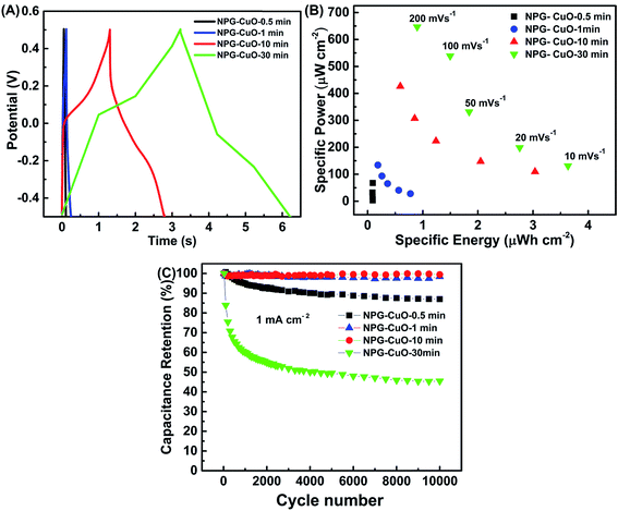
Galvanostatic charge–discharge (GCD) procedure was used to evaluate the electrochemical performance of NPG–CuO micro-supercapacitor. Fig. 7A shows GCD curves of NPG–CuO micro-supercapacitors at 1 mA cm−2 in potential range of −0.5 V to 0.5 V. The discharge time is minimum in NPG–CuO-0.5 min and maximum in NPG–CuO-30 min micro-supercapacitor. The nonlinear shape of GCD curves (Fig. 7A) reveals the features of pseudocapacitance, which are in conformity with the result of the CV curves (Fig. 6A).24,29 The nonlinear shape of GCD curve increases with increase in the thickness of the CuO layer.
The energy density and power density are very important parameters for any energy storage device. Ragone plots are used to illustrate the relation between energy density and power density of energy storage devices. The specific energy (EA) and specific power (PA) calculated by using eqn (2) and (3).9,45,46
 | (2) |
 | (3) |
 is scan rate of the micro-supercapacitor. NPG–CuO micro-supercapacitors shows maximum specific energy and specific power at 10 mV s−1 and 200 mV s−1, respectively (Fig. 7B). NPG–CuO-30 min micro-supercapacitor shows maximum specific energy 3.6 μW h cm−2 and specific power 646 μW cm−2, which is higher than specific energy 55 μW h cm−3 (ca. 2.024 nW h cm−2, using 368 nm) and specific power 3.4 W cm−3 (ca. 125 μW cm−2, using 368 nm) of NPG–MnO2 nanowires.34
Galvanic charge–discharge was performed to test the cyclic stability of NPG–CuO micro-supercapacitor for all NPG–CuO micro-supercapacitors. Cyclic stability of the NPG–CuO micro-supercapacitor was evaluated during continuous 10![[thin space (1/6-em)]](https://www.rsc.org/images/entities/char_2009.gif) 000 cycles at current density 1 mA cm−2 as shown in Fig. 7C. After 10
000 cycles at current density 1 mA cm−2 as shown in Fig. 7C. After 10![[thin space (1/6-em)]](https://www.rsc.org/images/entities/char_2009.gif) 000 cycles, the capacitance retention values for the 0.5, 1 min, 10 min and 30 min NPG–CuO micro-supercapacitors are 87%, 98%, 98% and 45%, respectively. The 1 min and 10 min NPG–CuO micro-supercapacitors demonstrating excellent cyclic stability with a high degree of reversibility in a repetitive charge/discharge process compare to 30 min sample. The capacitance retention of 30 min NPG–CuO micro-supercapacitor decreases within initial 2000 cycles, which might be due to degradation of electrode material during charging and discharging at high rate. However, after about 3000 cycles the loss factor is reduced.49,50 The capacitance retention of 30 min NPG–CuO micro-supercapacitor (with CuO layer thickness ca. 390 nm) is very less and is comparable reported value for bulk CuO electrodes, such as nanostructure CuO – 23% retention up to 2000 cycles,51 CuO nanoparticles – 49% retention up to 1000 cycles26 and bulk CuO – 51% retention up to 3000 cycles.42
000 cycles, the capacitance retention values for the 0.5, 1 min, 10 min and 30 min NPG–CuO micro-supercapacitors are 87%, 98%, 98% and 45%, respectively. The 1 min and 10 min NPG–CuO micro-supercapacitors demonstrating excellent cyclic stability with a high degree of reversibility in a repetitive charge/discharge process compare to 30 min sample. The capacitance retention of 30 min NPG–CuO micro-supercapacitor decreases within initial 2000 cycles, which might be due to degradation of electrode material during charging and discharging at high rate. However, after about 3000 cycles the loss factor is reduced.49,50 The capacitance retention of 30 min NPG–CuO micro-supercapacitor (with CuO layer thickness ca. 390 nm) is very less and is comparable reported value for bulk CuO electrodes, such as nanostructure CuO – 23% retention up to 2000 cycles,51 CuO nanoparticles – 49% retention up to 1000 cycles26 and bulk CuO – 51% retention up to 3000 cycles.42
4. Conclusions
In this work, we have successfully developed an all-solid-state on-chip micro-supercapacitor based on electrodeposition of pseudocapacitive CuO on interdigitated nanoporous gold electrodes. A simple two-step fabrication technique was followed to minimize the process steps as in rigorous lithography, while enhancing the functionality of the micro-devices produced. The as-prepared nanoporous gold substrate not only provides good electronic/ionic conductivity but also high intrinsic surface area for the electrodeposition of CuO, which forms a novel hybrid electrode. The microscale design of the device along with NPG substrate allows easy access of ions to the active material. Besides, the strong interfacial bonding between NPG and CuO minimizes electrical resistance in the device. This novel multiphase hybrid design of NPG–CuO micro-supercapacitor delivers high areal capacitance of 26 mF cm−2, specific energy 3.6 μW h cm−2 and specific power 646 μW cm−2, which are better than most of the reported carbon or oxide based micro-supercapacitors. The NPG–CuO micro-supercapacitors show excellent cyclic stability (98% capacitance retention after 10![[thin space (1/6-em)]](https://www.rsc.org/images/entities/char_2009.gif) 000 cycles at 1 mA cm−2) which is very important for their implementation in irreplaceable micro-electronic devices. The work demonstrates high surface area NPG with electrodeposited CuO is a unique approach to increase the capacitance and energy density of micro-supercapacitors.
000 cycles at 1 mA cm−2) which is very important for their implementation in irreplaceable micro-electronic devices. The work demonstrates high surface area NPG with electrodeposited CuO is a unique approach to increase the capacitance and energy density of micro-supercapacitors.
Acknowledgements
This work was supported by financial assistance from the DST (SERB, grant no. SB/S3/ME/057/2013). The authors acknowledge the funding for the dual beam FIB-SEM under the FIST programme (SR/FST/ETII-023/2012C), DST, GoI. Accesses to experimental facilities at Centre of Excellence in Nanoelectronics (CEN) and Centre for Research in Nanotechnology & Science (CRNTS), IIT Bombay, are highly acknowledged.References
- Z. L. Wang and W. Wu, Angew. Chem., Int. Ed., 2012, 51, 11700–11721 CrossRef CAS PubMed.
- Z. L. Wang, Adv. Mater., 2012, 24, 280–285 CrossRef CAS PubMed.
- J. Chmiola, C. Largeot, P. Taberna, P. Simon, J. Chmiola, C. Largeot, P. Taberna, P. Simon and Y. Gogotsi, Science, 2010, 328, 480–483 CrossRef CAS PubMed.
- A. Tyagi, K. M. Tripathi and R. K. Gupta, J. Mater. Chem. A, 2015, 3, 22507–22541 CAS.
- M. Beidaghi and Y. Gogotsi, Energy Environ. Sci., 2014, 7, 867–884 CAS.
- B. Song, L. Li, Z. Lin, Z.-K. Wu, K.-S. Moon and C.-P. Wong, Nano Energy, 2015, 16, 470–478 CrossRef CAS.
- D. Pech, M. Brunet, T. M. Dinh, K. Armstrong, J. Gaudet and D. Guay, J. Power Sources, 2013, 230, 230–235 CrossRef CAS.
- C. Soc, G. Wang and J. Zhang, Chem. Soc. Rev., 2012, 797–828 Search PubMed.
- Z.-S. Wu, K. Parvez, X. Feng and K. Müllen, Nat. Commun., 2013, 4, 2487 Search PubMed.
- M. F. El-Kady and R. B. Kaner, Nat. Commun., 2013, 4, 1475 CrossRef PubMed.
- D. Pech, M. Brunet, P. L. Taberna, P. Simon, N. Fabre, F. Mesnilgrente, V. Condéréa and H. Durou, J. Power Sources, 2010, 195, 1266–1269 CrossRef CAS.
- M. Beidaghi, W. Chen and C. Wang, J. Power Sources, 2011, 196, 2403–2409 CrossRef CAS.
- D. Pech, M. Brunet, H. Durou, P. Huang, V. Mochalin, Y. Gogotsi, P. Taberna and P. Simon, Nat. Nanotechnol., 2010, 5, 651–654 CrossRef CAS PubMed.
- B. Hsia, M. S. Kim, M. Vincent, C. Carraro and R. Maboudian, Carbon, 2013, 57, 395–400 CrossRef CAS.
- M. Beidaghi and C. Wang, Electrochim. Acta, 2011, 56, 9508–9514 CrossRef CAS.
- J. Han, Y.-C. Lin, L. Chen, Y.-C. Tsai, Y. Ito, X. Guo, A. Hirata, T. Fujita, M. Esashi, T. Gessner and M. Chen, Adv. Sci., 2015, 2, 1–7 Search PubMed.
- T. Mai, A. Achour, S. Vizireanu, G. Dinescu, L. Nistor, K. Armstrong, D. Guay and D. Pech, Nano Energy, 2014, 10, 288–294 CrossRef.
- X. Wang, Y. Yin, X. Li and Z. You, J. Power Sources, 2014, 252, 64–72 CrossRef CAS.
- X. Wang, B. D. Myers, J. Yan and G. Shekhawat, Nanoscale, 2013, 5, 4119–4122 RSC.
- W. Si, C. Yan, Y. Chen, S. Oswald, L. Han and O. G. Schmidt, Energy Environ. Sci., 2013, 6, 3218–3223 CAS.
- E. Eustache, R. Frappier, R. L. Porto, S. Bouhtiyya, J. F. Pierson and T. Brousse, Electrochem. Commun., 2013, 28, 104–106 CrossRef CAS.
- D. Kim, J. Yun, G. Lee and J. S. Ha, Nanoscale, 2014, 6, 12034–12041 RSC.
- Y. G. Zhu, Y. Wang, Y. Shi, J. I. Wong and H. Y. Yang, Nano Energy, 2014, 3, 46–54 CrossRef CAS.
- G. a. M. Ali, L. L. Tan, R. Jose, M. M. Yusoff, K. F. Chong, B. Vidhyadharan, I. I. Misnon, R. A. Aziz, K. P. Padmasree, M. M. Yusoff, R. Jose, M. Kumar, A. Subramania and K. Balakrishnan, J. Mater. Chem. A, 2014, 2, 6578–6588 Search PubMed.
- D. Mohapatra, S. Badrayyana and S. Parida, RSC Adv., 2016, 6, 14720–14729 RSC.
- A. Pendashteh, M. F. Mousavi and M. S. Rahmanifar, Electrochim. Acta, 2013, 88, 347–357 CrossRef CAS.
- V. D. Patake, S. S. Joshi, C. D. Lokhande and O. S. Joo, Mater. Chem. Phys., 2009, 114, 6–9 CrossRef CAS.
- V. Senthilkumar, Y. S. Kim, S. Chandrasekaran, B. Rajagopalan, E. J. Kim and J. S. Chung, RSC Adv., 2015, 5, 20545–20553 RSC.
- B. Heng, C. Qing, D. Sun, B. Wang, H. Wang and Y. Tang, RSC Adv., 2013, 3, 15719–15726 RSC.
- G. Wang, J. Huang, S. Chen, Y. Gao and D. Cao, J. Power Sources, 2011, 196, 5756–5760 CrossRef CAS.
- L. Y. Chen, J. L. Kang, Y. Hou, P. Liu, T. Fujita, A. Hirata and M. W. Chen, J. Mater. Chem. A, 2013, 1, 9202 CAS.
- C. Zhang, J. Xiao, L. Qian, S. Yuan, S. Wang and P. Lei, J. Mater. Chem. A, 2016, 4, 9502–9510 CAS.
- Z. Zeng, H. Zhou, X. Long, E. Guo and X. Wang, J. Alloys Compd., 2015, 632, 376–385 CrossRef CAS.
- Z. Zeng, X. Long, H. Zhou, E. Guo, X. Wang and Z. Hu, Electrochim. Acta, 2015, 163, 107–115 CrossRef CAS.
- S. Parida, D. Kramer, C. A. Volkert, H. Rösner, J. Erlebacher and J. Weissmüller, Phys. Rev. Lett., 2006, 97, 4–7 CrossRef PubMed.
- R. Liquid, J. Erlebacher, M. J. Aziz, A. Karma and N. Dimitrov, Nature, 2001, 410, 5–8 Search PubMed.
- W. Zhao, W. Fu, H. Yang, C. Tian, M. Li, Y. Li, L. Zhang, Y. Sui, X. Zhou, H. Chen and G. Zou, CrystEngComm, 2011, 13, 2871 RSC.
- D. A. Svintsitskiy, T. Y. Kardash, O. A. Stonkus, E. M. Slavinskaya, A. I. Stadnichenko, S. V. Koscheev, A. P. Chupakhin and A. I. Boronin, J. Phys. Chem. C, 2013, 117, 14588–14599 CAS.
- I. Platzman, R. Brener, H. Haick and R. Tannenbaum, J. Phys. Chem. C, 2008, 112, 1101–1108 CAS.
- D. S. Kozak, R. A. Sergiienko, E. Shibata, A. Iizuka and T. Nakamura, Sci. Rep., 2016, 6, 21178 CrossRef CAS PubMed.
- D. Mohapatra, S. Badrayyana and S. Parida, RSC Adv., 2016, 6, 14720–14729 RSC.
- S. E. Moosavifard, M. F. El-Kady, M. S. Rahmanifar, R. B. Kaner and M. F. Mousavi, ACS Appl. Mater. Interfaces, 2015, 7, 4851–4860 CAS.
- D. P. Dubal, D. S. Dhawale, R. R. Salunkhe, V. S. Jamdade and C. D. Lokhande, J. Alloys Compd., 2010, 492, 26–30 CrossRef CAS.
- I. E. Rauda, V. Augustyn, B. Dunn and S. H. Tolbert, Acc. Chem. Res., 2013, 46, 1113–1124 CrossRef CAS PubMed.
- M. D. Stoller and R. S. Ruoff, Energy Environ. Sci., 2010, 3, 1294–1301 CAS.
- S. Zhang and N. Pan, Adv. Energy Mater., 2015, 5, 1–19 Search PubMed.
- W. Gao, N. Singh, L. Song, Z. Liu, A. L. M. Reddy, L. Ci, R. Vajtai, Q. Zhang, B. Wei and P. M. Ajayan, Nat. Nanotechnol., 2011, 6, 496–500 CrossRef CAS PubMed.
- C. C. Liu, D. S. Tsai, D. Susanti, W. C. Yeh, Y. S. Huang and F. J. Liu, Electrochim. Acta, 2010, 55, 5768–5774 CrossRef CAS.
- S. M. Pawar, J. Kim, A. I. Inamdar, H. Woo, Y. Jo, B. S. Pawar, S. Cho, H. Kim and H. Im, Sci. Rep., 2016, 6, 1–9 Search PubMed.
- D. P. Dubal, G. S. Gund, R. Holze and C. D. Lokhande, J. Electroanal. Chem., 2014, 712, 40–46 CrossRef CAS.
- M. B. Gholivand, H. Heydari, A. Abdolmaleki and H. Hosseini, Mater. Sci. Semicond. Process., 2015, 30, 157–161 CrossRef CAS.
Footnote |
| † Electronic supplementary information (ESI) available. See DOI: 10.1039/c6ra19744b |
| This journal is © The Royal Society of Chemistry 2016 |


