Novel ibuprofenate- and docusate-based ionic liquids: emergence of antimicrobial activity

Clarissa P. Frizzo*a, Keli Wusta, Aniele Z. Tiera, Thaíssa S. Becka, Leticia V. Rodriguesa, Rodrigo A. Vaucherb, Leandro P. Bolzanb, Silvio Terrac, Felix Soaresc and Marcos A. P. Martinsa
aNUQUIMHE, Federal University of Santa Maria, Santa Maria, RS, Brazil. E-mail: clarissa.frizzo@gmail.com; Fax: +55 5532208756; Tel: +55 5532208756
bLab. of Microbiology, Centro Universitário Franciscano, Santa Maria, RS, Brazil
cDepart. of Molecular Biology, Federal University of Santa Maria, Santa Maria, RS, Brazil

Received 5th September 2016 , Accepted 13th October 2016

First published on 14th October 2016


Abstract

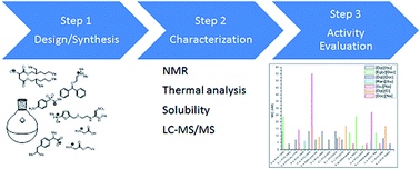
Six new ionic-liquid-based active pharmaceutical ingredients (IL-APIs) were prepared and their molecular structures characterized. Solubility and thermal properties was determined and compared with the salt precursors. Antifungal and antibacterial activities were also investigated. Some of the IL-APIs demonstrated limited water solubility and high thermal stability when compared with the salt precursor. The most interesting observation was that the combination of two pharmacologically active ingredients in an IL-API results in antifungal activity that was not present in the precursors. The antibacterial activities were very promising considering that one of them was active against Staphylococcus, which is a bacterium resistant to penicillin and methicillin. The study of the pharmacological properties of synthesized IL-APIs is essential for evaluating how a drug may interfere with the pharmacological properties of another drug, thus furnishing knowledge about the advantages or disadvantages related to the association of APIs.

Introduction

The potential pharmaceutical applications of ionic liquids (ILs) were initially demonstrated via determination of their cytotoxicity and antimicrobial activity.1 The applications have now expanded to: the formation of microemulsion droplets for the transport and release of drugs; stabilizing agents for active ingredients, additives, and polymers;2 and the development of ionic-liquid-based active pharmaceutical ingredients (IL-APIs).3

More than 50% of drugs on the market today are sold as salts.3 Ion-pair formation enhances the transport of various ionic drugs through the skin and across the absorbing membrane.3 Conversion of a drug into a salt is a crucial step in drug development and can have a huge impact on its properties; for example, solubility, dissolution rate, hygroscopic, stability, and particle characteristics.4 Conversion of a drug in IL (organic liquid salt) is an innovative approach that could eliminate many problems in crystalline formulations associated with delivery mechanisms (dissolution, transport, and bioavailability)5,6 or poor control over polymorphism, which can dramatically change properties, such as solubility,7–9 while retaining pharmacological activity and neutralizing improved or unwanted side effects. In other words, cations and anions could be selected to: (i) act pharmacologically synergic, (ii) act pharmacologically independent,3,10,11 or (iii) improve pharmacokinetic properties.12 Thus, highly ion-associated and pharmaceutically active ILs would be highly beneficial forms of the original pharmaceutically active salts.3 Previous work has shown that imidazolium-based ILs can be used as multifunctional material in coatings for titanium surfaces, due to them having desirable characteristics such as low toxicity,13 antimicrobial activity,13 affinity to titanium surfaces,14,15 and protecting the titanium surface against physical damage and wear. Efforts are increasing to develop ILs focused on increasing titanium bioactivity to improve osseointegration or to provide antimicrobial activity to prevent biofilm formation.

In this context, the aim of this work was to develop the synthesis as well as physical, thermal, and spectral characterization of novel ibuprofenate- and docusate-based IL-APIs, and to investigate their cytotoxicity as well as their antioxidant and antimicrobial activity, in order to show the potential of these two anions in the development of new drugs or new titanium coatings. Cations and anions were firstly selected, based on compounds that have been successful in the synthesis of IL-APIs,5 and new combinations were then produced. In order to achieve the aim of this work, we combine our know-how in organic synthesis,16 ILs as reaction media in organic reactions,17,18 and pharmacological evaluations of heterocycles such as pyrazoles,19–23 pyrimidines,24–27 quinolines28 and imidazoles.29

Experimental section

Synthesis and characterization

All ionic liquids derived from ibuprofen and docusate were synthesized according to the following methodology: in a 200 mL round-bottomed flask charged with a magnetic stir bar, (10 mmol) of sodium ibuprofen or sodium docusate was dissolved in 70 mL solution of ketone[thin space (1/6-em)]:[thin space (1/6-em)]water (8[thin space (1/6-em)]:[thin space (1/6-em)]2 v/v). To this stirred mixture, one 10 mmol portion of ranitidine, diphenhydramine, glycine, or glycine ethyl hydrochloride was added. The suspension was then stirred for 24 h at 40 °C, and after this time the precipitate was removed by gravity filtration, and then the solvent was removed in vacuo. All residues were extracted with 50 mL of dichloromethane and washed with water to remove any inorganic salt. The presence of chloride anion was monitored via the silver nitrate test. The solvent was removed using a rotary evaporator to furnish the product. Ranitidine sulfacetamide: was synthesized according methodology described above. However, the reaction product was washed with diethyl ether.

Solubility

A saturated solution of each IL-API was stirred for 24 h. The solutions were then left to rest for a further 24 h, and after this, 100 mL of the supernatant was collected. The solvent was evaporated at 40 °C, and the mass of the compounds was determined gravimetrically.

Thermal analysis

The DSC experiments were performed using an MDSC Q2000 apparatus (T-zeroTM DSC technology, TA Instruments Inc., New Castle, DE, USA). Dry high purity (99.999%) nitrogen gas was used as the purge gas (50 mL min−1). The instrument was calibrated using the onset temperatures for melting indium (156.60 °C). The heat capacity calibration was done by running a standard sapphire (α-Al2O3). The heating rate used for all the samples was 10 °C min−1. Each IL was subjected to three cycles of heating and cooling in the temperature range of −80 to 200 °C. Samples were crimped into hermetic aluminum pans with lids (Sartorius, M 500 P, Goettingen, Germany). TGA was done using a TGA Q5000 apparatus (TA Instruments Inc., USA). The heating rate was 10 °C min−1 and the N2 flow rate was 25 mL min−1 from 298.15 to 973.15 K. The sample mass was 2–5 mg. The TGA equipment was calibrated using CaC2O4·H2O (99.9%).

NMR analysis

1H and 13C NMR spectra were recorded on a Bruker Avance III 600 MHz (1H at 600 MHz and 13C at 150 MHz) in 5 mm sample tubes with DMSO-d6, using TMS as internal reference. The temperature of the samples was 298 K and the spectral digital resolution was ±0.01 ppm.

Mass spectrometry (ESI-MS)

Electrospray ionization mass spectra (ESI-MS) were acquired with an Agilent Technologies 6460 Triple quadrupole 6460 apparatus (LC/MS-MS) (Santa Clara, CA, USA), operated in the positive-ion mode. The gas temperature was 300 °C, the flow of the drying gas was 5 L min−1, and the nebulizer was set to 45 psi. The voltage of the capillary was 3500 V, while for the fragmentor it was 0 V. The ionic liquid solutions in H2O were introduced at a 5 μL min−1 flow rate. Nitrogen was used as nebulization gas and argon as collision gas. Molecular ions were detected using positive mode, in which the m/z ratio is given for one cation and one anion.

Elemental analysis

The elemental analyses for compounds were performed on a Perkin Elmer 2400 CHN analyser, on the Instituto da Química, USP, São Paulo.

Chlorine residual determination

Residual content of chloride was determined by a ICP-AES spectrometer (Spectro Ciros CCD, Spectro Analytical Instruments, Germany) equipped with a dual-step mist chamber (Scott type) and a cross-flow nebulizer. The plasma power was 1550 W and the argon flow was 12 L min−1 (plasma), 1 L min−1 (nebulizer), and 1 L min−1 (auxiliary). A wavelength of 134.724 nm was used for chloride.

Spectral data

[Ran][Ibu]. C26H40N4O5S, MW: 520.68 g mol−1; brown oil, 79%. Tg: −5.76 °C; 1H NMR (600 MHz, DMSO-d6): δ 0.85 (d, 6H, 2CH3), 1.34 (d, 3H, CH3), 1.80 (m, 1H, CH), 2.14 (s, 6H, 2CH3), 2.18 (s, 3H, CH3), 2.41 (d, 2H, CH2), 2.68 (m, 2H, CH2), 3.40 (s, 2H, CH2), 3.61 (q, 1H, CH), 3.81 (s, 2H, CH2), 6.19 (d, 1H, CH), 6.49 (d, 1H, CH-Ar), 7.09 (d, 2H, 2CH-Ar), 7.18 (d, 2H, CH-Ar), 9.99 (d, 1H, CH-Ar). RMN 13C 600 MHz (d6-DMSO): δ 18.49 (CH3), 22.09 (2CH3), 29.53 (CH), 44.15–44.32 (2CH3, CH, CH2), 96.89 (CH-Ar), 127.02 (2CH-Ar), 128.85 (2CH-Ar), 138.54 (C-Ar), 139.40 (C-Ar), 150.63 (C-Ar), 151.54 (C-Ar), 175.51 (CO2). Anal. calc. for C26H40N4O5S: C, 59.26; H, 7.56; N, 11.06. Found: C, 58.83; H, 7.86; N, 10.92. MS m/z molecular ion (M+): 315.2.
[Dip][Ibu]. C30H39NO3, MW: 461.64 g mol−1; colorless oil, 79%. Tg: −36.38 °C; 1H NMR (600 MHz, DMSO-d6): δ 0.85 (d, 6H, 2CH3), 1.34 (d, 3H, CH3), 1.80 (m, 1H, CH), 2.20 (s, 6H, 2CH3-Ar), 2.40 (d, 2H, CH2), 2.56 (t, 2H, CH2), 3.49 (t, 2H, CH2), 3.60 (q, 1H, CH), 5.45 (s, 1H, CH), 7.08 (d, 2H, 2CH-Ar), 7.19 (d, 2H, 2CH-Ar), 7.22 (t, 2H, 2CH-Ar), 7.31 (t, 4H, 4CH-Ar), 7.36 (d, 4H, 4CH-Ar). RMN 13C 600 MHz (d6-DMSO): δ 18.64 (CH3), 22.10 (2CH3), 29.55 (CH), 44.18 (CH2), 44.62 (2CH3), 45.05 (CH), 57.95 (CH2), 66.27 (CH2), 82.49 (CH), 126.45 (4CH-Ar), 127.04 (2CH-Ar), 127.15 (2CH-Ar), 128.22 (4CH-Ar), 128.80 (2CH-Ar), 138.90 (C-Ar), 139.34 (C-Ar), 142.41 (2C-Ar), 175.72 (CO2). Anal. calc. for C30H39NO3: C, 78.05; H, 8.52; N, 3.03. Found C, 76.72; H, 9.14; N, 3.45. MS m/z molecular ion (M+): 256.2.
[Gly][Doc]. C22H43NO9S, MW: 497.64 g mol−1; colorless oil, 70%. Tg: −26.22 °C; 1H NMR (600 MHz, DMSO-d6): δ 0.82–0.88 (m, 12H, CH3), 1.24–1.38 (m, 16H, CH2), 1.50 (m, 2H, CH), 2.78–2.82 (dd, 1H, CH2), 2.89–2.94 (dd, 1H, CH2), 3.63–3.66 (dd, 1H, CH), 3.70 (s, 2H, CH2), 3.87–3.91 (m, 4H, CH2), 8.06 (2H, NH). RMN 13C 600 MHz (d6-DMSO): δ 10.69–13.83 (4CH3), 22.29–29.65 (8CH2), 34.02 (CH2), 38.05 (2CH), 61.33 (CH), 65.95–66.08 (2CH2), 168.27 (CO2H), 168.98 (CO2R), 170.97 (CO2R). Anal. calc. for C22H43NO9S: C, 53.10; H, 8.71; N, 2.81. Found C, 52.76; H, 8.62; N, 2.28. MS m/z molecular ion (M+): 74.1.
[EGly][Doc]. C24H47NO9S, MW: 525.70 g mol−1; colorless oil, 76%. Tg: −44.52 °C; 1H NMR (600 MHz, DMSO-d6): δ 0.82–0.88 (m, 12H, CH3), 1.23–1.34 (m, 19H, CH2–CH3), 1.50 (m, 2H, CH), 2.77–2.81 (dd, 1H, CH2), 2.88–2.93 (dd, 1H, CH2), 3.62–3.64 (dd, 1H, CH), 3.81 (s, 2H, CH2), 3.86–3.91 (m, 4H, CH2), 4.21 (q, 2H, CH2), 8.17 (bs, 3H, NH). RMN 13C 600 MHz (d6-DMSO): δ 10.70 (CH3), 13.81–13.89 (CH3, CH2), 22.32–29.67 (CH2, CH3), 34.00 (CH2), 38.04–38.11 (CH), 61.35–61.50 (CH, CH2), 65.98–66.14 (CH2), 167.61 (CO2R), 168.26 (CO2R), 170.95 (CO2R). Anal. calc. for C24H47NO9S: C, 54.83; H, 9.01; N, 2.66. Found C, 54.29; H, 8.78; N, 3.02. MS m/z molecular ion (M+): 104.1.
[Doc][Dip]. C37H59NO8S, MW: 677.63 g mol−1; colorless oil, 68%. Tg: −37.19 °C; 1H NMR (600 MHz, DMSO-d6): δ 0.81–0.88 (m, 12H, CH3), 1.23–1.37 (m, 16H, CH2), 1.49 (m, 2H, CH), 2.82 (s, 6H, CH3), 2.79–2.84 (dd, 1H, CH2), 2.91–2.98 (dd, 1H, CH2), 3.37 (t, 2H, CH2), 3.65–3.69 (m, 3H, CH2, CH), 3.84–3.94 (m, 4H, CH2), 5.58 (s, 1H, CH), 7.26 (t, 2H, CH-Ar), 7.35 (t, 4H, CH-Ar), 7.41 (d, 4H, CH-Ar), 9.23 (bs, 1H, NH). RMN 13C 600 MHz (d6-DMSO): δ 10.71–13.84 (4CH3), 22.33–29.68 (8CH2), 34.07 (CH2), 38.08 (2CH), 42.77 (2CH3), 56.03 (CH2), 62.60 (CH), 66.03 (2CH2), 82.81 (CH), 126.59 (4CH-Ar), 172.45 (2CH-Ar), 128.34 (4CH-Ar), 141.67 (2C-Ar), 168.33 (CO2R), 170.98 (CO2R). Anal. calc. for C37H59NO8S: C, 65.55; H, 8.77; N, 2.07. Found C, 64.83; H, 8.70; N, 1.84. MS m/z molecular ion (M+): 256.2.
[Sulf][Ran]. C21H32N6O6S2, MW: 528.65 g mol−1; yellow oil, 50%. Tg: 25.13 °C; 1H NMR (600 MHz, DMSO-d6): δ 1.88 (s, 3H, CH3), 2.19 (6H, CH3), 2.70 (s, 3H, CH3), 3.46 (s, 2H, CH2), 3.82 (s, 2H, CH2), 6.11 (bs, 2H, NH2), 6.24 (d, 2H), 6.51 (d, 1H, CH-Ar), 6.62 (d, 2H, CH-Ar), 7.27 (d, 1H, CH-Ar), 7.54 (d, 2H, CH2-Ar); RMN 13C 600 MHz (d6-DMSO): δ 23.22 (CH3), 27.08 (CH2), 30.58 (CH2), 44.06 (2CH3), 54.42 (CH2), 96.99 (CH-Ar), 108.38 (CH), 112.21 (2CH-Ar), 129.61 (2CH-Ar), 150.79 (C-Ar), 153.40 (C-Ar), 168.56 (NCO). Anal. calc. for C19H30N6O5S2: C, 46.90; H, 6.21; N, 17.27. Found C, 43.80; H, 6.53; N, 13.22. MS m/z molecular ion (M+): 315.2.

Antifungal assay

Disk diffusion testing was done strictly in accordance with CLSI standard M44-A2 (2008), using Mueller–Hinton agar plus 2% glucose and 0.5 μL mL−1 methylene blue dye. Filter paper discs were placed on the surface of the medium and inoculated with 10 μL of the solutions of the IL-API (100 mg mL−1). The plates were incubated at 37 °C and the inhibition zone was recorded after 24 h and 48 h of incubation. The microdilution technique was executed according to the CLSI M27-A3 protocol (2008). The assay was done in 96-well microtiter plates—each well was inoculated in 100 μL of RPMI-1640 supplemented with MOPS. Subsequently, 100 μL of the IL-API solution was pipetted in the first well and a serial dilution was conducted to the desired concentration. After dilution, 10 μL of inoculum was inserted into each well. The plates were incubated for 48 h and the reading was done by microscopy to determine the presence or absence of yeast in the wells. Negative and positive controls were performed in the test.

Antibacterial assay

Each sample was mixed with an inoculum prepared in the same medium at a density adjusted per tube to 0.5 on the McFarland scale (1.5 × 10⁻⁸ CFU mL−1). With sterile swabs, bacterial suspensions were plated on the surfaces of Petri plates containing about 15 mL of Mueller–Hinton agar with a thickness of approximately 4 mm. Disks with a diameter of 6 mm, and sterilized filter paper containing 10 μL of the IL-API were placed on the surface of the plates in contact with the inoculum of the microorganism. The plates were incubated at 37 °C, and the inhibition zone was recorded after 24 h of incubation. The minimal inhibitory concentration (MIC) was determined by microdilution techniques in Mueller–Hinton broth (disk) for the Paenibacillus species (CLSI, 2008). The assay was done in 96-well microtiter plates. Each sample was mixed with an inoculum prepared in the same medium—at a density adjusted per tube to 0.5 on the McFarland scale (1.5 × 10⁻⁸ CFU mL−1)—and diluted at a ratio of 1[thin space (1/6-em)]:[thin space (1/6-em)]10 for the broth microdilution procedure. Microtiter trays were incubated at 37 °C, and the MICs were recorded after 24 h of incubation. The MIC was defined as the lowest concentration of compounds that inhibits bacterial growth. This test was performed in triplicate, on separate occasions. The 2,3,5-triphenyltetrazolium chloride was used as an indicator of bacterial growth.

Total antioxidant capacity assay

The total antioxidant potential of the IL-APIs was evaluated via the phosphomolybdenum method, which has already been described.30 A sample aliquot in ethanol (0.3 mL) solution was combined in a vial with reagent solution (0.6 M sulfuric acid, 28 mM sodium phosphate, and 4 mM ammonium molybdate, 3 mL). The compounds were tested at concentrations of 100, 250, 500, and 1000 μM. The vials were capped and incubated in a water bath at 95 °C for 90 min. After cooling the mixture to room temperature, the absorbance was measured at 695 nm against a blank control.

DPPH radical scavenging method

The radical scavenging activities of the IL-APIs were determined in accordance with the method previously described by Brand-Williams et al.31 Each compound was tested at 100, 250, 500, and 1000 μM in 1% DMSO. DPPH˙ (diluted in ethanol) was added to the final concentration of 0.3 mM and allowed to react at room temperature for 30 min in dark conditions. The absorbance was measured at 518 nm using Spectra Max Plate Reader® M3 (Molecular Devices, Sunnyvale, California, USA). Individual dependent variable data were analyzed statistically by one-way analysis of variance (ANOVA), followed by Bonferroni's multiple comparison test, when appropriate. Differences between groups were considered to be significant when p < 0.05. Data are expressed as mean ± SEM, and each experimental procedure was performed in at least four individual experiments with three replicates each.

Cytotoxicity evaluation

Culture of lymphocytes. Peripheral blood samples were obtained by venipuncture, from discarded samples of the Clinical Analysis Laboratory School, using a top Vacutainer® (BD Diagnostics, Plymouth, UK) and heparin tubes. The research was approved by the Human Research Ethics Committee of the Centro Universitário Franciscano (CAAE: 31211214.4.0000.5306). The Histopaque-1077® (Sigma-Aldrich, St. Louis, MO) density gradient was used to separate lymphocyte cells, using 20 mL blood samples. After further centrifugation for 30 min at 2500 × g, the cells were transferred to a culture medium containing 5 mL RPMI 1640 with 10% fetal bovine serum, 1% penicillin, and 1% streptomycin. The cells were cultured in a 96-well microplate at an initial density of 2 × 10⁵ cells, and at 37 °C in a 5% humidified CO2 atmosphere, in order to assess cell viability. The cells were then treated with two concentrations of Gly, Doc, Bup, Ibu, Lip, Dip, and Ran; and the ILs [Gly][Doc], [EGly][Doc], [Dip][Ibu], [Dip][Doc], [Ran][Ibu], and [Ran][Suf] (100 and 1000 μM) were dissolved in the medium and further incubated for 72 h. Subsequently, the cytotoxicity in the lymphocytes was determined via the MTT assay.
MTT assay. Cell viability was evaluated via the MTT assay, which is a colorimetric assay that measures the reduction of yellow 3-(4,5-dimethythiazol-2-yl)-2,5-diphenyl tetrazolium bromide (MTT) by mitochondrial succinate dehydrogenase, as described by Mosmann32 with modifications. Initially, 20 μl of MTT (5 mg mL−1) in phosphate-buffered saline was added to each well and the plate was incubated at 37 °C for 4 h. The medium was then removed, and 100 μl of DMSO was added to each well. After 10 min of incubation at 37 °C, the plate was read at 570 nm using a TP-Reader microplate reader (Thermoplate, China). As a positive control, 10% of SDS was used. Additionally, a negative control—control wells without treatment—was employed and prepared under the same experimental conditions. All treatments were done in triplicate (in the same 96-well plate) and repeated two times in independent experiments. The cell viability percentage was calculated as: (absorbance of test/absorption of the control) × 100.33 Data were expressed as the mean ± standard deviation (SD) for three independent determinations for each experimental point. Data were analyzed using the GraphPad Prism 4.00 software package for Windows (GraphPad Software, San Diego-CA, USA). All data from this study were submitted to analysis of variance (oneway ANOVA) followed by the Tukey test (p < 0.05).

Results and discussion

Synthesis and characterization

The IL-APIs ranitidinium ibuprofenate, diphenhydraminium ibuprofenate, glycinium docusate, ethyl ester glycinium docusate, diphenhydraminium docusate, and ranitidinium sulfacetamide were prepared from their respective commercial salts via metathesis reactions.4,12 All of the IL-APIs were isolated at moderate yields (50–84%) and high purity (Table 1). All of the salts used in the synthesis of the IL-APIs were solid and displayed crystal characteristics, except for docusate sodium. The IL-APIs obtained are liquid at room temperature (Table 2). Ibuprofenate and docusate salts were clear, slightly yellow viscous liquids at room temperature, except for [Ran][Ibu], which was light brown. The residual chloride content was determined by chloride ion quantitative analysis, and the results showed 0.0073, 0.0091, 0.00021, 0.003, and 0.0077 mol kg−1 of the chloride impurity in [Ran][Ibu], [Dip][Ibu], [Dip][Doc], [EGly][Doc], and [Ran][Sulf] respectively. For [Gly][Doc], the impurity was below the detection limit. The sodium or hydrochloride salts and their corresponding ibuprofenate and docusate salts were analyzed by 1H and 13C nuclear magnetic resonance (NMR), electrospray ionization mass spectrometry (ESI-MS), and elemental analysis. Additionally, the compounds were characterized by thermogravimetric analysis (TGA) and differential scanning calorimetry (DSC). Changes in chemical shift after IL-API formation were more sensitive in the 1H NMR spectrum than in the 13C NMR. In general, 1H NMR signals from the ibuprofen in the IL-API showed a downfield shift in relation to sodium ibuprofenate. The largest changes in chemical shift after the formation of [Ran][Ibu] and [Dip][Ibu] were observed in the methyl and methine adjacent to the carboxylate (δΔ = 0.4–1.0) of the ibuprofen. When cations in the IL-API were analyzed, 1H NMR signals indicated an upfield shift. The largest changes in chemical shift after the formation of [Ran][Ibu] and [Dip][Ibu] were observed in the methyl and methine adjacent to the amino (δΔ = 0.4–0.6) of the ranitidine and diphenhydramine. It is important to note that the signal at 11.06 and 11.15 ppm of the hydrogen of the amino group, in the [Ran][Cl] and [Dip][Cl] spectrum, respectively, disappears upon IL-API formation. Docusate-based ionic liquid did not show significant changes in chemical shifts in the 1H NMR signals when compared to the precursor. For example, the hydrogen of the amino group of the glycine and glycine ethyl ester in the IL-API showed an upfield shift (δΔ = 0.4–0.6), while the 1H signal of the amino group (diphenhydramine) showed a downfield shift. The hydrogen signal of the methylene group attached to N showed an upfield shift. In the case of [Ran][Sulf], all the hydrogens of the sulfacetamide showed a downfield shift, while the hydrogens of the ranitidine showed an upfield shift when compared with precursor salts.
Table 1 Structure, common name, and abbreviation of the IL-APIs
Structure Common name Abbreviation
image file: c6ra22237d-u1.tif Ranitidinium ibuprofenate [Ran][Ibu]
image file: c6ra22237d-u2.tif Diphenhydraminium ibuprofenate [Dip][Ibu]
image file: c6ra22237d-u3.tif Glycinium docusate [Gly][Doc]
image file: c6ra22237d-u4.tif Ethylglycinium docusate [EGly][Doc]
image file: c6ra22237d-u5.tif Diphenhydraminium docusate [Dip][Doc]
image file: c6ra22237d-u6.tif Ranitidinium sulfacetamide [Ran][Sulf]


Table 2 Thermal propertiesa of IL-APIs
IL-API Tmb (°C) Tgc (°C) T5% onsetd (°C) Tde (°C)
a Phase transitions were evaluated from DSC (second cycle) at 5 °C min−1. Decomposition temperatures were determined at a TGA heating rate of 10 °C min−1 under N2 flux. Samples were initially heated and then cooled to −80 °C.b Tm is the melting temperature.c Tg is the glass transition temperature.d T5% onset is the 5 wt% mass loss temperature.e Td is the total mass loss temperature.f Indicates the Td for multiple decomposition steps.
[Ran][Ibu] −6 164 219
[Dip][Ibu] −36 162 229
[Gly][Doc] −26 200 1st: 221; 2nd: 246; 3rd: 320f
[EGly][Doc] −45 233 1st: 245; 2nd: 326f
[Dip][Doc] −37 240 1st: 263; 2nd: 291f
[Ran][Sulf] 25 181 207


As a method of soft ionization, electrospray ionization is a technique used in the analysis of non-volatile molecules from the liquid phase, and it enables molecules linked by non-covalent interactions to be detected. Thus, it is an essential tool in the characterization of the structures of IL-APIs. The formation of [Ran][Ibu], for example, was monitored by the ESI-MS spectra in the positive and negative mode. In the positive mode, the ion was observed with m/z 315.2, which corresponds to the ranitidinium cation; while in the negative mode the ion was observed with m/z 205.1, which corresponds to the ibuprofenate anion. Likewise, the formation of the other IL-APIs was monitored using the ESI-MS spectra—see ESI for data on molecular formula, molecular weight, and monoisotopic mass, as well as mass spectrometry data for the IL-APIs (Table S1).

DSC analysis of the IL-APIs indicated either a glass transition and melting point at a negative temperature or no melting point (Table 2). For example, [Ran][Ibu] and [Ran][Sulf] had a glass transition at −5.56 and 25.13 °C, respectively, and no melting point. Thus, it was possible to prepare two ILs which do not crystallize and, consequently, will not suffer from polymorphism, which is one of the main problems of ranitidine.4

TGA provides information that allows comparison of the thermal stability of the IL-API with the precursor, and from this data one can judge if each IL-API is more or less stable. In general, TGA data suggested a one-step decomposition temperature with good thermal stabilities (>155 °C) for all IL-APIs derived from ibuprofen sodium (Table 2). [Ran][Ibu], for example, had a T5% onset of 165 °C, which is higher than that of ranitidine hydrochloride (154 °C) and ibuprofen sodium (54 °C). The T5% onset of docusate sodium was 225 °C in one-step decomposition. The IL-API derivatives of this anion had one, two, or three decomposition steps with thermal stabilities ≥200 °C. In other words, the cations contribute to an increase in the number of decomposition steps, as well as higher thermal stability of the IL-APIs.

Finally, [Ran][Sulf] also had good thermal stability, with a T5% onset of 181 °C, which is significantly higher than that of sulfacetamide sodium (155 °C) and ranitidine hydrochloride (154 °C).

Our results are supported by previous results, described by Bica et al.,34 which state that the liquid glass-like state by itself is not necessarily related to reduced thermal stability. On the other hand, the long-term stability of IL-APIs—similar to that for any other salts—is directly related to the stability of the ions. Successful formation of a stable IL requires stable ions.

Solubility and ionicity

The maximum water solubility of the IL-APIs and the respective precursors (sodium or hydrochloride salts) was determined. The solubility assay indicated that, in general, the water solubility of the new IL-APIs was similar to that of their precursors (Table S2). Ibuprofen is a common non-steroidal anti-inflammatory drug used to treat arthritis symptoms and fever, and it can also be used as an analgesic.4 Sodium salt is readily available and it is primarily used to increase the water solubility of ibuprofen, which is less than 1 mg mL−1 in free acid form. Combined with the ibuprofenate anion to prepare a dual action IL, as shown in Table 1, it was possible to obtain compounds with improved solubility compared to ibuprofen in free-acid form. The maximum water solubility of the IL-API derivatives of ibuprofen was determined for [Ran][Ibu] (145 ± 7 mg mL−1), which was similar to that observed for sodium ibuprofen (140 ± 10 mg mL−1). The ranitidine anion has higher hydrophobicity than the sodium cation, which can lead to similar or reduced solubility of the IL-API in comparison with sodium salt. Sodium docusate has emollient properties and was used by Rogers12 in the development of an IL with hydrophobic characteristics. The salt form of this API has low solubility in water (20 mg mL−1), due to its long alkyl chains. The IL-APIs [Gly][Doc] and [Dip][Doc] have solubilities similar to or lower than those of bupivacaine, diphenhydramine, and glycine hydrochloride. Thus, it can be seen that docusate is responsible for the decreased solubility of IL-API, due its hydrophobic character. Sulfacetamide is generally prescribed in salt form—it exhibits solubility problems in its free-acid form.4 [Ran][Sulf] showed low solubility in water and moderate solubility in organic solvents, and it was less soluble than sodium sulfacetamide, but slightly more soluble than ranitidine hydrochloride.

The degree of ionicity is critical, especially for pharmaceutical properties. Ionicity affects properties of the ILs, which are sometimes defined at least in part by their ionicity.35 Thus, quantifying ionicity is highly desirable to satisfy this growing concern.36,37 The ionicity of [Dip][Ibu], [Dip][Doc], and [Gly][Doc] was determined via the percentage of the complex (proton transfer) in the API combinations, using 1H NMR experiments.38 The chemical shifts of selected 1H nuclei in base free (B), conjugated acid (BH+), and base in API (BAPI) were well resolved and they were used to determine the % free base (Table 3).38 In general, pKa values of APIs should differ by at least 3 units for effective proton transfer.39 As expected, for [Dip][Ibu]—whose ΔpKa = 4.1—the proton transfer was not total; while for docusate-based API-IL—whose ΔpKa = 10—the proton transfer was effective. The percentage of the base in the complex (% B) shows that the proton transfer was not total for [Dip][Ibu], because only 3% was in the ionized form and 97% was in the form that is not totally ionized (the hydrogen was not totally transferred). This equilibrium is given by K = 0.03. In the case of [Dip][Doc], 98% was in the ionized form and 2% was in the form that is not totally ionized as a proton transfer compound. In the case of the [Gly][Doc], despite ΔpKa being equal to 10, the percentages for the ionized form and the form that is not totally ionized were about the same, resulting in K = 1.

Table 3 1H NMR data and the percentage of complex (proton transfer compound) found in IL-APIs
API combination Proton NMR δB (ppm) δBIL (ppm) δBH+ (ppm) % B % salt K ΔpKa
a Experiment performed in DMSO-d6.b Experiment performed in D2O.
[Dip][Ibu]a –CH2N(Me)2 2.49 2.52 3.34 97 3 0.03 4.1
–N(CH3)2 2.16 2.18 2.76 97 3 0.03
[Dip][Doc]a –CH2N(Me)2 2.49 3.34 3.36 2 98 0.02 10
–N(CH3)2 2.16 2.76 2.81 8 92 0.1
[Gly][Doc]b NH2CH2CO2H 3.57 3.71 3.88 55 45 1.0


Antimicrobial assay

All of the synthetized IL-APIs were subjected to antibacterial and antifungal evaluation. The minimum inhibitory concentrations (MIC) found during the antifungal evaluation are shown in Fig. 1a and b. Although [Na][Ibu], [Dip][Cl], and [Na][Doc] do not exhibit inherently antimicrobial, antibacterial, or antifungal activities, they were evaluated and the results were included in Fig. 1a and b for comparison. The results show that the IL-APIs' ibuprofenate derivatives were active against all Candida tested.
image file: c6ra22237d-f1.tif
Fig. 1 MIC values (mM) for the antifungal assay of the IL-API derivative of (a) the ibuprofenate anion and (b) the docusate anion. Abbreviations on the X-axis are: C. kf. = C. kefyr; C. gd. = C. guilhermondii; C. a. = C. albicans; C. d. = C. dubliniensis; C. gl. = C. glabrata; C. p. = C. parapsilosis; C. l. = C. lusitaneae; C. kr. = C. krusei; C. t. = C. tropicalis; C. c. = C. catenulata; C. gc. = C. geocharles; C. n. = C. neoformans; C. m. f. = C. membrana faciens; C. l. = C. lusitaneae; and C. gc. = C. geocharles. Testing was performed in triplicate. As a positive control, the growth culture medium over the microbial inoculum was used.

Generally, the activity observed was at a higher level than that of the API precursors. [Dip][Ibu] showed better activity than the precursors [Dip][Cl] and [Ibu][Na] against the vast majority of the Candidas tested. [Ran][Ibu] showed antifungal activity against some of the Candidas tested, and for these Candidas it was more active than its precursor. It is worth noting that [Ran][Ibu] showed activity against C. krusei, while its precursors were inactive. This result is important because the combination of ranitidine and ibuprofen leads to the absence of activity in precursors.

In general, the antifungal activity of the docusate anion's IL-API derivative against Candida was either similar to that of its precursors or was absent. [Dip][Doc], for example, was inactive in almost all cases, or it lost the activity that had been observed for its precursors. [EGly][Doc] was only active against C. kefyr, while its precursor, [Gly][Cl], was not active against any of the fungi tested.

Among the ibuprofenate derivatives with antibacterial activity, [Dip][Ibu] was the most promising. Precursors [Ibu][Na] and [Dip][Cl] did not show activity against E. aerogenes, E. coli ATCC, P. vulgaris, or S. typhimurium; while [Dip][Ibu] furnished a satisfactory MIC, indicating that the combination of these drugs results in antimicrobial activity (Fig. 2a and b). [Dip][Ibu] was also active against Escherichia, Enterobacter, and Pseudomonas and had a lower MIC against Shigella sonnei than [Ibu][Na] did. Moreover, [Dip][Ibu] has antimicrobial activity against Acinetobacter baumannii, which was not observed for any of its precursors. Among the docusate derivatives, [Egly][Doc] had the best antimicrobial activity. Antibacterial activity was observed against: Enterobacter aerogenes, Streptococcus sp., Staphylococcus aureus PNCQ, Staphylococcus aureus, and Listeria monocytogenes. The combination of [Egly] and [Doc] led to the development of activity against Enterobacter aerogenes and Staphylococcus aureus PNCQ, because the API precursors are not active against these organisms. One of most important results was the activity of [Egly][Doc] against MRSA (methicillin-resistant Staphylococcus aureus), which is resistant to the antibiotics penicillin and methicillin. [Dip][Doc], in turn, showed antimicrobial activity against: E. coli, Citrobacter freundii, and Klebsiella pneumoniae. The combination of ranitidine and sulfacetamide to form [Ran][Sulf] is a good example of an IL-API with pharmacological potential, since the combination of the two drugs used in antimicrobial therapy is expected to enhance this activity in relation to the salt precursors. However, an unexpected result was observed: although sodium sulfacetamide and ranitidine hydrochloride have known microbicidal activity, the [Ran][Sulf] showed no growth-inhibiting activity against any of the bacteria tested.


image file: c6ra22237d-f2.tif
Fig. 2 MIC values (mM) for the antibacterial assay of IL-API, (a) in which: E. a. = Enterobacter aerogenes ATCC 13048; E. c. ATCC 25922 = Escherichia coli ATCC 25922; S. b. = Shigella boydii serotype 10 NCTC 9358 ATCC-IAL 07199; A. b. = A. baumannii ATCC 19606; P. m. = Proteus mirabilis ATCC 25933; P. a. = Pseudomonas aeruginosa PNQC proex 340; P. v. = Proteus vulgaris ATCC 39882; S. t. = Salmonella typhimurium ATCC 14028; (b) E. c. ATCC 35218 = Escherichia coli ATCC 35218; E. c. ATCC 8739 = Escherichia coli ATCC 8739; S. sp. = Streptococcus sp. (clinical isolate); S. a. PNCQ = Staphylococcus aureus PNCQ; S. a. ATCC 00039 = Staphylococcus aureus ATCC 00039; S. s. = S. sonnei (clinical isolate); L. m. = Listeria monocytogenes ATCC 7644; C. f. = Citrobacter freundii ATCC 8090; and K. p. = Klebsiella pneumoniae ATCC 700603. Testing was performed in triplicate. As a positive control, the growth culture medium over the microbial inoculum was used.

Antioxidant assay

The production of reactive species (RS) can develop oxidative stress and various metabolic diseases,40–42 and because of this, novel potential drugs are being developed to prevent the generation of RS. Additionally, several biological and physiological variables can be accessed and measured via antioxidant assays, including metabolism and bioavailability.43,44 Thus, we consider it important to evaluate the antioxidant activity of the novel IL-APIs presented here. In this work, two methods are used to evaluate the antioxidant activity of the IL-APIs: the phosphomolybdenum method (for total antioxidant activity) and free radical scavenging (DPPH˙). The total antioxidant capacity (TAC) of the compounds as evaluated via the phosphomolybdenum assay, as previously described,30 with a few modifications. Each compound was tested at 100, 250, 500 and 1000 μM. Butylhydroxytoluene (BHT) was used—at 100, 250, 500 and 1000 μM—as positive control. The TAC was expressed in relation to the BHT 100 μM absorbance (control 100%) and calculated from the following equation: % TAC = [(Abs. of sample − Abs. of blank) × 100/(Abs. of control − Abs. of blank)].

The results demonstrated that four compounds had a TAC% that was concentration dependent (Fig. 3). Thus, it is possible to affirm that only [Dip][Ibu] (1000 μM) had higher antioxidant capacity than BHT (100 μM), and all the other compounds had antioxidant activity lower than BHT (100 μM). The results obtained for the IL-API [Dip][Ibu] showed that the combination of ibuprofen with cation [Dip] increased the antioxidant activity of ibuprofen only at the highest concentration (1000 μM). However, the [Dip][Doc] showed antioxidant activity that was not seen in the precursor salts. Moreover, the compounds [Ran][Sulf] and [Ran][Ibu] demonstrated significant antioxidant activity at the lowest concentration tested and above. The results showed that the combination of ibuprofen and sulfacetamide with ranitidine did not decrease the antioxidant activity of the ranitidine hydrochloride. It can be seen that some IL-API had an effect on the total antioxidant assay. This indicates that IL-APIs are able to reduce the Mo6+ to Mo5+, probably due to the electron donation.45 It can be hypothesized that the antioxidant effect with IL-API created by ranitidine, sulfacetamide, and docusate occurs due to the presence of the sulfur atom in their chemical structure.46


image file: c6ra22237d-f3.tif
Fig. 3 Total antioxidant capacity (%) of the IL-APIs and precursors. Scavenger activity was measured via the phosphomolybdenum assay and is expressed as an inhibition percentage (%). Values are mean ± S.D. and they indicate statistical differences compared by one-way ANOVA followed by Tukey post hoc tests. The lowercase letters indicate that p < 0.05 when compared to the corresponding control group. The averages followed by the same letter are not statistically different from each other.

In DPPH˙ scavenging test, the IL-APIs had antioxidant activity, while their precursors were inactive (Fig. 4). The IL-API [Ran][Sulf], for example, had an antioxidant effect at 1000 Mm, while [Ran][Ibu] had an antioxidant effect at concentrations of 500–1000 μM. The results suggest that the combination of ibuprofen or sulfacetamide with ranitidine led to the development of compounds with potential antioxidant activity. The effect of antioxidant compounds on DPPH radical scavenging is due to their ability to donate a hydrogen atom47 IL-APIs studied here had less activity in DPPH˙ scavenging, which is probably due to their chemical structure not allowing hydrogen donation.


image file: c6ra22237d-f4.tif
Fig. 4 Antioxidant capacity of free radical scavenging (%) of the IL-APIs and precursors. Scavenger activity was measured by the scavenging of DPPH radicals and it is expressed as an inhibition percentage (%). Values are mean ± S.D. and indicate statistical differences compared by one-way ANOVA followed by Tukey post hoc tests. The lowercase letters indicate that p < 0.05 when compared to the corresponding control group. The averages followed by the same letter are not statistically different from each other.

Cytotoxicity assay

The cytotoxicity of lymphocytes was evaluated by using the MTT assay after treatment. Two different concentrations of IL-API and IL-API precursor (100 and 1000 μM) were tested, and the results are shown in Fig. 5 and 6. As expected, at all concentrations, the IL-API precursors showed no significant decreased cell viability in comparison with the control cells. For some IL-APIs, the lymphocyte viability increased compared to the control. At lower concentration, the IL-APIs [Gly][Doc] and [Egly][Doc] had cell viability similar to that of the control cells. [Dip][Ibu], [Dip][Doc], and [Ran][Ibu] had similar behaviour with less significant decreased cell viability. At higher concentrations, only [Dip][Doc] and [Ran][Ibu] had significantly decreased lymphocyte viability. In all cases, the lymphocyte viability was higher than that in H2O2 (negative control). A correlation between the antibacterial and antifungal activity and the cytotoxicity assay showed that the conflict between cytotoxicity and antimicrobial activity does not occur for all IL-APIs. The IL-API concentration in MIC bars is in a range of 10–20 mM, while cytotoxicity starts to appear for some IL-APIs at concentrations of 100 μM. Thus, the IL-API can be considered to be a strong candidate for biological applications. This means that, in contact with both bacteria and host cells, the IL is able to limit bacterial growth but not host cell proliferation.
image file: c6ra22237d-f5.tif
Fig. 5 Lymphocyte viability evaluated by MTT assay after treatment with IL-API precursors. Counter-ions are sodium for the anions [Ibu] and [Doc], and chloride for cations (μg mL−1). Values are mean ± S.D. and indicate statistical differences compared by one-way ANOVA followed by Tukey post hoc tests. The lowercase letters indicate that p < 0.05 when compared to the corresponding control group. The averages followed by the same letter are not statistically different from each other.

image file: c6ra22237d-f6.tif
Fig. 6 Lymphocyte viability evaluated by MTT assay after treatment with IL-APIs (μg mL−1). Values are mean ± S.D. and indicate statistical differences compared by one-way ANOVA followed by Tukey post hoc tests. The lowercase letters indicate that p < 0.05 when compared to the corresponding control group. The averages followed by the same letter are not statistically different from each other.

Conclusion

In this paper, results concerning the synthesis, characterization, solubility, thermal stability, and antimicrobial activity of new six IL-APIs are reported. Thermal analysis showed that, in general, the IL-APIs were thermally more stable than their precursors. The solubility assay indicated that the water solubility of the new IL-APIs was similar to that of their precursors. The results for antimicrobial activity indicated that the derivatives of the IL-API ibuprofenate were active against the Candida tested and their activity was greater than that of the precursors. On the other hand, docusate derivatives were less active than their precursors against the Candida tested. The most interesting observation was that the combination of two pharmacologically active ingredients in an IL-API resulted in antifungal activity that was not present in the precursors. The most promising example of this was observed for [Egly][Doc], which was active against MRSA (methicillin-resistant Staphylococcus aureus)—a major cause of hospital-acquired infections, which are becoming increasingly difficult to combat because of emerging resistance to all current antibiotic classes. The results presented indicated that the pharmacological properties of the new synthesized IL-APIs are essential for evaluating how a drug may interfere with the pharmacological properties of another drug, thus furnishing knowledge about the advantages or disadvantages of the association of APIs. The results reported here are valuable for a better rationalization towards synthesizing ILs for use as drugs. Additionally, some of the IL-APIs developed can be used as multifunctional materials (titanium coatings), due to them having low toxicity and high antimicrobial activity.

Acknowledgements

The authors are grateful for the financial support from: The National Council for Scientific and Technological Development (CNPq)—Universal/Proc. 474895/2013-0; the Rio Grande do Sul Foundation for Research Support (FAPERGS)—Proc. 2262-2551/14-1 and 2290-2551/14-1; and the Coordination for Improvement of Higher Education Personnel (CAPES/PROEX). The fellowship from CNPq (M. A. P. M. and C. P. F.) is also acknowledged.

References

  1. E. B. Carter, S. L. Culver, P. A. Fox, R. D. Goode, I. Ntai, M. D. Tickell, R. K. Traylor, N. W. Hoffman and J. H. Davis Jr, Chem. Commun., 2004, 630–631 RSC.
  2. M. Moniruzzamana, N. Kamiya and M. J. Goto, J. Colloid Interface Sci., 2010, 352, 136–142 CrossRef PubMed.
  3. J. Stoimenovski, D. R. MacFarlane, K. Bica and R. D. Rogers, Pharm. Res., 2010, 27, 521–526 CrossRef CAS PubMed.
  4. W. L. Hough and R. D. Rogers, Bull. Chem. Soc. Jpn., 2007, 80, 2262–2269 CrossRef CAS.
  5. A. Balk, U. Holzgrabe and L. Meinel, Eur. J. Pharm. Biopharm., 2015, 94, 291–304 CrossRef CAS PubMed.
  6. A. Balk, J. Wiest, T. Widmer, B. Galli, U. Holzgrabe and L. Meinel, Eur. J. Pharm. Biopharm., 2015, 94, 73–82 CrossRef CAS PubMed.
  7. D. Schuster, C. Laggner and T. Langer, Curr. Pharm. Des., 2005, 11, 3545–3559 CrossRef CAS PubMed.
  8. S. Datta and D. J. W. Grant, Nat. Rev. Drug Discovery, 2004, 3, 42–57 CrossRef CAS PubMed.
  9. L. Yu, S. M. Reutzel and G. A. Stephenson, Pharm. Sci. Technol. Today, 1998, 1, 118–127 CrossRef CAS.
  10. S. V. Malhotra, Ionic Liquid Applications: Pharmaceuticals, Therapeutics, and Biotechnology, American Chemical Society, Washington DC, 2010 Search PubMed.
  11. C. P. Frizzo, I. M. Gindri, A. Z. Tier and L. Buriol, in Ionic Liquids – New Aspects for the Future, ed. Jun-ichi Kadokawa, InTech, 2013, vol. 21, pp. 557–579, ISBN 978-953-51-0937-2 Search PubMed.
  12. W. L. Hough-Troutman, M. Smiglak, S. Griffin, W. M. Reichert, I. Mirska, J. Jodynis-Liebert, T. Adamska, J. Nawrot, M. Stasiewicz, R. D. Rogers and J. Pernak, New J. Chem., 2009, 33, 26–33 RSC.
  13. I. M. Gindri, D. A. Siddiqui, P. Bhardwaj, L. C. Rodriguez, K. L. Palmer, C. P. Frizzo, M. A. P. Martins and D. C. Rodrigues, RSC Adv., 2014, 4, 62594–62602 RSC.
  14. I. M. Gindri, C. P. Frizzo, C. R. Bender, A. Z. Tier, M. A. P. Martins, M. A. Villetti, G. Machado, L. C. Rodriguez and D. C. Rodrigues, ACS Appl. Mater. Interfaces, 2014, 6, 11536–11543 CAS.
  15. I. M. Gindri, D. A. Siddiqui, C. P. Frizzo, M. A. P. Martins and D. C. Rodrigues, ACS Appl. Mater. Interfaces, 2015, 7, 27421–27431 CAS.
  16. M. A. P. Martins, C. M. P. Pereira, A. P. Sinhorin, A. F. C. Flores, H. G. Bonacorso and N. Zanatta, Curr. Org. Synth., 2004, 1, 391–403 CrossRef CAS.
  17. M. A. P. Martins, C. P. Frizzo, D. N. Moreira, N. Zanatta and H. G. Bonacorso, Chem. Rev., 2008, 108, 2015–2050 CrossRef CAS PubMed.
  18. L. Buriol, C. P. Frizzo, L. D. T. Prola, D. N. Moreira, M. R. B. Marzari, E. Scapin, N. Zanatta, H. G. Bonacorso and M. A. P. Martins, Catal. Lett., 2011, 141, 1130–1135 CrossRef CAS.
  19. S. M. Oliveira, C. Gewehr, G. D. Dalmolin, C. A. Cechinel, A. Wentz, R. V. Lourega, R. C. Sehnem, N. Zanatta, M. A. P. Martins, M. A. Rubin, H. G. Bonacorso and J. Ferreira, Basic Clin. Pharmacol. Toxicol., 2008, 104, 122–129 CrossRef PubMed.
  20. P. D. Sauzem, G. S. Sant'Anna, P. Machado, M. M. M. F. Duarte, J. Ferreira, C. F. Mello, P. Beck, H. G. Bonacorso, N. Zanatta, M. A. P. Martins and M. A. Rubin, Eur. J. Pharmacol., 2009, 616, 91–100 CrossRef CAS PubMed.
  21. G. Trevisan, M. F. Rossato, C. I. B. Walker, S. M. Oliveira, F. Rosa, R. Tonello, C. R. Silva, P. Machado, A. A. Boligon, M. A. P. Martins, N. Zanatta, H. G. Bonacorso, M. L. Athayde, M. A. Rubin, J. B. Calixto and J. Ferreira, Neuropharmacology, 2013, 73, 261–273 CrossRef CAS PubMed.
  22. H. G. Bonacorso, S. Cavinatto, P. T. Campos, L. M. F. Porte, J. Navarini, G. R. Paim, M. A. P. Martins, N. Zanatta and C. Z. Stuker, J. Fluorine Chem., 2012, 135, 303–314 CrossRef CAS.
  23. D. F. Ramos, G. Fiss, C. P. Frizzo, M. A. P. Martins, H. G. Bonacorso, N. Zanatta and P. E. A. Silva, Int. J. Antimicrob. Agents, 2014, 43, 481–483 CrossRef CAS PubMed.
  24. N. Zanatta, S. S. Amaral, J. M. Santos, D. L. Mello, L. S. Fernandes, H. G. Bonacorso, M. A. P. Martins, A. D. Andricopulo and D. M. Borchhardt, Bioorg. Med. Chem., 2008, 16, 10236–10243 CrossRef CAS PubMed.
  25. P. D. Sauzem, P. Machado, M. A. Rubin, G. S. Sant'Anna, H. B. Faber, A. H. Souza, C. F. Mello, P. Beck, R. A. Burrow, H. G. Bonacorso, N. Zanatta and M. A. P. Martins, Eur. J. Med. Chem., 2008, 43, 1237–1247 CrossRef CAS PubMed.
  26. J. Milano, S. M. Oliveira, M. F. Rossato, P. D. Sauzem, P. Machado, P. Beck, N. Zanatta, M. A. P. Martins, C. F. Mello, M. A. Rubin, J. Ferreira and H. G. Bonacorso, Eur. J. Pharmacol., 2008, 581, 86–96 CrossRef CAS PubMed.
  27. M. A. P. Martins, P. Machado, L. A. Piovesan, A. F. C. Flores, M. M. A. Campos, C. Scheidt, H. G. Bonacorso and N. Zanatta, Monatsh. Chem., 2008, 139, 985–990 CrossRef CAS.
  28. W. Cunico, C. A. Cechinel, H. G. Bonacorso, M. A. P. Martins, N. Zanatta, M. V. N. Souza, I. O. Freitas, R. P. P. Soares and A. U. Krettli, Bioorg. Med. Chem. Lett., 2006, 16, 649–653 CrossRef CAS PubMed.
  29. J. G. Villarinho, K. V. Pinheiro, F. V. Pinheiro, S. M. Oliveira, P. Machado, M. A. P. Martins, H. G. Bonacorso, N. Zanatta, R. Fachinetto and J. Ferreira, Prog. Neuro-Psychopharmacol. Biol. Psychiatry, 2013, 44, 136–142 CrossRef CAS PubMed.
  30. P. Prieto, M. Pineda and M. Aguilar, Anal. Biochem., 1999, 269, 337–341 CrossRef CAS PubMed.
  31. W. Brand-Williams, M. E. Cuvelier and C. Berset, LWT--Food Sci. Technol., 1995, 28, 25–30 CrossRef CAS.
  32. T. Mosmann, J. Immunol. Methods, 1983, 65, 55–63 CrossRef CAS PubMed.
  33. R. A. Vaucher, A. S. da Motta and A. Brandelli, Cell Biol. Int., 2010, 34, 317–323 CrossRef CAS PubMed.
  34. K. Bica, C. Rijksen, M. Nieuwenhuyzen and R. D. Rogers, Phys. Chem. Chem. Phys., 2010, 12, 2011–2017 RSC.
  35. D. R. MacFarlane and K. R. Seddon, Aust. J. Chem., 2007, 60, 3–5 CrossRef CAS.
  36. M. Yoshizawa, W. Xu and C. A. Angell, J. Am. Chem. Soc., 2003, 125, 15411–15419 CrossRef CAS PubMed.
  37. H. Tokuda, S. Tsuzuki, M. A. B. H. Susan, K. Hayamizu and M. Watanabe, J. Phys. Chem. B, 2006, 110, 19593–19600 CrossRef CAS PubMed.
  38. D. N. Moreira, F. Nieves, R. Perez-Fernández, C. P. Frizzo, P. Goya, C. Marco, M. A. P. Martins and J. Elguero, Tetrahedron, 2015, 71, 676–685 CrossRef CAS.
  39. R. J. Bastin, M. J. Bowker and B. J. Slater, Org. Process Res. Dev., 2004, 427–435 Search PubMed.
  40. G. Benzi and A. Moretti, Neurobiol. Aging, 1995, 16, 661–667 CrossRef CAS PubMed.
  41. P. C. Keane, M. Kurzawa, P. G. Blain and C. M. Morris, Parkinson's Dis., 2011, 15, 716871–716888 Search PubMed.
  42. B. Halliwell and J. M. C. Gutteridge, Free Radicals in Biology and Medicine, Oxford University Press, Incorporated, 2007 Search PubMed.
  43. (a) N. Martins, L. Barros and I. C. F. R. Ferreira, Trends Food Sci. Technol., 2016, 48, 1–12 CrossRef CAS; (b) J. C. Espín, M. T. García-Conesa and F. A. Tomás-Barberan, Phytochemistry, 2007, 68, 2986–3008 CrossRef PubMed.
  44. B. Holst and G. Williamson, Curr. Opin. Biotechnol., 2008, 19, 73–82 CrossRef CAS PubMed.
  45. S. T. Stefanello, A. S. Prestes, T. Ogunmoyole, S. M. Salman, R. S. Schwab, C. R. Brender, L. Dornelles, J. B. T. Rocha and F. A. A. Soares, Toxicol. In Vitro, 2013, 27, 1433–1439 CrossRef CAS PubMed.
  46. L. Orian and S. Toppo, Free Radical Biol. Med., 2014, 66, 65–74 CrossRef CAS PubMed.
  47. A. S. Prestes, S. T. Stefanello, S. M. Salman, A. M. Pazini, R. S. Schwab, A. L. Braga, N. B. de Vargas Barbosa and J. B. Rocha, Mol. Cell. Biochem., 2012, 365, 85–92 CrossRef PubMed.

Footnote

Electronic supplementary information (ESI) available: Values of inhibition halos (mm), MIC values of antimicrobial assay and total antioxidant capacity (%) of the compounds by phosphomolybdenum method. See DOI: 10.1039/c6ra22237d

This journal is © The Royal Society of Chemistry 2016