Rituparna Das,
Sourav Ghosh
and
Milan Kanti Naskar*
Sol-Gel Division, CSIR-Central Glass and Ceramic Research Institute, Kolkata 700 032, India. E-mail: milan@cgcri.res.in; Fax: +91 33 24730957
First published on 28th September 2016
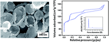
Hollow collapsed kippah-shaped silicalite-1 was synthesized by an emulsion based steam assisted conversion (ESAC) method employing water, ethyl ether, tetraethylorthosilicate (TEOS), tetrapropylammonium hydroxide (TPAOH) and cetyltrimethylammonium bromide (CTAB) as starting materials. Hierarchical silicalite-1 exhibited bimodal pore size distributions with unusual double hysteresis loops, revealing two distinct pore systems around the mesopore regime.
There are several approaches which have been applied to modify zeolitic crystal from their intrinsic structure and morphology by controlling gel composition, crystallization temperature and time or by using polymer and organosilanized groups etc. Choi et al. have reported amphiphilic organosilane-directed synthesis of crystalline zeolite with sponge like morphology and tunable mesoporosity.10 Hierarchical mesoporous zeolite was synthesized in the presence of mesoscale cationic polymer,11 block co-polymers,12 ionic liquids13 and many others soft templating agents. An extensive interest have been paid for the fabrication of hollow spherical zeolites by means of diverse methodologies, e.g., the carbon black and silica microspheres hard template self-assembly combined with hydrothermal crystallization process,12 layer-by-layer (LBL) self-assembly technique,13,14 and several others. Even, morphologically tuned hollow spherical silicalite-1 has been successfully synthesized by polystyrene sphere based templating technique.15 Recently, steam-assisted conversion (SAC) method has drawn an enormous interest due to higher yields, less water and low template consumption. In this method, initial dry gel containing organic templating agent remains in close contact to steam followed by crystallization of the dry gel. Zhu et al. synthesized nanocrystalline mesoporous MFI zeolite by solvent evaporation assisted novel technique.16 Zhang et al. reported the synthesis of hierarchical porous ZSM-5 based on steam assisted crystallization approach.17 Pure silica ZSM-22 zeolite was quietly prepared by ionic liquid-directed dry-gel conversion method.18 Alfaro et al. also demonstrated fabrication of silicalite-1 using dry gel conversion method.19 Recently solvent evaporation technique has been recommended for the designing of hierarchically porous ZSM-11.20
Herein, we report the synthesis of hierarchical hollow collapsed kippah-shaped silicalite-1 in water–ether medium in the presence of tetraethylorthosilicate (TEOS), tetrapropylammonium hydroxide (TPAOH) and cetyltrimethylammonium bromide (CTAB) followed by the steam assisted dry-gel crystallization method. In this study, the formation of hollow collapsed kippah-shaped silicalite-1 with controllable hierarchically by emulsion based steam assisted conversion method (ESAC) is reported for the first time to the best of our knowledge. Stirring time dependent formation mechanism has been proposed for the generation of collapsed kippah-like silicalite-1 particles. The effect of stirring times on the microstructure and textural properties of synthesized product has also been investigated in this study.
All the chemicals used in this experiment were obtained from commercial sources and used without further purification. Deionized (DI) water was used throughout the experiment. Tetraethylorthosilicate (TEOS), tetrapropylammonium hydroxide (TPAOH), diethyl ether, cetyltrimethylammonium bromide (CTAB) were supplied by Sigma-Aldrich.
In a typical experiment, 8 mL H2O, 40 mL ethyl ether and 3.51 g of TPAOH were mixed under vigorous stirring in a closed Teflon container at room temperature for 5 min. Then 3 mL of TEOS was added dropwise to the mixture under stirring condition. After 3 h, again the same amount of TEOS (3 mL) was added dropwise to this mixture followed by addition of 1.5 g of CTAB. The mix solutions thus prepared were continuously stirred for 20 h at room temperature. Then, the white turbid dispersions were poured into Petri dishes, and kept at room temperature (35 °C) for 48 h to dryness.
The as-prepared dry gel powder (0.5 g) was transferred to a Teflon cup, placed in a Teflon-lined autoclave (100 mL) containing 40 mL of water at the bottom of the autoclave so that water could not be in contact with the sample. The autoclave was sealed and crystallization was carried out at 170 °C for 72 h. After the steam assisted synthesis (autoclaving), the products were dried at 100 °C followed by calcination at 550 °C for 6 h. To investigate the formation mechanism, the stirring times were varied for 3 h and 12 h maintaining the other procedures same. The calcined samples were designated as Z-3, Z-12 and Z-20 for 3 h, 12 h and 20 h stirring times, respectively.
Powder X-ray diffraction (PXRD) studies of the samples were performed by Philips X'Pert Pro PW 3050/60 powder diffractometer using Ni-filtered Cu-Kα radiation (λ = 0.15418 nm) operated at 40 kV and 30 mA. The characteristic vibration bands of the products were confirmed by FTIR Spectrometer (PerkinElmer, Spectrum two) with KBr pellet at a resolution of 4 cm−1. Nitrogen adsorption–desorption measurements were conducted at 77 K with a Quantachrome (ASIQ MP) instrument. The surface area was obtained using Brunauer–Emmett–Teller (BET) method, and the pore size distributions were calculated by Barrett–Joyner–Halenda (BJH) method and density functional theory (DFT) method. The nitrogen adsorption volume at the relative pressure (p/po) of 0.99 was used to determine the pore volume. The morphology of the particles was examined by FESEM (Model: Zeiss, Supra™ 35VP, Germany) operating with an accelerating voltage of 10 kV, and TEM using a Tecnai G2 30ST (FEI) instrument operating at 300 kV. Fig. 1a shows the XRD pattern of silicalite-1 particles for the sample synthesized at 170 °C/72 h via emulsion based steam assisted conversion method.
![]() | ||
| Fig. 1 (a) XRD pattern and (b) FTIR spectra of silicalite-1 prepared with the stirring time of 20 h via ESAC method at 170 °C/72 h. | ||
The major crystalline peaks with 2θ values at 7.98°, 8.82°, 23.18°, 24.02° and 24.46° were assigned to (101), (020), (501), (151) and (303) lattice planes, respectively which are in good agreement of crystalline silicalite-1 zeolite structure.21 The FTIR spectra (Fig. 1b) of the samples show an intense pentasil vibration band at 550 cm−1 of silicalite.22 The appearance of absorption bands at 462 and 795 cm−1 agreed with Si–O–Si rocking, and symmetric stretching and bending vibrations, respectively. The internal and external asymmetric stretching modes of vibrations are observed at 1105 and 1230 cm−1, respectively. Combining both XRD pattern and FTIR spectra, it is attributed that the calcined sample was solely silicalite-1 in the absence other impurities.
Fig. 2a and b shows TEM images of silicalite-1 crystals synthesized by ESAC method at 170 °C/72 h. It reveals collapsed kippah-shaped microstructure of silicalite-1 particles of the size range 300–800 nm. From the higher magnification image of the particle it is obvious that the large number of nano-particles (50–100 nm) self-assembled together to form hierarchical microstructure. The interior structures of the hollow spherical silicalite-1 particles were also confirmed by TEM images of the broken kippah-shaped microstructure (Fig. S1, ESI†). A significant contrast difference between the hollow and dense solid parts of the kippah-shaped microstructure was noticed. Indeed the rims of kippah architecture were significantly darker due to reduced electron transmission through the rims.23
![]() | ||
| Fig. 2 (a and b) TEM images, (c–e) FESEM images, (f) EDS of silicalite-1 prepared with the stirring time of 20 h via ESAC method at 170 °C/72 h. | ||
Further, the tilting angles of darker region on the wall for all the particles were not similar because of the random position and tilting angles of collapsed kippah particles. The FESEM images (Fig. 2c–e) of silicalite-1 crystal indicates that how hierarchical hollow sphere was formed through the self-assembly of nano-particles. It renders the formation of hierarchical collapsed kippah-shaped morphology of silicalite-1 via layer by layer self assembly of nanoparticles. The collapsed region can be witnessed perfectly as the darker region within kippah shaped interior. However, the lighter rims of collapsed kippah particles were also clearly obtained due to stronger secondary electrons from the rims. Fig. S2, (ESI†) shows the side view of the kippah architecture as hemi-spherical shape with hollow microstructure. The growth mechanism of the hierarchical hollow silicalite-1 particles composed of nanoparticle assembly has been explained shortly. The elemental analysis of the relevant sample studied by EDX analysis (Fig. 2f) indicates the Si/O ratio, which is close to the stoichiometric composition of silicalite-1 zeolite.
To investigate the formation mechanism of the synthesized hierarchical collapsed kippah-shaped silicalite-1, time dependent stirring was performed for 3 h and 12 h during dry gel powder synthesis. The formation of silicalite-1 for 3 h and 12 h stirring was successfully characterized by XRD and FTIR analysis (Fig. S3, ESI†). The morphological evolution of the products with increase in stirring time is shown in FESEM images (Fig. 3).
![]() | ||
| Fig. 3 FESEM images of silicalite-1 prepared with the stirring time of 3 h (a and b), 12 h (c and d) and 20 h (e and f) via ESAC method at 170 °C/72 h. | ||
For the 3 h of stirring time, hollow microspherical silicalite-1 was formed of the size range 8–12 μm, and in each microsphere, collapsed kippah-like nanoparticles (150–350 nm) were indeed self-assembled. Further increase in stirring time for 12 h, hollow microspherical silicalite-1 (5–9 μm) was retained along with collapsed kippah-like nanoparticles (200–600 nm). Finally after 20 h stirring, submicron sized collapsed kippah-like particles was formed with the coexistence of very few solid nanospheres. It was observed that the collapsing of silicalite-1 particles just started forming at 3 h of stirring time.
Fig. 4 shows the TEM images of the silicalite-1 particles prepared with the stirring times of (a, b) 3 h, (c, d) 12 h and (e, f) 20 h during dry gel powder synthesis. It renders that with increase in stirring time from 3 h to 20 h, kippah-like nanoparticles were further formed from “soft” hollow microsphere.20 For the 3 h of stirring time, larger microsphere with little depressions on the surfaces was observed. However, depressions around the microsphere surfaces further aggregated to multishell wrinkle for 12 h stirring time due to excessive amount of membrane undulation of longer wavelengths. Finally, hollow collapsed kippah-shaped silicalite-1 was formed from multishell wrinkle. It is worthy to mention that the volume of the centre occupied major proportion of the total volume of the hollow microsphere (Fig. 4e and f). It further reveals that at the initial stage of reaction, the growth of the particles was still incomplete, which started growing towards a symmetrical hollow collapsed kippah-shaped silicalite-1 with significant amount of mesoporosity connecting the inner and outer-shell of microsphere. The microstructure of synthesized product has been further investigated for the stirring time of 30 h. However, variation of stirring time from 20 h to 30 h, the morphology and crystallization of silicalite-1 samples remained practically unchanged (Fig. S4, ESI†).
![]() | ||
| Fig. 4 TEM images of silicalite-1 prepared with the stirring time of 3 h (a and b), 12 h (c and d) and 20 h (e and f) via ESAC method at 170 °C/72 h. | ||
The crystalline phase (XRD) and microstructure (FESEM) of the 20 h stirred sample at the different synthesis stages, i.e., before and after the autoclaving have also been investigated (Fig. S5, ESI†). In the XRD patterns, signature amorphous peak of SiO2 along with crystalline peaks of CTAB (JCPDS # 48-2454) was revealed for 20 h stirred sample before autoclaving (Fig. S5a,† sample ID: S-20 g). However, after autoclaving, the characteristic peak of silialite-1 was obtained (Fig. S5d,† sample ID: Z-20 g). The solvothermal and calcination steps could not significantly influence the morphology of silicalite-1 samples, i.e., microstructure could not alter distinctly (Fig. S5b, c, e and f†). The size of particles was not uniform throughout the sample. However, the particles obtained after calcination were less agglomerated compared to those obtained before and after the autoclaving in the pre-calcination stage. Interestingly, no such distinct sharp edge of kippah like microstructure was noticed in the pre-calcination stage. It is noteworthy that experimental steps like solvothermal and calcination are important parameters to obtain such a sharp multishell kippah shaped microstructure of silialite-1.
Fig. 5 shows (a) N2 adsorption–desorption isotherms, and pore size distributions (PSDs) by (b) BJH and (c) DFT methods of hollow collapsed kippah-shaped silicalite-1 synthesized by ESAC method at 170 °C/72 h for 20 h of stirring time. The isotherm exhibits an unusual behaviour with double hump hysteresis loops appeared at around p/po of 0.1–0.5 and 0.5–0.99 indicating the existence of two different types of pores with narrow pore size distribution.24 From the isotherm, it is evident that at lower relative pressure H2 type hysteresis loops appeared indicating ink-bottle like pores, while at higher relative pressure H3 type hysteresis loops resulted showing slit-like mesopores.25,26 Further, PSDs curve reveal that the one type of pores is generated at around 18 Å just below mesopore range, and other type of pores appeared at around 40 Å. It is interesting to mention that the CTAB and ethyl ether molecules played as templating and co-templating roles,27 respectively which acted as porogen to render porosity within higher micropore and nearly lower mesopore regions. The characteristic structural micropores (∼5.7 Å) of silicalite-1 were confirmed by DFT method. The BET isotherms and PSDs of the 3 h and 12 h stirring time samples are shown in Fig. S6 and S7 (ESI†), respectively. For 3–12 h stirring time, the isotherms exhibited rare behavior of double hysteresis loops at lower p/po at around 0.1–0.2 and higher p/po at around 0.5–0.95.28,29 For both of the samples, a steep rise in the isotherm occurred at lower relative pressure, around p/po = 0.1, which indicated an abundance of microporosity in the samples. The textural properties (BET surface area, total pore volume, and pore size) of the samples are shown in Table 1. It indicates that BET surface is drastically increased with increasing stirring time from 3 h to 12–20 h, and the increase was mainly contributed by the micropore surface area. Further, the BET isotherms and PSDs of the silicalite-1 sample for starring time of 30 h (Fig. S8, ESI†) have been investigated. It is to be noted that comparing 20 h stirring time, 30 h stirring time could not have any significant influence on the textural property of silicalite-1 samples, i.e., total BET surface area (409 m2 g−1), microporous surface area (351 m2 g−1), total pore volume (0.246 cm3 g−1) and average pore size (24 Å) remained almost comparable to those obtained for 20 h stirring time. However, the disappearance of double hysteresis loops within N2 adsorption–desorption isotherm inhibited the existence of mesopore around 18 Å. It was further evidenced by BJH and DFT pore size distributions of 30 h stirred silicalite-1 sample. It is worth stating that total specific surface area is determined by BET method. The microporous contribution is examined by the difference between the BET surface area and the external surface area i.e., the mesoporous surface area (derived from the slope of the t (statistical thickness)-plot).22 For microporous silicalite-1, the linear BET region occurs at p/po of 0.1, while the linear t-plot range is obtained for higher p/po. However, for all the samples bi-modal pore size distribution was obtained within mesopore regime. The significant enhancement of total surface area along with micropore contribution could be due to gasification of ethyl ether through exothermic hydrolysis of TEOS.23 Furthermore, autoclaving and calcinations effect towards textural property of 20 h stirred sample was investigated by N2 adsorption–desorption isotherm study. The BET isotherms and PSDs of the samples obtained before autoclaving (S-20 g) and after autoclaving (Z-20 g) are revealed in Fig. S9 and S10 (ESI†), respectively. It exhibits that BET surface area and microporous surface area decreased in the order of Z-20 > Z-20 g > S-20 g. The increment of BET surface area along with microporous surface area from the sample S-20 g to Z-20 g could be attributed to the contribution of zeolitic micropore under solvothermal condition. The formation of silicalite-1 was evidenced by XRD (Fig. S5d, ESI†). Moreover, it can be concluded that the characteristic structural micropores (∼5.7 Å) of silicalite-1 resulted due to the solvothermal treatment, which was further evidenced by DFT pore size distributions (Fig. S10c, ESI†). Significant increase of total BET surface area along with microporous surface area from the sample Z-20 g to Z-20 could be due to removal of template during calcination. The textural properties (BET surface area, total pore volume, and pore size) of the samples are shown in Table S1.† It shows that the BET surface area along with microporous surface area is significantly increased due solvothermal and calcination steps for 20 h stirred silicalite-1 sample. It is worth mentioning that the templating effect of CTAB is also an important parameter towards textural property of silicalite-1 sample. Fig. S11 (ESI†) shows the BET isotherms and PSDs of the 550 °C calcined as-prepared sample with stirring time of 20 h before autoclaving (sample ID S-20). Large BET surface area (1017 m2 g−1) along with microporous surface area (528 m2 g−1) clearly rendered the templating effect of CTAB in the as-prepared silica sample. Fig. S12a and b (ESI†) show the XRD and FESEM image of the as-prepared (before-autoclaving) calcined sample.
![]() | ||
| Fig. 5 (a) N2 adsorption and desorption isotherms, pore size distributions (PSD) by (b) BJH and (c) DFT method of silicalite-1 prepared with the stirring time 20 h via ESAC method at 170 °C/72 h. | ||
| Sample ID | SBETa (m2 g−1) | Smicroporeb (m2 g−1) | Sexternalc (m2 g−1) | Vp-totald (cm3 g−1) | dPe (Å) |
|---|---|---|---|---|---|
| a Where aBET surface area, bmicropore surface area, cexternal surface area, dtotal pore volume, eaverage pore size. | |||||
| Z-3 | 200 | 140 | 60 | 0.206 | 41 |
| Z-12 | 407 | 346 | 61 | 0.234 | 23 |
| Z-20 | 404 | 336 | 67 | 0.243 | 24 |
Fig. 6 shows schematic growth mechanism for the fabrication of hierarchical hollow collapsed kippah-shaped silicalite-1. Initially an emulsion was formed in the presence of water, TPAOH, TEOS and ethyl ether. TPAOH and TEOS molecules further oriented at the interface of water and ethyl ether layers. After the addition of the surfactant, CTAB with excess TEOS, a cooperative assembly was acquired in which CTAB generated micelles.30 The silicate species in solution self-assembled around TPA+ cations and polycondensed.31 Indeed water could not move through the hydrophobic channels but ethyl ether could. However, ethyl ether (boiling point 34 °C) continuously gasified due to exothermic hydrolysis of TEOS.24 The escaped ethyl ether was in competition with the silica condensation process. Therefore, hollow sphere started to collapse at early stage of reaction. Moreover, the escaped ethyl ether could pass through the shells of condensed silica forming nanochannels, self-assembling of condensed silica species was further promoted by CTAB and generated collapsed kippah-like nanoparticles. Kim et al. reported the exceptional advantages of this dynamic templating technique for the fabrication of hierarchical nanocapsules.32,33 The ethyl ether co-template finally gasified completely forming collapsed kippah-shaped silicalite-1 microstructure. The formation of such microstructure would result from dynamic cross-coupling of two processes: dynamical gasification of ethyl ether and stabilizing process of condensation and self-assembly. Although ethyl ether is slightly dissolvable in water (ca. 8 g/100 mL of water) and exerts co-templating as well as co-solvating role.24 It is noteworthy that both ethyl ether and CTAB played as dynamical template and stabilizing agent, respectively for the fabrication of collapsed kippah shaped silicalite-1.
Footnote |
| † Electronic supplementary information (ESI) available. See DOI: 10.1039/c6ra19640c |
| This journal is © The Royal Society of Chemistry 2016 |