Manoj Vermaa,
Abhitosh Kediab,
M. Boazbou Newmaia and
P. Senthil Kumar*a
aDepartment of Physics & Astrophysics, University of Delhi, Delhi-110007, India. E-mail: duplasmonics@gmail.com; Tel: +91 9717623716
bDepartment of Physics, Uka Tarsadia University, Bardoli, Surat-394350, Gujarat, India
First published on 16th August 2016
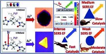
We have systematically utilized the simple yet uncommon XRD measurements together with FTIR data for not only quantifying the phase purity of as-synthesized Au nanoparticles, but also for meticulously interpreting the differential role of PVP in conjunction with its halide modified counterpart in the in situ fine-tuning of the nanoparticle growth. We thereby illustrate the robustness of the present synthetic protocol, further substantiating our relentless quest in achieving size/shape tunable metal nanostructures with high precision/yield by solely utilizing the full potential of the versatile polymer, polyvinyl pyrrolidone (PVP). Sincere efforts were undertaken in corroborating the optical plasmonic signatures of the different as-prepared Au nanostructures with the corresponding TEM measurements. Further, the comparative catalytic (4-nitroaniline to para-phenylenediamine) as well as surface enhanced raman scattering (of crystal violet dye molecules) investigations distinctly demonstrate a constructive structure–property correlation among the different Au nanostructures stabilized by the same polymer, the varied surface conformations of which solely dictate the physico-chemical properties in an illustrious manner, thereby establishing a new paradigm for better quantification of complex metal nanoarchitectures in general.
A number of shapes ranging from rods23–25 to cubes,26,27 disks, and nanostars28,29 can be routinely obtained by solution based methods with high monodispersity in size/shape. The relatively recent addition to this vast list from the past decade are the Au nanotriangles (NTs), owing to their intriguing plasmonic response.30 However, the yield percentage and shape/size monodispersity in Au NTs in any of the conventional synthesis protocols still lags behind that of Au nanorods or nanostars.31,32 A large volume of literature is available for the gold nanorod synthesis33,34 including a reasonably well laid out growth mechanism; in comparison, methods for the synthesis of metal nanotriangles are relatively limited. Novel routes broadly based on three categories such as the seed mediated approach,33,35 polyol method and the photochemical/biogenic processes,36,37 were routinely utilized in synthesizing prismatic, plate like nanostructures (such as the nanoprisms, nanotriangles, nanoplates or the nanodisks), by way of suitable adjustment/control with the experimental reaction parameters,38 so as to have size/shape monodispersity along with high yield. Indeed, in the existing synthetic protocols the NTs usually have relatively large lateral dimensions and synthetic protocols for the preparation of Au NTs below 60 nm with high yield are very rare in the literature. The yield of common synthetic methods for Au NTs is usually below 30 to 40%, and the particles often show high polydispersity.31,32
Recently, it has been reported that halide ions have a strong tendency to adsorb on metallic surfaces, thus directing the (over)growth of certain crystal facets resulting in fine tuning of the as-formed nanoparticle shapes.39,40 Earlier, iodine/iodide ions were used in the synthesis of largely polydispersed gold NTs,39 which has been normalized with the seed mediated approach.33 In the present study, we illustrate a simple single step solution-phase chemical reduction process utilizing bromine ions, which not only confronts the conformational variation of polyvinylpyrrolidone (PVP) in methanol, as revealed by FTIR, but also diligently differentiates the gold nanotriangle/prism growth mechanism from conventional sphericity through careful XRD measurements, significantly identifying their anisotropic lateral size. In contrast with many other similar synthetic routes for gold NTs, our present synthetic protocol produces no other anisotropic/branched nanostructures, elucidating its versatile robust nature. Our detailed investigation for the formation and growth mechanism of Au NTs through the interaction between halide ions and PVP lead to interesting conclusions, which were not explored till now to best of our knowledge. Although a complete comprehension of the growth mechanism is beyond the scope of this work.
Though bulk gold is unmistakably a poor catalyst,41 it has been well demonstrated in the last two decades that nanosized Au particles have excellent catalytic activity,42,43 mainly attributing to their large surface-to-volume ratio and the existence of various special adsorption sites on their surface. The reduction of aromatic nitro compounds to their amino derivatives using gold nanoparticles are well reported in the literature.44,45 Furthermore, the enhanced catalytic reduction of 4-nitroaniline to p-phenylenediamine in the presence of Au nanoprisms were systematically compared with that of Au nanospheres of approximately same size (comparison for the same size but with different geometry are made for the first time) resulting in the simplistic yet effective determination of nanoparticle size and shape on catalysis. Also, the SERS ability of these anisotropic gold nanostructures synthesized were calculated and compared. It is well known that SERS enhancement factor of metal nanoparticles is strongly dependent on the size as well as anisotropic geometry of nanoparticles.46 This dependence relies on the fact that sharp corners on the surface of nanoparticles are able to create a greater localized electric field in comparison to rounded ones.47 This enhancement due to sharp edges/tips is referred to as the lightning rod effect because similar to a pointed lightning rod, the electric field induced at the sharp edges/tips will be much stronger than other areas on the surface.48 The influence of the lightning rod effect can be observed near high curvature points on many different shapes.49 This lightning rod effect allows nanostructures to act as optical antenna providing an enhanced electromagnetic field.50
Physico-chemical properties of metal nanoparticles also depend on the type of surfactant that stabilizes them and the surrounding chemical environment (pH, solvent, presence of by-products etc.).51,52 Thus, for exclusively contrasting the shape/size effect of nanoparticles on their distinct properties requires similar stabilizers as well as chemical environment around them. Some research groups had beautifully correlated the role of shape and size on the properties of nanoparticles,43 but synthesized from different protocols, thereby not confirming the exact shape/size contribution. In terms of overcoming this routine persistent problem, we had synthesized all our present nanoparticles by the same synthesis protocol utilizing PVP as the simultaneous reductant/stabilizer in the same solvent/pH, except varying the halide ion concentration at ambient conditions, exquisitely yielding the perfect shape/size contribution in unraveling the various intrinsic properties of metal nanoparticles.
In a typical synthesis of gold pseudo-spherical nanoparticles, 0.27 mM aqueous solution of hydrochloroauric acid (HAuCl4·3H2O) was mixed with 15 mL of 10 mM polyvinylpyrrolidone (PVP average MW = 10000) solution in methanol and kept undisturbed at room temperature. The transformation from pale yellow solution to ruby red solution within two hours of reaction signifies the formation of gold pseudo-spherical nanoparticles. For the synthesis of gold nanotriangles, different concentrations of NaBr were added to the PVP–methanol system prior to the addition of HAuCl4. Effect of seeds, pH were investigated by adding appropriate seeds and NaOH to the solution of PVP–Br–methanol prior to the addition of HAuCl4 respectively. Different molecular weights of PVP were used to see the effect of chain length of PVP. Different halides were also used instead of NaBr for comparison purpose. For synthesis of smaller sized nanotriangles, PVP–Br mixture in methanol was refluxed at a constant temperature of 65 °C with continuous stirring maintaining the volume of solution throughout for a couple of hours and then gradually cooled to RT, in which copious amount of HAuCl4 is added.
For a typical catalysis reaction, 600 μL of 1 mM stock 4-NA solution was taken in 4 mL of triple distilled water and mixed thoroughly. 600 μL of freshly prepared ice cold NaBH4 was added and mixed well. Finally different volumes of Au NP solution (spheres, large nanotriangles, small nanotriangles) were added separately to the reaction mixture, keeping the number of Au particles approximately the same in all cases. The kinetically monitored reduction reaction, once completed, turns from the light yellowish color of 4-NA solution to a colorless one due to the formation of the reduced product p-PDA, which corresponds to the change in the optical absorption spectrum as well.
SERS substrates were made by simply making a monolayer of five-fold centrifuged gold nanostructures dispersed in ethanol onto silicon substrates, followed by the drop cast deposition of the 10−6 M of the crystal violet dye molecules.
All of the spectroscopic data were baseline corrected individually and manually stacked one above the other in a single plot, just for the sake of visual comparison only and should not be confused for overall normalization. Solvent peaks were multiplied by appropriate factor before subtracting it from all of our FTIR spectra by way of baseline correction with free solvents, which greatly simplifies the problem in analysis of FTIR spectra.
TEM samples were prepared by drying the 5-fold centrifuged samples in ethanol at around 6500 rpm (to remove excess PVP) on carbon Formvar coated copper grids and the images were acquired using the FE-Technai G2 system operated at an accelerating voltage of 300 kV.
The crystal structure parameters ware evaluated by analysing the X-ray diffraction (XRD) spectra collected with Cu-Kα radiation using a Bruker D-8 Advance X-ray diffractometer using the five-fold centrifuged nanoparticle samples drop casted on a glass substrate.
Surface enhanced Raman scattering (SERS) spectra were obtained using a Renishaw inVia Raman Spectrometer equipped with a CCD system with 785 nm He–Ne laser source, objective 50×, spot size of 1 μm, exposure time 10 seconds with a laser power of 30 mV.
Simple addition of gold ions to PVP in alcoholic methanol medium in the PVP/Au ratio of ∼3333 results in the formation of pseudospherical/spherical gold nanoparticles (solution color changes from pale yellow to ruby red within 2 h), indicated by the presence of strong surface plasmon resonance (SPR) peak at 524 nm (Fig. 1(a)) in the optical absorption spectra. TEM images of as-synthesized spherical nanoparticles and their size distribution histogram is shown in Fig. 1(b) and S1(a).† Average size calculated from the histogram is found to be 40 ± 3.5 nm (Fig. S1(a)†). But, the addition of 1.5 mM NaBr to the above mentioned pristine PVP–Au solution results in the solution color change from pale yellow to colorless to light blue to violet in 7 days, as illustrated by the presence of two SPR peaks, one at 535 nm and another at 750 nm, attributed in general to the transverse and longitudinal oscillations of the electron cloud on the surface of the nanoparticles, asserting the presence of anisotropic gold nanoparticles. The TEM images clearly demonstrate the presence of gold nanotriangles as well as spherical particles (Fig. 1(d) and S1(a)†). Average size calculated from the histogram is found to be 86 ± 12 nm for nanotriangles and 37 ± 3 nm for nanospheres (Fig. S1(b)†). In this case, only a small percentage (a maximum of 40–50%) of triangles was obtained, the statistical polydispersity data of different shapes present in the as-prepared sample is shown in the inset of Fig. 1(d).
Single particle HRTEM images of nanosphere and nanotriangle and their corresponding SAED pattern (Fig. 1(b)) clearly shows their strong crystalline nature. Hexagonal SAED pattern of a single nanotriangular structure emphasizes the dominance of (111) crystallographic facet as is well-known. We had synthesized gold spherical nanoparticles and gold nanotriangles only by introducing NaBr to the system which also only interacts with the PVP not to the gold nanoparticles (which we will show later in the manuscript). So, in this way we had synthesized two different shape-size particles by same synthesis protocol and hence both types of particles have same stabilizer (PVP) and same chemical environment around them.
I. Action of bromine on PVP to change its conformation in the solution and thus form say PVP*.
Br− + PVP = PVP* | (1) |
II. Reduction of Au3+ to Au0 by PVP
Au3+ + nPVP = Au0 | (2) |
III. Capping of Au0/Au atoms clusters by PVP and PVP* which leads to form spherical and nanotriangles respectively during early stages of growth as shown in Fig. 2.
![]() | ||
Fig. 2 Scheme for the formation of Au seeds in the solution, PVP and PVP* plays a key role in formation of pseudospherical and nanotriangles respectively by capping their surfaces. |
Intriguingly, the illustrious combinatorial effect of these above mentioned reactions and their rates dictate the formation and percentage yield of nanotriangles in the solution. In the case of spherical nanoparticles, as Br− is absent in the reaction mixture i.e., no PVP* formation take place and the pristine PVP would reduce all the Au3+ ions to Au0 atoms, thereby signifying the spherical shape formation due to the large PVP/Au ratio. In the presence of Br ions, both the formation of PVP* (reaction/process (1)) and the nucleation of gold atoms (reaction/process (2)) takes place concomitantly, while the final concentration of PVP* would decide the yield of nanotriangles i.e. higher the concentration of PVP* in the solution (at which the nanoparticle growth starts), higher the yield of nanotriangles. Formation of PVP* and its equilibrium constant (by reaction (1)) customarily depends on the factors like pH, temperature, concentration of bromide ion.
To investigate the effect of different halide ions, we replace the bromide ion in the reaction mixture with iodide or chloride ions, which unequivocally results in the formation of quasi-spherical and defect structured gold nanoparticles, the optical absorption spectra of which are shown in Fig. 3(a) and the corresponding TEM images are shown in Fig. S3,† confirming the fact that bromide ions are highly essential and plays a key role in the growth of gold nanotriangles.
![]() | ||
Fig. 3 (a) Absorption spectra of nanoparticles synthesized with different halide ions. (b) Absorption spectra of nanoparticles synthesized by replacing NaBr with CTAB. |
To further rule out the possibility of direct capping of the nanoparticle surface by bromide ions or their etching effect, we utilized a different bromide ion source such as CTAB (instead of NaBr) in our reaction mixture, as it is well known that both PVP and CTAB have minimal/negligible interaction with each other under immaculate ambient conditions. This results in the formation of polydisperse Au nanostructures, with rod-like and spherical particles dominating the entire lot (Fig. 3(b)), which again is in tune with the literature reports.54 Thus, we safely conclude that free and abundant bromide ion source incites the growth and stable formation of Au nanotriangles under our present ambient synthetic conditions.
In terms of identifying the effect of polymer chain length on the formation of Au nanotriangles, we tried the same reaction with different molecular weights of PVP, the optical spectra and TEM images of which are shown in Fig. S2,†clearly illustrating consistent change in the nanoparticle morphology with PVP molecular weight. For 8k PVP, polydisperse nanostructures were formed; with 10k PVP, polydisperse nanostructures with a large share of triangles were seen; with 25k PVP, few spherical and more polyhedral shapes are seen and with 40k PVP, large sized polyhedral shapes dominate, thereby consistent with our general strategy to utilize PVP-10k in all our reactions. This in turn corroborates the fact that higher the PVP molecular weight, slower is the rate of nucleation leading to the formation of large sized polyhedral shapes, whereas the lower molecular weights fasten the rate of nucleation, thus generating smaller sized nanostructures.55,56 However, changing the molecular weight of PVP yield different size of nanostructures but major yield in each case was triangles which further confirm the necessity of PVP–Br interaction in formation of nanotriangles.
Next, we examined the effect of concentration of HAuCl4 as shown by the absorption spectra in Fig. 4(a), depicting the corollary that for the same concentration of PVP* (from reaction (1)), small variation in the concentration of HAuCl4 does not affect/alter the formation of nanotriangles, though low in number density, but a higher concentration of HAuCl4 significantly alters the reaction rate as well as the ensuing nanoparticle morphology.
![]() | ||
Fig. 4 (a–d) Shows the absorption spectra of nanoparticles synthesized by varying the concentration of HAuCl4, NaBr, NaOH and seed respectively. |
Further, we studied the effect of NaBr concentration (Fig. 4(b)), which constructively demonstrate that NaBr accelerates reaction (1), drastically increasing the PVP* concentration in the reaction mixture, leading to the increased yield of nanotriangles. Nevertheless, an upper limit of NaBr concentration (≤2 mM) is fixed, as it induces a huge deficiency in the pure PVP concentration, necessary for the dynamic reduction of Au3+ to Au0, thereby restricting the reaction even for months under ambient conditions.
Again, by the pH variation of the reaction medium (Fig. 4(c)), we intend to play with the two most significant aspects of our reaction mixture, one the reducing ability of PVP and the other is the speciation of the gold salt, both of which are appreciably higher in the basic medium for the well-known reasons. Though both effects take place simultaneously in our reaction mixture, acceleration of reaction (2) (process of conversion of Au3+ to Au0), as confirmed by the blue shift of the absorption peak (Fig. 4(c)), dominates over the effect of reaction (1), thus a faster seed nucleation along with the presence of lower PVP* concentration results in the meager yield of nanotriangles.
For the sake of comparison, we tried the seed mediation strategy as well (Fig. 4(d)). Seed mediation surely fastens the reaction kinetics (nucleation as well as growth), but as the reaction (1) remains unchanged, producing same amount of PVP*, which in turn leads to the formation of isotropic particles only, as confirmed from Fig. 4(d).
In order to increase the formation of PVP* in the solution, we tweaked the synthesis protocol by preheating the PVP–Br mixture in methanol for 1 hour at reflux temperature prior to the addition of gold salt, essentially allowing the reaction (1) to generate considerable amount of PVP*, so as to stabilize uniform Au nanotriangles within 6 h, as shown in the absorption spectra (Fig. 5(a)). The SPR peak at 640 nm due to longitudinal oscillation of the electron cloud consolidates that the edge length of Au nanotriangles formed are quite small58 in accordance with the TEM images (Fig. 5(b)) confirming an edge length of about 30 nm. By this simple tweaking, we got a very high percentage yield of nanotriangles and reaction completes within 6 h of heating. Though higher the tendency for the PVP* formation with higher concentration of bromide ions, the external heating phenomenally enhances the reducing ability of pristine PVP present in the reaction mixture, thus ensuing complete reduction as well as increased stabilization of Au nanotriangles.
The affinity for the stabilization of smaller Au triangles at high reaction temperatures arise from the fact that higher the rate of reduction, faster the creation of fresh, tiny Au nuclei consuming precursor gold ions, resulting in the rapid stabilization of optimal sized Au nanotriangles with ease, as illustrated in Fig. 6. In case of room temperature synthesis, the slower reduction kinetics encourages the jumbled aggregation of precursor gold ions onto the surface of existing depreciated seed nuclei, which on delayed growth evidently supports the formation of polycrystalline spherical nanoparticles (in the absence of halide ions) and a collective mixture of spherical as well as larger triangles (in presence of Br− ions), as shown in Fig. 6. These simple yet thought provoking measurements highlight the significance of PVP–Br or PVP–halide ion interaction with reference to pristine PVP in the synthesis of noble metal nanostructures in general, which forms the major theme of our present work. From our spectroscopic measurements, it is safely confirmed that the prolonged preheating of PVP–bromine mixture maximizes the dynamic PVP* formation in the solution (responsible for the easy stabilization of Au nanotriangles), whereas the untouched pure PVP concentration constantly maintains the gold seed formation. Thus, an optimal ratio of PVP to PVP* is THE necessary and sufficient condition for the formation of monodisperse Au nanotriangles.
![]() | ||
Fig. 6 Schematic representation of the size dependence of Au nanotriangles on the nucleation rate modified by the reaction temperature. |
The XRD patterns obtained for different gold nanostructures (spherical, large and small triangles) as shown in Fig. 7 illustrate three prominent peaks, corresponding to the (111), (200), (220) planes of fcc Au in agreement with the d-values reported for gold nanocrystals (JCPDS card number-04-0784) (as tabulated in Table S1†) along with the average crystallite size calculated from the (111) peak full width at half maximum (FWHM) based on the Debye–Scherrer equation. The crystallite size is assumed to be the size of a coherently diffracting domain and it is not necessarily the same as particle size. However, the trend in increase or decrease of crystallite size of different nanoparticles is the same as particle size measured from TEM observations.
In all the three cases, the (111) plane is more intense than the other planes. The close examination of the XRD profile shows that the ratio of intensities, (200)/(111) (as given in Table S2†), decreases in the order of spherical particles, larger nanotriangles and finally smaller nanotriangles, significantly delineating the fact that the planar surface of the nanotriangles as well as spherical nanoparticles predominantly encompass the (111) plane, implying that the gold atoms tend to always prefer minimum symmetry positions.
Corresponding strain calculations59 along different crystallographic planes show exciting results, such as the strain on the (220) plane is less in the case of larger triangles than in the case of spherical particles (Table S2†), which conveniently imply that growth takes place along the (220) plane during the formation of large triangles.
On the contrary, in the case of small triangles, strain on both planes, i.e., (220) as well as (200) decrease considerably, suggesting simultaneous growth along both the planes, thus significantly restricting the size of the nanotriangles to 30 nm. Furthermore, the strain on the crystallographic planes (220) and (200) are less than that on the (111) axis, due to which the atoms tend to deposit along the lateral direction having atomically flat (111) surface for both smaller and larger nanotriangles. Strain variations along different crystallographic axis of nanoparticles were plotted against Nelson–Riley function (NRF) (see Fig. 8), which clearly suggests the induced strain variation among different (hkl) planes of different nanoparticles during the growth stage itself.
![]() | ||
Fig. 8 Strain vs. NRF plot for different crystallographic axis of spherical particles, large nanotriangles and small nanotriangles. |
Thus, XRD studies evidently reveal the predominant binding of bromine modified PVP* with that of the (111) plane than the corresponding pristine PVP [which differentially binds with reference to the planar surface energy; (111) > (100) > (110)], thus promoting growth along (220) and (200) crystallographic axis; whereas the strain distribution along these axes strictly determines the lateral dimension of the as-formed Au nanotriangles, demonstrating the novelty of our data analysis, the first of its kind in the literature.
Herein, we identify the conformational changes in PVP by the action of bromide ion with the help of FTIR data (Fig. 9). The conventional CO peak of the pyrrolidone ring at 1663 cm−1 remains unchanged even after NaBr addition, reminiscent of the fact that in case of methanol as the solvent medium, bromine ion doesn't really alter the C
O of pyrrolidone ring. There are several other peaks in the range 1380 to 1500 cm−1, due to the CH2 scissor, C–H bending, CH2 twist etc. also remains unaffected by NaBr addition, essentially underlining the fact that the bromide ion conclusively attaches with the (N–C
O) group to form energy activated PVP i.e., PVP*, thereby drastically altering the mild reducing as well as strong capping abilities of pristine PVP.
Hitherto unexplored changes were demonstrated in the combined group of N–CO bending/C–C chain vibrational peaks (as magnified in the inset of Fig. 9), unequivocally emphasizing the collective role of the solvent as well as the bromide ions in the formation of respective size/shaped metal nanostructures. In general, the vibrational peaks between 650 and 690 cm−1 corresponds to N–C
O bending61 and multiple peaks (not shown in Fig. 9 for clarity) between 700 and 750 cm−1 corresponds to C–C chain, as previously noted by us.60
In the case of pristine PVP in methanol (black color spectra in Fig. 9), only two known vibrational peaks, 667 and 679 cm−1 are visible, which red-shifts in the case of Au–PVP (blue color), invariably invoking the polymer adsorption via the non-bonding electrons of the carbonyl group, leading to the formation quasi-spherical gold nanostructures, in tune with our present arguments.
Three hump-like features arise in the case of PVP–NaBr in methanol (red color) at around 667, 679 and 688 cm−1, displaying the intriguing coupling of bromine ion with both the solvent as well as PVP, thereby drastically altering the role of non-bonding electrons of the carbonyl group, which in turn, restricts their easy availability to bind with the metal ions/nanostructures, thus ensuing the stable kinetic formation of Au nanotriangles with high yield in our present synthesis protocol, utilizing PVP–NaBr–Au (green color). The transmission intensity ratios of the vibrational peaks at around 679 and 688 cm−1 serves as a vital analytical tool in inherently attributing the binary shape distribution (extremely in coincidence with the optical absorption spectra in Fig. 1(a) and TEM images Fig. 1(d)) in the case of non-uniform PVP* activation at RT and the unique monodisperse formation of Au nanotriangles with normalized PVP* formation at high temperatures (in coincidence with Fig. 5).
In the catalytic reduction of 4-NA with NaBH4, the optical absorption peak at 380 nm due to the intermolecular charge transfer of 4-NA decreases and a new peak at 229 nm starts increasing gradually due to formation of reduced product para-phenylenediamine (p-PDA) in the solution. In the absence of Au NPs, reduction of 4-NA with NaBH4 alone is very slow and characteristic band of 4-NA at 380 nm decreases only 4% after 2 days. The reduction reaction was greatly accelerated by the addition of Au nanoparticles and the measured increase in the absorption peak of p-PDA clearly signifies the complete conversion of 4-NA to p-PDA. In our present study, we propose that initially the 4-NA molecules adsorbed on to the surface of Au nanoparticles play an important role in the electron transfer process that takes place from negatively charged BH4− ions to 4-NA via the Au nanoparticles, which in turn initiates the reduction of 4-NA to p-PDA as depicted in Fig. S4.†
In the literature reports, catalysis properties of different sized Au spherical nanoparticles and their corresponding rate constants were found to decrease with increase in size.56 Further, among the different shaped nanoparticles, the rate constant is maximum for spheres followed by nanoprisms and nanorods.43 In our present work, we compared the rate constant of three Au nanoparticles in precise, 30 nm spheres, 120 and 30 nm triangles and found that the rate constant of nanospheres were always greater than that of large nanotriangles, consistent with literature reports. Surprisingly, the rate constant of spherical nanoparticles were found to be much lesser in comparison with nanotriangles of similar lateral size, strongly corroborating the significant shape contribution of nanoparticles on the catalytic efficiency. The UV-Visible absorption spectra for successive reduction of 4-NA with NaBH4 in the presence of different Au nanostructures and their rate constant calculations are shown in Fig. 10.
The calculated rate constants of larger triangles, spherical particles and smaller triangles were 1.760 × 10⁻², 2.674 × 10⁻² and 4.684 × 10⁻² min−1 respectively (as derived from Fig. 10). The lower rate constants of the nanoparticles in comparison with some previously reported values were solely attributed to the long chain polymer PVP stabilized on the metal nanoparticle surface, which significantly decrease the number of active sites on the surface of nanoparticles and also the room temperature catalysis studies further affects the rate constant to a large extent. Although our calculated rate constants were lesser than the literature reports,42 aesthetically they reveal the exact contribution of shape and size of gold nanoparticles, as all our nanoparticles were synthesized by the same protocol via PVP as the reductant/stabilizer.
Higher catalysis activity of gold nanotriangles in comparison with the corresponding equivalent sized nanospheres were ascribed to the presence of edges and flat surfaces of triangles, which contains atoms of low coordination number having more affinity to bind with reactant molecules, thus accelerating the reaction kinetics. Such active regions of the nanotriangle surfaces might act as effective catalytic sites for the nitro compound reduction. Those well-defined edges and corners of the nanotriangles are very important as they promote better understanding of the physical properties during the electron transfer process.63 The robust stability of our Au nanoparticles withstanding the catalytic activity has been duly illustrated in Fig. 10.
We also compute the catalysis activity since the activity can be assumed to be directly proportional to the reaction rate constant, the active gold sites on the small nanotriangles increased by a factor of 175%.
Carbonium carbon (Fig. S5†) of crystal violet having positive charge have tendency to bind with charged nanoparticles and then amine groups are expected to bind with the SERS substrate. In the measured range, crystal violet shows various Raman modes at different wavenumbers shown in Fig. S5.† Lower concentrations (<10−3 M) of CV could not be detected without the assistance of a plasmonic SERS substrate. SERS data at a concentration of 10−6 M of analyte CV coated on the monolayer surface of different nanostructures is shown in Fig. 11. It is exciting to see that for all nanoparticles solution dye upto a concentration of 10−6 M can be easily detected with prepared SERS substrate. Even for small triangles the counts was as high as 23500 so that we can even detect 10 nM concentration considering 250 counts as threshold for detection.
The SERS enhancement factor was calculated by comparing the SERS signal from crystal violet adsorbed on the gold nanoparticles and the Raman signal of crystal violet (the detailed procedure of which is given at the end of ESI†). The representative band 1170 cm−1 due to ring C–H bend was selected to calculate the enhancement factor. We obtained the numerical values of EF as tabulated in Table 1 for different type of nanoparticles.
Nanoparticles | Enhancement factor |
---|---|
Spherical | 1.87 × 10⁴ |
Large triangles | 5.03 × 10⁴ |
Small triangles | 9.39 × 10⁴ |
Assuming that all the CV molecules contribute equally, it is alluring to observe that the SERS signal intensity of adsorbed CV dye molecule is typically weak for quasi-spherical particles, while it increases greatly for large nanotriangles and reaches the maximum in case of small nanotriangles (as tabulated in Table 1 and Fig. 10), due to the visible structural differences of the NPs (see Fig. S6†). Evidently speaking, nanotriangles (either small or large in size) should have greater EF than spherical ones owing largely to their anisotropic shape and thus have more SERS hotspots.64 However, the simultaneous comparison of SERS enhancement of respective small and large Au nanotriangles with the quasi-spherical Au nanoparticles inherently reveal the intriguing fact that though for the same shape, the EF significantly increases with decrease in size, thus clearly attributing the SERS signal enhancement to both size as well as the nanoparticle shape, which promotes highly efficient SERS-active hotspots, wherein the analyte/dye molecules preferentially adsorb, thereby making the anisotropic nanoparticles very attractive SERS labels for chemical/bio-analytical sensing/imaging.65 Our SERS enhancement factors were appropriately compromised, as though the polymer PVP on the surface of these nanoparticles contaminate the SERS signals more than ionic stabilizers like citrate ions, they give extreme stability to these particles against various chemical environments (as some dyes imparts a partial aggregation of gold nanoparticles66), which makes these particles ideal for sensing a large number of dyes and for their reusability. On the other hand, unlike previous reports,47–50 our present data reveal the exact contribution of shape/size of gold nanoparticles on their SERS enhancement factor, as all our nanoparticles were synthesized by the same protocol employing PVP as the reductant/stabilizer.
Moreover, it has already been well formulated that the SERS enhancement factor increases with decrease in strain.67 In the present case, the mechanical strain of our Au nanoparticle samples decreases along the 〈220〉 axis, which is indeed well correlated with the optical absorbance characteristics (refer Fig. 5 and 8), corroborating the fact that manipulating the local curvature of the nanoparticle surface without inter-particle aggregation, itself increases the number and strength of local field hot spots with ease (see Fig. S6†). Thus, apart from the density of size/shaped nanoparticles and their corresponding coupling effect of aggregation, the active surface area for analyte adsorption also would invariably affect the SERS response, forms the very basis of our present work and would be further explored in detail in our future communications.
Thus, for the first time to the best of our knowledge, we have synergistically identified the exact role of size/shape of gold nanostructures synthesized/stabilized by the polymer, PVP and its strategic effect on their properties, namely, SERS and catalysis. The central role of PVP in not only providing steric hindered colloidal stability of the as-prepared gold nanostructures, but also in dictating their SERS and catalysis activities in a meticulous manner has been well-established analytically in a clear cut manner beyond doubt. Our present foolproof experimental measurements should further encourage refined quantum mechanical calculations for illustrating the exact role of PVP in constructing a cohesive structure–property correlation in noble metal nanostructures, which should be of prime importance in understanding the aesthetic aspects of complex metal and semiconductor nanostructures in great detail.
Footnote |
† Electronic supplementary information (ESI) available. See DOI: 10.1039/c6ra18345j |
This journal is © The Royal Society of Chemistry 2016 |