
A functional oligonucleotide probe from an encapsulated silver nanocluster assembled by rolling circle amplification and its application in label-free sensors†
Yuna Guoa,
Yu Wangb,
Su Liu*c,
Jinghua Yua,
Qianqian Peia,
Xueqi Lengc and
Jiadong Huangab
aKey Laboratory of Chemical Sensing & Analysis in Universities of Shandong, School of Chemistry and Chemical Engineering, University of Jinan, Jinan 250022, P. R. China. E-mail: chm_huangjd@ujn.edu.cn; Fax: +86-531-82769122; Tel: +86-531-89736122
bSchool of Biological Sciences and Technology, University of Jinan, Jinan 250022, P. R. China
cSchool of Resources and Environment, University of Jinan, Jinan 250022, P. R. China
First published on 12th September 2016
Abstract
A novel label-free, low cost electrochemical biosensor for highly sensitive and selective detection of E. coli has been developed based on rolling circle amplification (RCA) coupled silver nanoclusters (AgNCs) as an effective electrochemical probe. This strategy is to our knowledge the first approach where the combination of RCA with the efficient catalytic property of AgNCs has been applied in bioanalysis. Our biosensor relies on sandwich type immuno-recognition and an in situ RCA reaction, which results in the enrichment of functional oligonucleotide probe (FOP) encapsulated AgNCs. Due to the high electrocatalytic activity of AgNCs toward H2O2 reduction, stable and sensitive electrochemical signals can be achieved from the biological recognition events. The results reveal the calibration plot obtained for E. coli O157:H7 is approximately linear from 3.7 × 10 to 3.7 × 10⁶ cfu mL−1 with a limit of detection of 31 cfu mL−1. This label-free biosensor has advantages over conventional electrochemical assays in its significantly improved sensitivity, remarkably widened dynamic range, and substantially lowered background signal. Moreover, the proposed biosensor has been demonstrated to allow accurate quantitative assay of E. coli O157:H7 in milk samples. By virtue of these merits, our RCA coupled AgNCs-based electrochemical biosensor might create a useful and valuable tool for the detection of E. coli O157:H7 and related molecular diagnostics in food safety analysis.
1. Introduction
Bacterial contamination of food is a human health threat of global proportion.1,2 Escherichia coli O157:H7 (E. coli O157:H7), as one of the most dangerous food borne pathogenic bacteria, is of most concern today since it can cause severe and even life-threatening illnesses, especially in cases involving children and the elderly.3 In most countries, E. coli O157:H7 identification has been regarded as an essential term in food quality inspection to protect people from bacterial infections.Owing to the significance of E. coli O157:H7 in food safety analysis and clinical diagnosis, a variety of strategies have been reported for the detection of E. coli, including the conventional culture-based method,4 polymerase chain reaction (PCR),5 quartz crystal microbalance (QCM) system,6 antimicrobial peptides array (APA),7 electrochemical impedance spectroscopy (EIS),8 and chemiluminescence (CL)-based assay9 etc. Despite these methods has been broadly applied, they are usually time-consuming, cumbersome and require laborious pretreatments of samples, sophisticated instrumentation and trained technical personnel.
To circumvent these problems mentioned above, antibody-based immunoassay methods such as enzyme-linked immunosorbent assay (ELISA)10 have been reported for E. coli detection. The conventional ELISA technique possesses the advantages of simplicity and accuracy. However, they might not meet the requirements of high sensitivity and low cost in real analysis, particularly when analysis is limited by the number of available sample or the low level of antigen. Fortunately, the emergence of nucleic acid aptamer has considerable potential for resolving these issues effectively.11,12 Rolling circle amplification (RCA), an isothermal DNA amplification procedure, can generate a linear concatenated DNA molecule containing up to 1000 complementary copies of the circular DNA in 1 h.13,14 Because of the high sensitivity, inherent simplicity, portability, and low cost derived from RCA techniques, the incorporation of the RCA in an electrochemical immunoassay might hold great promise for ultrasensitive cell determination in decentralized studies and point-of-care diagnosis.15,16 Several RCA-based assay methods have been reported by using peroxidase-mimicking DNAzyme,17 or alkaline phosphatase (ALP)-labeled probe18 as signal reporters. However, these methods may suffer from some drawbacks such as inherent instability of the enzyme, requirement of adding additional labile reagents, and the undesirable background signal.19 Therefore, it is highly desirable to develop simple and label-free electrochemical biosensing strategies for quantifying E. coli with inherent stability and a low background signal.
DNA-based nanomaterials provide a promising sensing platform, because of the high loading of receptor molecules for synergistic amplification of the target response, the high surface areas for improved mass transport, and unique optical, electronic, and catalytic properties for translating of the biorecognition events to an electrochemical or spectroscopic response.20,21 The synthesis and applications of DNA-based nanomaterials, particularly nucleic acid encapsulated silver nanoclusters (AgNCs), have recently attracted substantial research efforts.22–24 With regard to their luminescence, the AgNCs represent a new exceptional fluorophores for label of bioimaging and chemical/biological detection.25–27 However, few researches have been dedicated to the application of catalytic activity of AgNCs in bioanalysis. The use of metal nanoparticles or nanoclusters as a catalyst is analogous to the way of enzymes and might overcome some of the problems related to the inherent thermal and environmental instability of the enzyme.28,29
Herein, a novel label-free electrochemical biosensing strategy for ultrasensitive detection of E. coli O157:H7 has been developed using RCA coupled AgNCs as effective electrochemical probes. To the best of our knowledge, this work is the first time that the combination of RCA with the high electrocatalytic activity of AgNCs has been applied in bioanalysis. Our method features with several significant aspects. First, RCA coupled AgNCs has been utilized to function as the effective electrochemical probes such that the ultrasensitive electrochemical signals can be achieved owing to the high catalytic capability of AgNCs in response to H2O2 reduction. Compared with existing techniques for E. coli detection, such as ELISA and other previous methods reported in the literature, our RCA coupled AgNCs-based biosensing strategy offered improved sensitivity and widened linear range. Second, the use of sandwich type-based immunoreaction offers high specificity in biological recognition events, which improves binding efficiency and enhances the selectivity of the biosensor. Third, functional oligonucleotide probe-stabilized AgNCs is used as catalytic tag, which makes our method technically label-free, and eliminates electrochemical labeling steps and additional labile reagents as well. In view of these advantages, our RCA coupled AgNCs-based electrochemical biosensor might create a useful and valuable tool for the detection of E. coli O157:H7 and related molecular diagnostics in food safety analysis.
2. Experimental section
2.1 Reagents and materials
Oligonucleotides used in this work were HPLC-purified and synthesized by Sangon Biotechnology Co. Ltd. (Shanghai, China). The sequences of oligonucleotides are listed in Table S1 (ESI†). T4 DNA ligase, phi29 DNA polymerase, exonuclease I (Exo I), exonuclease III (Exo III) and deoxyribonucleoside triphosphates (dNTPs) were obtained from New England Biolabs Inc. (Ipswich, MA, USA). Rabbit anti-E. coli O157:H7 antibody was purchased from Shanghai Prajna Biology Technique Ltd. (Shanghai, China). E. coli O157:H7, Salmonella, Bacillus subtilis and Listeria were obtained from Institute of Microbiology Chinese Academy (Beijing, China). Silver nitrate (AgNO3), hydrogen peroxide (H2O2), sodium borohydride (NaBH4), thioglycolic acid (MACA, ≥98%), N-(3-dimethylaminopropyl)-N′-ethylcarbodiimide (EDC, ≥97.0%) and N-hydroxysuccinimide (NHS) were purchased from Sigma Aldrich Chemical Co. (St. Louis, MO, USA). Other chemicals were of analytical grade and were used without further purification. Ultrapure water obtained from a Millipore filtration system was used throughout.2.2 Preparation of the circular template
The circular DNA template was prepared as follows. First, 1 μL of the phosphorylated padlock probe (100 μM) and 1 μL ligation probe (100 μM) were hybridized in the T4 ligase buffer. Second, 1 μL T4 DNA ligase (60 U μL−1) was added, and the mixture was allowed to incubate overnight at 16 °C. After deactivation of T4 ligase by incubation at 65 °C for 15 min, Exo I (1 μL, 20 U μL−1) and Exo III (0.5 μL, 100 U μL−1) were added to digest the leftover ssDNA and dsDNA to yield the circular DNA. The enzymes were denatured by heating at 85 °C for 10 min. Finally, the product was purified by ethanol precipitation and verified by gel electrophoresis and stored at 4 °C until use.2.3 Preparation of functional oligonucleotide probe (FOP) encapsulated AgNCs
FOP encapsulated AgNCs were prepared by the classical NaBH4 reduction route.22 Briefly, 15 μL of 100 mM FOP was mixed with 76 μL of 20 mM phosphate (pH 7.0) buffer, and then 4.5 μL of 2 mM AgNO3 solution was added to provide a Ag+-to-DNA molar ratio of 6![[thin space (1/6-em)]](https://www.rsc.org/images/entities/char_2009.gif) :1. After cooling at 4 °C for 15 min, this mixture was reduced quickly by 4.5 μL of 2 mM NaBH4, followed by vigorously shaking for 1 min. The reaction was kept in the dark at 4 °C for 6 h before use.
:1. After cooling at 4 °C for 15 min, this mixture was reduced quickly by 4.5 μL of 2 mM NaBH4, followed by vigorously shaking for 1 min. The reaction was kept in the dark at 4 °C for 6 h before use.
2.4 Label-free and amplified electrochemical detection of E. coli O157:H7
Self-assembled monolayer modified Au electrodes were fabricated by immersing pretreated electrodes in 1.0% MACA ethanol solution for 24 h. Terminal carboxylic acid groups were converted to NHS ester groups by the subsequent treatment of the electrodes with a solution containing 0.4 M EDC and 0.1 M NHS for 1 h. After that, the electrodes were incubated with 10 μL of 20 μg mL−1 E. coli O157:H7 antibody solution at 37 °C for 1 h. Different concentrations of E. coli O157:H7 were incubated in antibody modified electrodes for 60 min. After the electrodes were washed with PBS solution, 10 μL of the 100 nM aptamer–primer in PBS solution was applied to the electrodes surface and incubated at 37 °C for 60 min. Subsequently, RCA reaction solution containing 1 μL phi29 DNA polymerase buffer, 0.5 μL 10 mM dNTPs, 0.2 μL 0.1 mg mL−1 BSA, 1 μL 10 U μL−1 phi29 DNA polymerase, and 10 μL 120 nM circular template was introduced into electrodes, and the electrodes were incubated at 37 °C for 2 h. Then, in the process of assembly, AgNCs were bound to the periodic sequences of the RCA product. After rinsed and dried, the modified electrodes were then incubated with the reaction product of AgNCs for 1 h at room temperature to capture the AgNCs on the Au sensing electrodes by their complementary sequences through DNA hybridizations. After washed three times by PBS, the electrodes were immersed into the working solution to perform electrochemical measurements.3. Results and discussion
3.1 Design principle of the proposed electrochemical E. coli O157:H7 detection using RCA coupled AgNCs as effective electrochemical probes
The proposed electrochemical biosensor based on RCA coupled AgNCs as effective electrochemical probe was illustrated in Scheme 1. A functional oligonucleotide probe (FOP) was designed to consist of C-rich sequence as the template for in situ synthesis of AgNCs, and a recognition sequence, which was used for hybridization with long repetitive DNA strands synthesized from RCA. A primer-linked aptamer probe (PAP) containing anti-E. coli O157:H7 aptamer sequence and the primer sequence at 3′ could hybridize with the circular probe and activate RCA reaction with the help of phi29 and dNTPs. In this approach, the circular probe, which contains the sequence just the same as the recognition sequence of FOP, was first prepared by using a ligation probe and a padlock probe through T4 DNA ligase-catalyzed ligase reaction. FOP encapsulated AgNCs (FOP AgNCs) was presynthesized independently by a classical NaBH4 reduction method. Our biosensor was constructed by the immobilization of anti-E. coli O157:H7 polyclonal antibody on gold electrode (GE) surface using carbodiimide method.30 Followed by the successive addition of E. coli O157:H7 and PAP, a sandwich type-based recognition generated via the specific binding between target with antibody and aptamer. Subsequently, the prepared circular probe hybridized with PAP and a RCA reaction was initiated in the presence of phi29 and dNTPs, producing a single-stranded tandem repeated copy of the circular probe. Thus, a great amount of FOP AgNCs could combine to the long RCA products and brought to the electrode surface, which resulted in a very strong peak current signal because of the efficient catalytic property of FOP AgNCs toward H2O2 reduction. In general, combining exceptional metal mimic enzyme properties of FOP AgNCs together with RCA, a novel and label-free electrochemical biosensor for ultrasensitive detection of E. coli O157:H7 was successfully constructed. | ||
| Scheme 1 Illustration of the ultrasensitive and label-free electrochemical assay for the detection of E. coli O157:H7 based on RCA coupled FOP AgNCs as effective electrochemical probes. | ||
3.2 Characterization of the RCA and FOP AgNCs
Direct proof of the RCA mechanism could be acquired through gel electrophoresis analysis, as shown in Fig. 1A, two bright bands with the same mobility and intensity were observed on the image before (lane 2) and after (lane 3) the circular probe treatment with Exo I and Exo III, indicating the successful preparation of the circular probe and its resistance to enzymatic digestion. The anticipated high molecular weight of the RCA product was confirmed in lane 4. After RCA, a strong fluorescence band corresponding to DNA with length in excess of 15000 bp of the marker was observed. These results give immediate evidence for the high molecular weight of these products, indicating that the proposed method could provide enormous signal amplification in immunoassay.
 | ||
| Fig. 1 (A) Gel electrophoresis image obtained for different samples. Lane 1, DNA marker; lane 2, circular probe; lane 3, circular probe incubated with Exo I and Exo III; lane 4, the large-molecule-weight RCA products. (B) Typical fluorescence spectra of FOP with (a) and without (b) the C-rich regions. (C & D) Typical TEM images of the AgNCs (C) and RCA coupled AgNCs (D). | ||
It was also known that FOP AgNCs exhibit excellent fluorescence properties. The fluorescence spectra of FOP AgNCs was further employed to prove the successful synthesis of the FOP AgNCs. The FOP 5 with/without the C-rich regions followed by further incubation with AgNO3 and NaBH4 was monitored by fluorescent spectrophotometer. According to Fig. 1B, upon excitation at 560 nm, the FOP AgNCs showed a maximum emission at 615 nm. The FOP 5 without the C-rich regions shows minimal fluorescent emission (curve b) while the FOP 5 with C-rich regions exhibits strong fluorescent emission at 615 nm (curve a), which is consistent with previous reports.22,26 However, no change in the intensity or peak positions was observed when the solutions were centrifuged, indicating that the spectra cannot be attributed to nanoparticles.
Transmission electron microscopy (TEM) and high-resolution transmission electron microscopy (HRTEM) were employed to investigate the morphology and microstructure of the as-prepared FOP AgNCs and RCA coupled AgNCs. Fig. 1C revealed that the AgNCs were monodispersed and the average diameter was approximate 4 nm. It could be also easily found that a narrow size distribution of AgNCs was obtained, which is useful for bioanalysis. In the presence of the long enzymatic backbone, plenty of monodisperse AgNCs were stacking on the RCA products forming self-assembled AgNCs nanowires (Fig. 1D).
3.3 Electrochemical characterization of the proposed label-free electrochemical biosensor
To verify the feasibility of designed RCA coupled AgNCs-based strategy for E. coli O157:H7 assay, differential pulse voltammetry (DPV) measurements were performed in 10 mM PBS containing 3 mM H2O2 and those obtained in a series of control experiments were depicted in Fig. 2A. When the electrode was incubated in the presence of E. coli O157:H7, a significantly strong peak current appeared in the DPV curves (curve a). This was attributed that a substantial number of AgNCs binding to the electrode surface to form mimic enzyme, thus generating extremely strong catalytic ability toward the reduction of H2O2 to give an amplified current signal. In contrast, a very weak peak current was observed after the incubation in the absence of E. coli O157:H7, suggesting almost no AgNCs was assembled on electrode surface (curve b). For the proof-of-concept experiment, we performed further control experiments to verify the specificity of the biosensor. When anti-Salmonella antibody was immobilized on electrode surface in place of E. coli O157:H7 antibody, a negligible peak current was observed in the DPV curve (curve c). Similarly, almost no obvious peak current was achieved after incubation with ligation probe in place of PAP (curve d). Additionally, a very weak DPV peak was observed in the presence of non-target pathogenic bacteria Salmonella instead of E. coli O157:H7 (curve e). This implied the RCA reaction and assembled of AgNCs was target E. coli O157:H7 dependent, rather than induced by other non-specific interactions. On the basis of these results, it was reasonably concluded that the proposed approach can be potentially employed to detect low abundance of E. coli O157:H7. | ||
| Fig. 2 (A) Typical DPV responses of the biosensor obtained upon analyzing 3.7 × 10⁶ cfu mL−1 E. coli O157:H7 (a). Curves (b) to (e) are for the control experiments which are performed without target E. coli O157:H7 (b), Salmonella antibody in place of E. coli O157:H7 antibody (c), ligation probe in place of PAP (d), and non-target pathogenic bacteria Salmonella in place of E. coli O157:H7 (e). (B) EIS obtained for the bare GE (a), antibody/GE (b), E. coli O157:H7/BSA/antibody/GE (c), PAP/E. coli O157:H7/BSA/antibody/GE (d), RCA products-linked PAP/E. coli O157:H7/BSA/antibody/GE (e), AgNCs/RCA products-linked PAP/E. coli O157:H7/BSA/antibody/GE (f). | ||
3.4 Electrochemical characterization of the modification of the electrode
The modification of the sensing interface of the electrode was of great importance to the achievable magnitude of E. coli O157:H7 response. Electrochemical impedance spectroscopy (EIS) is an effective and facile electrochemical technique for monitoring the changes in the surface features of the modified electrodes in the assembly processes.31 Each step of the sensing interface modification was monitored by EIS, and the results were shown in Fig. 2B. Prior to the immobilization of the capture antibody, the Ret of the bare electrode was quite small due to the direct transfer of electrons in the sensing system (curve a). After the immobilization of the E. coli O157:H7 antibody on the electrode surface, it was obvious that the diameter of the semicircle increased, indicating the successful immobilization of the antibody (curve b). Followed by the addition of target E. coli O157:H7, the diameter of the semicircle was increased compared with that of curve b, indicating E. coli O157:H7 was captured on electrode and further hindered the electron transfer (curve c). Similarly, after the modification of PAP (curve d), RCA product (curve e) and AgNCs (curve f), the Ret was further increased respectively. This increase of the Ret after immobilization of DNA probes could be attributed to the negatively charged phosphate skeleton of the ssDNA, which could block the electron transfer of negatively charged [Fe(CN)6]3−/4− from the electrode surface. All above results suggested the construction of the sensing interface for E. coli O157:H7 and the specific target recognition were successfully achieved. The corresponding cyclic voltammetry (CV) spectras could further verify the above results (Fig. S1, ESI†).3.5 Optimization of conditions for the proposed label-free electrochemical biosensor
Considering the important role of FOP in the formation of AgNCs and the electron transfer between AgNCs and H2O2, we also investigated the effect of the sequence of FOP on the current response. As shown in Fig. S2,† five FOPs (FOP 1 to 5) were designed and the properties of prepared AgNCs were evaluated through the electrochemical (Fig. S2A†) and fluorescence measurements (Fig. S2B†). It was found that the fluorescence intensities and current signal achieved the maximum value with FOP 5. So, FOP 5 was adopted as the optimum FOP sequences and employed for all other investigations.Fig. S2C† depicted the effect of the RCA reaction time on the DPV current response in the presence of 3.7 × 10⁶ cfu mL−1 E. coli O157:H7. Theoretically, the longer the reaction time expected the higher peak current for E. coli O157:H7. However, as shown in Fig. S2C,† in practice the peak current increased at the early stage of the reaction with increasing reaction time up to 80 min, where a maximum peak current was obtained. Beyond an RCA reaction time of 80 min, the DPV response obtained for E. coli O157:H7 did not exhibit further increase. The possible reason for this phenomenon could be attributed to the exhaustion of the available substrates for RCA. Consequently, a reaction time of 80 min was adopted as the optimum and employed for all other investigations.
3.6 Analytical performance of the proposed label-free electrochemical biosensor
Under optimal conditions, the electrochemical response of the biosensor to H2O2 reduction was adopted for the determination of a series of different concentrations of E. coli O157:H7. As shown in Fig. 3A, the amplified electrochemical signal increased gradually with the increasing concentration of E. coli O157:H7. The plot of the current intensity versus the logarithm value of target concentration displayed a linear relationship in the range from 3.7 × 10 to 3.7 × 10⁶ cfu mL−1 (Fig. 3B), and with the limit of detection (LOD) as low as 31 cfu mL−1 at four times the standard deviation of the control. Besides, the analytical performance of the proposed biosensor in the quantitative assay of E. coli O157:H7 was compared with that of some previously reported methods, and the results were shown in Table 1. It was observed that our biosensor offered improved sensitivity and widened linear range. | ||
| Fig. 3 (A) Typical DPV responses of the biosensor to different concentrations of E. coli O157:H7 (from curve a to h: 0, 3.7 × 10, 3.7 × 10², 3.7 × 10³, 3.7 × 10⁴, 3.7 × 10⁵, 3.7 × 10⁶, 3.7 × 10⁷ cfu mL−1). (B) Typical calibration curve for E. coli O157:H7 detection. Error bars are standard deviations across three repetitive experiments. | ||
| Detection methods | Detection range (cfu mL−1) | Detection limit (cfu mL−1) | Reference |
|---|---|---|---|
| QCM | 5.0 × 10⁴ to 5.0 × 10⁷ | 1.7 × 10⁴ | 6 |
| APA | 104 to 107 | 104 | 7 |
| EIS | 1.5 × 10² to 1.0 × 10⁷ | 1.5 × 10² | 8 |
| CL-based assay | 4.3 × 10³ to 4.3 × 10⁵ | 1.2 × 10³ | 9 |
| ELISA | 1.0 × 10³ to 1.0 × 10⁷ | 1.0 × 10³ | 10 |
| The proposed RCA coupled AgNCs-based biosensor | 3.7 × 10 to 3.7 × 10⁶ | 31 | This work |
To estimate the reproducibility of the proposed RCA coupled AgNCs-based electrochemical biosensor, six assays were performed following identical processing steps. Their responses toward 3.7 × 10⁶ cfu mL−1 target bacteria gave an average stripping peak current of 12.6 μA with a relative standard deviation of 4.32%, indicating that the biosensor could be constructed and used for analysis with excellent reproducibility.
Selectivity is one of the important parameters for practical application of biosensor, which was also evaluated by using three non-target bacteria, including Salmonella, Bacillus subtilis and Listeria, respectively. As shown in Fig. 4, only the target E. coli O157:H7 induced an obvious variation of the peak current, while the current variations induced by the other bacteria at the 10-fold concentrations could be ignored. This indicated the proposed biosensor exhibited superb specificity towards E. coli O157:H7. This satisfactory selectivity could be attributed to the good selectivity and high affinity of both antibody and aptamer.
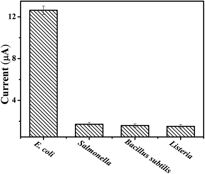 | ||
| Fig. 4 Plot of DPV peak current intensity obtained with different bacteria. The concentration of E. coli O157:H7 is 3.7 × 10⁶ cfu mL−1. The concentration of non-target bacteria is 3.7 × 10⁷ cfu mL−1. Error bars are standard deviations across three repetitive experiments. | ||
3.7 Real sample analysis
To demonstrate the validity of our biosensor to real samples, the quantitative assay of spiked milk samples was considered. Different concentrations of E. coli O157:H7 were spiked into the samples and detected directly without any pretreatments except for dilution with a buffer solution. The spiked samples were further quantified by a classic plate count method and compared with results obtained with the biosensor. From the results in Table S2,† it was observed that the results obtained via our method were consisted with those of plate count method, and the discrepancies between two methods were all smaller than 9.6%. Besides, the recovery of the proposed method was in the range of 93.4–102.7%. These data clearly demonstrated the potential of our method for applications in complicated samples.4. Conclusion
In summary, we have developed a high sensitive and label-free electrochemical biosensing strategy for detection of E. coli O157:H7 based on RCA coupled AgNCs as effective electrochemical probe. This strategy is to our knowledge the first approach that the combination of RCA with the efficient catalytic property of AgNCs has been applied in bioanalysis. The results reveal the calibration plot obtained for E. coli O157:H7 is approximately linear from 3.7 × 10 to 3.7 × 10⁶ cfu mL−1 with the limit of detection of 31 cfu mL−1. This approach has the advantages over conventional electrochemical assays in its significantly improved sensitivity, remarkably widened dynamic range, and substantially lowered background signal. Moreover, AgNCs is used as catalytic tag, which makes our method technically label-free, and eliminates electrochemical labeling steps and additional labile reagents as well. Additionally, the proposed biosensor has been demonstrated to allow accurate quantitative assay of E. coli O157:H7 in milk samples. In virtue of these merits, our RCA coupled AgNCs-based electrochemical biosensor might create a useful and valuable tool for the detection of E. coli O157:H7 and related molecular diagnostics in food safety analysis.Acknowledgements
This work was supported Shandong Province Natural Science Funds for Distinguished Young Scholars (JQ201410), NSFC (1471644, 21405060), Promotive Research Fund for Excellent Young and Middle-aged Scientists of Shandong Province (BS2014SW033), and Shandong Province Natural Science Funds (ZR2015CM027).References
- M. R. Seyedsayamdost, G. Carr, R. Kolter and J. Clardy, J. Am. Chem. Soc., 2011, 133, 18343–18349 CrossRef CAS PubMed
.
- W. Gao, R. H. Fang, S. Thamphiwatana, B. T. Luk, J. Li, P. Angsantikul, Q. Zhang, C. M. Hu and L. Zhang, Nano Lett., 2015, 15, 1403–1409 CrossRef CAS PubMed
- Y. N. Guo, Y. Wang, S. Liu, J. H. Yu, H. Z. Wang, M. Cui and J. D. Huang, Analyst, 2015, 140, 551–559 RSC
- D. Wu, H. Cui, J. Zhu, X. Qina and T. Xie, J. Mater. Chem. B, 2016, 4, 2606–2613 RSC
- P. Wali and S. Toze, Environ. Sci. Technol., 2015, 49, 3084–3090 CrossRef PubMed
- F. Ma, A. Rehman, H. Liu, J. Zhang, S. Zhu and X. Zeng, Anal. Chem., 2015, 87, 1560–1568 CrossRef CAS PubMed
- M. S. Mannoor, S. Zhang, A. J. Link and M. C. McAlpine, Proc. Natl. Acad. Sci. U. S. A., 2010, 107, 19207–19212 CrossRef CAS PubMed
- Y. Wang, J. Ping, Z. Ye, J. Wu and Y. Ying, Biosens. Bioelectron., 2013, 49, 492–498 CrossRef CAS PubMed
- Y. Zhang, C. Tan, R. Fe, X. Liu, Y. Zhou, J. Chen, H. Chen, R. Zhou and Y. Hu, Anal. Chem., 2014, 86, 1115–1122 CrossRef CAS PubMed
- A. D. Pris, F. J. Mondello, R. J. Wroczynski, A. J. Murray, H. Boudries, C. M. Surman and T. L. Paxon, Anal. Chem., 2009, 81, 9948–9954 CrossRef CAS PubMed
- V. Ruzicka, W. Marz, A. Russ and W. Gross, Science, 1993, 260, 698–699 CAS
- E. R. Hendrickson, T. M. H. Truby, R. D. Joerger, W. R. Majarian and R. C. Ebersole, Nucleic Acids Res., 1995, 23, 522–529 CrossRef CAS PubMed
- Y. Q. Cheng, X. Zhang, Z. P. Li, X. X. Jiao, Y. C. Wang and Y. L. Zhang, Angew. Chem., Int. Ed., 2009, 48, 3268–3272 CrossRef CAS PubMed
- W. Zhao, M. Ali, M. A. Brook and Y. Li, Angew. Chem., Int. Ed., 2008, 47, 6330–6337 CrossRef CAS PubMed
- M. M. Ali, F. Li, Z. Q. Zhang, K. X. Zhang and D. K. Kang, Chem. Soc. Rev., 2014, 43, 3324–3341 RSC
- F. Li, H. Zhang, Z. Wang, A. M. Newbigging, M. S. Reid, X. F. Li and X. C. Le, Anal. Chem., 2015, 87, 274–292 CrossRef CAS PubMed
- L. Shi, Y. Yu, Z. Chen, L. Zhang, S. He, Q. Shi and H. Yang, RSC Adv., 2015, 5, 11541–11548 RSC
- L. Zhou, L. J. Ou, X. Chu, G. L. Shen and R. Q. Yu, Anal. Chem., 2007, 79, 7492–7500 CrossRef CAS PubMed
- Y. Y. Yu, Z. G. Chen, L. J. Shi, F. Yang, J. B. Pan, B. B. Zhang and D. P. Sun, Anal. Chem., 2009, 86, 8200–8205 CrossRef PubMed
- F. A. Aldaye, A. L. Palmer and H. F. Sleiman, Science, 2008, 321, 1795–1799 CrossRef CAS PubMed
- J. Li, C. Fan, H. Pei, J. Shi and Q. Huang, Adv. Mater., 2013, 25, 4386–4396 CrossRef CAS PubMed
- N. Enkin, F. Wang, E. Sharon, H. B. Albada and I. Willner, ACS Nano, 2014, 8, 11666–11673 CrossRef CAS PubMed
- E. G. Gwinn, P. O'Neill, A. J. Guerrero, D. Bouwmeester and D. K. Fygenson, Adv. Mater., 2008, 20, 279–283 CrossRef CAS
- C. I. Richards, S. Choi, J. C. Hsiang, Y. Antoku, T. Vosch, A. Bongiorno, Y. L. Tzeng and R. M. Dickson, J. Am. Chem. Soc., 2008, 130, 5038–5039 CrossRef CAS PubMed
- J. C. Hsiang, A. E. Jablonski and R. M. Dickson, Acc. Chem. Res., 2014, 47, 1545–1554 CrossRef CAS PubMed
- E. Sharon, N. Enkin, H. B. Albada and I. Willner, Chem. Commun., 2015, 51, 1100–1103 RSC
- C. Yang, K. Shi, B. Dou, Y. Xiang and Y. Chai, ACS Appl. Mater. Interfaces, 2015, 7, 1188–1193 CAS
- H. Yu, Q. Zhang, H. Liu, M. Dahl, J. B. Joo, N. Li, L. Wang and Y. Yin, ACS Nano, 2014, 8, 10252–10261 CrossRef CAS PubMed
- H. Dong, S. Jin, H. Ju, K. Hao, L. P. Xu, H. Lu and X. Zhang, Anal. Chem., 2012, 84, 8670–8674 CrossRef CAS PubMed
- M. Ali, W. Hong, S. Oren, Q. Wang, Y. Wang, H. Jiang and L. Dong, RSC Adv., 2016, 6, 67184–67195 RSC
- G. Zhang, S. Deng, W. Cai, S. Cosnier, X. Zhang and D. Shan, Anal. Chem., 2015, 87(15), 7738–7745 CrossRef PubMed
Footnote |
| † Electronic supplementary information (ESI) available. See DOI: 10.1039/c6ra18257g |
| This journal is © The Royal Society of Chemistry 2016 |