Optimized synthesis of CuInS2/ZnS:Al–TiO2 nanocomposites for 1,3-dichloropropene photodegradation

Lili Yana, Jiaolong Qina, Long Kongb, Huibo Zhic, Mingxing Sunc, Guoqing Shen*a and Liang Li*b
aSchool of Agriculture and Biology, Shanghai Jiao Tong University, 800 Dongchuan Road, Shanghai 200240, China. E-mail: gqsh@sjtu.edu.cn
bSchool of Environmental Science and Engineering, Shanghai Jiao Tong University, 800 Dongchuan Road, Shanghai 200240, China. E-mail: liangli117@sjtu.edu.cn
cShanghai Entry-Exit Inspection and Quarantine Bureau, 1208 Minsheng Road, Shanghai 200135, China

Received 15th July 2016 , Accepted 11th August 2016

First published on 11th August 2016


Abstract

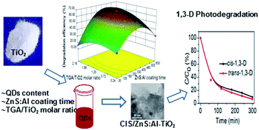
A novel and improved protocol was developed to prepare CuInS2/ZnS:Al-sensitized TiO2 (CIS/ZnS:Al–TiO2) nanocomposites for 1,3-dichloropropene (1,3-D) photodegradation. Response surface methodology was employed to model and optimize the operational parameters of this protocol. The CIS/ZnS:Al quantum dot (QDs) content, ZnS:Al coating time, and TGA/TiO2 molar ratio were chosen as independent variables at three levels by using the Box–Behnken design; their combined effects on the 1,3-D degradation efficiency were investigated. The synthesized CIS/ZnS:Al QDs and CIS/ZnS:Al–TiO2 nanocomposites were characterized by their photoluminescence intensity, UV-vis absorption spectra, X-ray diffraction, and transmission electron microscopy. Based on the experimental results, an empirical expression was established and subsequently applied to predict the 1,3-D degradation efficiency with the prepared photocatalysts. The predicted degradation efficiencies matched well with the experimental values (R2, 0.9874). The ANOVA results showed that the significance of the parameters was ZnS:Al coating time > QDs content > TGA/TiO2 molar ratio. The 3D response surface plots indicated that the optimum synthesis parameters were a CIS/ZnS:Al QDs content of 23%, a coating time of 418 min, and a molar ratio of 1.6. The 1,3-D degradation efficiency with the optimized photocatalyst reached 92% under an irradiation time of 5 h. The photodegradation kinetics of 1,3-D was well fitted with a pseudo-first-order kinetic equation. The experimental design and theoretical prediction methods in this work are of great significance in the design and development of high-performance CIS/ZnS:Al–TiO2 nanocomposites.


Introduction

Semiconductor photocatalysts have attracted much attention in the past few decades because of their applications in environmental purification.1–4 Among these photocatalysts, titanium dioxide (TiO2) has been extensively studied for environmental protection and is one of the most promising photocatalysts because of its high activity, non-toxicity, and low cost.5–8 However, given its large energy band gap (3.2 eV), TiO2 could only be excited by near UV region light, which limits the effective utilization of solar light.9 The combination of TiO2 with a narrow band gap semiconductor, such as quantum dots (QDs), has been proven to be effective for overcoming this limitation.10,11 CuInS2 (CIS) QDs, as the direct band gap I–III–VI nontoxic semiconductor compound, are of special interest because they could serve as a sensitizer in photocatalytic applications.12–15 However, CIS QDs suffered from poor photostability or chemical stability. Recently, the formation of self-passivation layer on the surface of the QDs improved the stability of CIS QDs, and such improvement has received much attention.16 However, the photocatalytic performance of this photocatalyst is highly influenced by the preparation conditions for pollutant photodegradation. Shen et al.17 reported that TiO2 nanoparticles were sensitized with CIS QDs by changing the content of CIS QDs. The optimum content was 16.7% to obtain the best photocatalytic activity of the methyl orange aqueous solution. Ge et al.18 showed that the doping amount of CdS QDs had a great influence on the photocatalytic activity of the QDs-sensitized photocatalyst. Therefore, the QDs content is an important factor that affects the photocatalytic activity of QDs-sensitized TiO2. In addition, Huang et al.19 found that the ability to undergo a photocatalytic reaction is related to the electron transfer efficiency. The optimized electron transfer efficiency and QDs stability can be realized by controlling the shell thickness.20 Therefore, coating time which controls the shell thickness is another important factor. Nevertheless, the coupling of QDs to TiO2 surfaces is not efficient enough; QDs are size-dependent and difficult to control. This drawback could be tackled, to some extent, by optimizing the preparation technology.21 Bifunctional linker molecules, such as thioglycolic acid (TGA), are commonly used to bridge the TiO2 surface and the QDs.22,23 Pan et al. found that the reduction of bifunctional molecule mercaptopropionic acid in QDs-sensitized solar cells could improve the electron transfer efficiency.24 Taken together, the photocatalytic activity of QDs-sensitized TiO2 nanocomposites depends on optimal preparation conditions.

In conventional optimization experiments, the one-variable-at-a-time approach is time consuming and work intensive, as well as lacks the representation of the effects of interactions between different factors.25 Therefore, an adequate experimental design is particularly important to optimize the preparation conditions for the fabrication of high-performance photocatalysts. Response surface methodology (RSM) is an experimental design strategy that is often used for process analysis, optimization, and modeling.26–30 The combined effects of different factors on the desired response can be evaluated with RSM; thus, an optimum condition can be easily achieved. RSM has been successfully applied to determine the optimal conditions for various processes. However, no publications to date have applied the RSM to optimize various affecting parameters for the synthesis of QDs-sensitized TiO2 as photocatalyst. RSM was first demonstrated to optimum the synthesis of CIS/ZnS:Al–TiO2 nanocomposites by changing three factors at three levels: content of CIS/ZnS:Al (QDs content, mass ratio of CIS to TiO2), ZnS:Al coating time in synthesis of QDs (ZnS:Al coating time), and TGA/TiO2 molar ratio.

The RSM results were used to guide the application of CIS/ZnS:Al–TiO2 nanocomposites in the photocatalytic degradation of 1,3-dichloropropene (1,3-D, which includes cis- and trans-1,3-D isomers) as a model for soil fumigants. 1,3-D is a major chemical soil fumigant that has replaced the banned broad-spectrum fumigant methyl bromide. However, 1,3-D is listed as a hazardous air pollutant under the Clean Air Act.31–33 Therefore, the effective photocatalytic degradation of 1,3-D is beneficial to protect the environment. In the present study, the desired response values (1,3-D degradation efficiency) were predicted by RSM. The optimum conditions were obtained for 1,3-D degradation. The effects of the interactions between different factors were also discussed.

Experimental

Materials

TiO2 (anatase, 25 nm), copper iodide (CuI, 99.95%), dodecanethiol (DDT, 98%), 1-octadecene (ODE, >90%), n-butylamine (>99%), zinc acetate (Zn(Ac)2, 99%), oleylamine (90%), and TGA (≥98%) were obtained from Aladdin Chemicals (Shanghai, China). Indium acetate (In(Ac)3, 99.99%) was provided by Alfa Aesar (Ward Hill, MA, USA). Oleic acid (OA, 90%) and aluminum isopropoxide (Al(IPA)3, ≥98%) were obtained from Sigma-Aldrich (St. Louis, MO, USA). Acetone (HPLC grade) was purchased from Merck. 1,3-D (96%; includes 50[thin space (1/6-em)]:[thin space (1/6-em)]50 cis- and trans-isomers) was obtained from J&K Scientific Ltd (Beijing, China). All chemicals were used without further purification.

Response surface methodology

RSM was used for the experimental design and optimization with the Design-Expert software (version 8.0.5, Stat-Ease, Inc., Minneapolis, MN). The influences of QDs content, ZnS:Al coating time, and TGA/TiO2 molar ratio on the synthesis of CIS/ZnS:Al–TiO2 nanocomposites were investigated in detail with a Box–Behnken design (BBD) by RSM. These factors were selected with coded values at 3 levels (−1, 0, and +1). The 1,3-D degradation efficiency was chosen as the response to the three factors. The experimental ranges and levels of variables are given in Table 1. Notably, the choice of the three factors was based on our preliminary experiments.
Table 1 Experimental ranges and levels of independent variables
Independent variables Ranges and levels
−1 0 1
QDs content (%) 5 20 35
ZnS:Al coating time (min) 30 330 630
TGA/TiO2 molar ratio 0.24 1.20 2.16


Photocatalyst preparation

Zn(OA)2 precursor was synthesized according to our recent paper.16 Zn(Ac)2 (13.8 g) was mixed with 45 mL of OA with the addition of 240 mL of ODE and 15 mL of n-butylamine in a three-neck round-bottom flask. The mixture was refluxed under an inert atmosphere at 120 °C to form an optically transparent solution. This solution was cooled to room temperature and stored as the stock solution. CIS core solution was synthesized via one pot synthesis from 1.9 g of CuI, 2.9 g of In(Ac)3, 10 mL of oleylamine, 50 mL of DDT, and 50 mL of ODE in a three-neck round-bottom flask. The mixture was degassed at 120 °C and heated to 230 °C for 40 min under a nitrogen flow.

To synthesize the CIS/ZnS:Al QDs, the CIS core solution was first diluted with ODE and without intermediate purification step. The solution was then degassed at 120 °C, and the reaction temperature was maintained at 230 °C under a nitrogen atmosphere. Zn(OA)2 solution (0.25 mmol mL−1) and DDT mixed with Al(IPA)3 (molar ratio of Al to Zn, 0.5) were injected to the CIS core solution through a syringe pump. Subsequently, the reaction mixture was cooled to the room temperature, precipitated with methanol, and dispersed in toluene.

CIS/ZnS:Al–TiO2 nanocomposites were prepared by using modification of the preparation reported by Pijpers et al.34 TiO2 (0.2 g) was first dispersed in 8 mL of ethanol followed by adding different amounts of TGA. The excess TGA and ethanol were removed by centrifugation. The sediment was dried in a vacuum for 8 h at 60 °C. Functionalized TiO2 nanoparticles were mixed with QDs in toluene at different concentrations. The resulting solution was magnetically stirred for several hours.

Characterization

Transmission electron microscopy (TEM) of the synthesized CIS/ZnS:Al QDs and CIS/ZnS:Al–TiO2 nanocomposites were obtained with a JEM-2100 microscope operated at 200 kV. X-ray diffraction (XRD) patterns were obtained with a Shimadzu XRD-6100 diffractometer. Fourier transform infrared spectroscopy (FTIR) was performed with a Nicolet 6700 spectrometer (Thermo Fisher Scientific). The UV-vis absorption spectra and photoluminescence (PL) intensity of CIS/ZnS:Al QDs were obtained with a UV-vis spectrophotometer (T6, Beijing Purkinje General Instrument Co., Ltd.) and a Gangdong F-380 fluorescent spectrophotometer, respectively. The UV-vis absorption spectra of CIS/ZnS:Al–TiO2 nanocomposites were obtained with a Perkin Elmer Lambda 750 UV-vis/NIR spectrometer.

Degradation of 1,3-D on photocatalyst

A headspace vial (21 mL) was added with a certain amount of photocatalyst and injected with 10 μL of 1,3-D (10 mM in acetone). Each vial was immediately capped with a Teflon-faced butyl-rubber septum and an aluminum cap (Agilent Technologies, Inc.). The bottom of the vial was placed on a quartz glass and irradiated under simulated solar light (Xenon lamp, CEL-S500, Aulight, China) with an average intensity of 90 mw cm−2. The light intensity was measured by using a CEL-NP2000 light power meter. No filter was used. The vials were moved to a freezer (−20 °C) and stored for analysis at certain time intervals. Subsequently, 5 mL of acetone was added into the frozen vials, which were immediately capped with Teflon-faced butyl rubber septa and aluminum caps. All vials were shaken for 1 h. The supernatants were filtered with a 0.22 μm nylon syringe filter, and an aliquot was transferred to a 2.1 mL vial for 1,3-D analysis on an Agilent 6890N gas chromatograph (GC) equipped with an Agilent 5973 mass selective detector (MS). Separation was performed with a DB-5 MS capillary column (30 m × 250 μm × 0.25 μm). The inlet temperature and MS source temperature were 250 °C and 230 °C, respectively. The initial oven temperature was set at 40 °C for 1 min and ramped to 100 °C at a rate of 15 °C min−1 and finally to 125 °C at a rate of 50 °C min−1. Under these conditions, the retention times of cis-1,3-D and trans-1,3-D were 3.1 and 3.4 min, respectively.

Kinetics evaluation

The pseudo-first-order model was applied to analyze the kinetic data with a kinetic equation.35 The equation is a simplification of the Langmuir–Hinshelwood kinetic expression, which is commonly used to explain the kinetics of heterogeneous photocatalysis.36 The equations are listed as follows:
 
image file: c6ra18081g-t1.tif(1)
 
image file: c6ra18081g-t2.tif(2)
where C0 is the initial 1,3-D concentration, C is the concentration at any time (t), k is the pseudo-first-order rate constant, and t1/2 is the half-life.

Results and discussion

Model establishment and analysis

Based on the experimental results in Table 2, the data were analyzed by the Design Expert software. Analysis of variance (ANOVA) was utilized to evaluate the significance of each factor and interaction term (Table 3). The response surface quadratic model was significant with F = 43.5 and p < 0.001. The F value for lack of fit (1.5) implied that the lack of fit was not significant, and p = 0.4189 suggested the excellent applicability of the model. In the table, A, B, and C, as well as the quadratic coefficients A2, B2, and C2 and the interaction coefficients BC and AC, were significant at a level less than 0.0500 except for AB. The interaction between QDs content (A) and ZnS:Al coating time (B) had no significant effect on the degradation of 1,3-D (p = 0.1546; p > 0.05). The effect of ZnS:Al coating time (B) on the degradation process was more considerable than that of QDs content (A) or TGA/TiO2 molar ratio (C), and this finding was obtained by comparing the p-values of the three independent variables. The significance of the parameters is in the following order: ZnS:Al coating time > QDs content > TGA/TiO2 molar ratio.
Table 2 Factors and responses values of BBD
Run Factor A Factor B Factor C Response
QDs content (%) ZnS:Al coating Time (min) TGA/TiO2 molar ratio Degradation efficiency (%)
1 35 30 1.20 21.16
2 5 330 2.16 36.16
3 20 30 0.24 26.46
4 20 30 2.16 15.57
5 20 330 1.20 68.09
6 5 30 1.20 8.66
7 20 630 2.16 52.94
8 20 330 1.20 62.69
9 20 330 1.20 63.21
10 35 330 0.24 37.57
11 5 630 1.20 38.80
12 35 330 2.16 59.05
13 5 330 0.24 34.84
14 35 630 1.20 39.84
15 20 630 0.24 30.26


Table 3 ANOVA for response surface quadratic model
Source Sum of squares dfa Mean square F Valueb p valuec Prob > F
a Degrees of freedom.b Test for comparing model variance with residual (error) variance.c Probability of seeing the observed F-value if the null hypothesis is true.
Model 4573.9 9 508.2 43.5 0.0003
A (QDs content) 191.7 1 191.7 16.4 0.0098
B (ZnS:Al coating time) 1012.3 1 1012.3 86.6 0.0002
C (TGA/TiO2 molar ratio) 149.6 1 149.6 12.8 0.0159
AB 32.8 1 32.8 2.8 0.1546
AC 101.6 1 101.6 8.7 0.0320
BC 281.7 1 281.7 24.1 0.0044
A2 670.5 1 670.5 57.3 0.0006
B2 2139.7 1 2139.7 183.0 <0.0001
C2 318.2 1 318.2 27.2 0.0034
Residual 58.5 5 11.7    
Lack of fit 40.7 3 13.6 1.5 0.4189
Pure error 17.7 2 8.9    
R2 0.9874        
Adjusted R2 0.9647        


Based on parameter estimation, the empirical relationship between the response variable and independent variables can be expressed as follows:

 
Y = −11.49 + 2.51X1 + 0.19X2 + 12.06X3 − 6.37 × 10−4X1X2 + 0.35X1X3 + 0.029X2X3 − 0.060X12 − 2.67 × 10−4X22 − 10.07X32 (3)
where Y is the degradation efficiency of 1,3-D under an irradiation time of 3 h. X1, X2, and X3 represent the actual values of QDs content, ZnS:Al coating time, and TGA/TiO2 molar ratio, respectively.

The degradation efficiencies of 1,3-D were predicted by eqn (3). The predicted degradation efficiencies matched well with the experimental results by plotting the predicted values against the actual values (Fig. 1a). In addition, the coefficient of determination (R2, 0.9874) was close to the adjusted coefficient of determination (adjusted R2, 0.9647), thereby further confirming the adaptability of this second-order polynomial model.


image file: c6ra18081g-f1.tif
Fig. 1 Predicted and actual degradation efficiencies of 1,3-D by photocatalysts (a) and residuals versus predicted values (b).

Fig. 1b shows the plot of the residuals (i.e., the difference between the values of response from experiment and prediction) versus the predicted responses. The residual points were randomly scattered without any systematic structure and obvious pattern. Therefore, the obtained model can be adequate to describe the relationship between the degradation efficiency and the synthetic factors.

Effects of the factors on the 1,3-D photodegradation efficiency

The 3D response surface plots were constructed for further illustrating the interaction effects of the factors on the 1,3-D photodegradation efficiency. An optimum solution for the synthesis of CIS/ZnS:Al–TiO2 nanocomposites can also be obtained. All plots are shown in Fig. 2, which depicts the interactions between the two variables when the third variable is kept constant at its zero level for 1,3-D degradation. Moreover, to further depict the relationship between the factors and the 1,3-D degradation efficiency, the XRD patterns, TEM images, PL intensity, UV-vis absorption spectra, and FTIR spectra of some typically prepared CIS/ZnS:Al–TiO2 nanocomposites and CIS/ZnS:Al QDs are displayed in Fig. 3–6.
image file: c6ra18081g-f2.tif
Fig. 2 Response surface plots of 1,3-D degradation efficiency by photocatalysts as the function of (a) QDs content and ZnS:Al coating time (TGA/TiO2 molar ratio, 1.2), (b) ZnS:Al coating time and TGA/TiO2 molar ratio (QDs content, 20%), and (c) QDs content and TGA/TiO2 molar ratio (ZnS:Al coating time, 330 min). Experimental conditions: solar light intensity, 90 mw cm−2; irradiation time, 3 h.

image file: c6ra18081g-f3.tif
Fig. 3 XRD patterns (a) and absorption spectra (b) obtained over CIS/ZnS:Al–TiO2 of variable QDs contents (0%, 5%, 20%, and 35%). (*) TiO2 and (+) CIS/ZnS:Al.

image file: c6ra18081g-f4.tif
Fig. 4 Evolution of TEM images of CIS/ZnS:Al QDs with different QDs contents: 0% (a), 5% (b), 20% (c), and 35% (d). ZnS:Al coating time, 330 min; TGA/TiO2 molar ratio, 1.2.

image file: c6ra18081g-f5.tif
Fig. 5 Evolution of PL spectra (a), absorption spectra (b), XRD patterns (c), and TEM images (d) of CIS QDs with different sizes during the growth of a ZnS:Al shell. ZnS:Al coating time, 0, 30, 330, and 630 min. Scale bar, 20 nm.

image file: c6ra18081g-f6.tif
Fig. 6 FTIR spectra of TiO2, TiO2 + TGA (TGA/TiO2 molar ratio, 1.2), CIS/ZnS:Al–TiO2 (QDs content, 20%; ZnS:Al coating time, 330 min; TGA/TiO2 molar ratio, 1.2).

Fig. 2a shows the simultaneous influence of QDs content and ZnS:Al coating time on the 1,3-D degradation efficiency (TGA/TiO2 molar ratio, 1.2). As the QDs content increased from 5% to 23%, the degradation efficiency increased from 47.61% to 65.32% and peaked at 65.32%. With an additional increase to 35%, the degradation efficiency decreased to 55.52%. A proper explanation can be obtained from the characteristic results in Fig. 3 and 4. The major diffraction peaks observed at 25.3°, 37.8°, and 48.0° in Fig. 3a can be indexed to the (101), (004), and (200) reflection directions, respectively, of the anatase TiO2 structure (JCPDS 21-1272). Compared with the bare TiO2 nanoparticles (0%), the addition of 5% QDs to the TiO2 nanoparticles produced new weak peaks. Further addition of QDs generated strong peaks at 28.0°, 46.8°, and 55.2°, which can be attributed to the reflection direction of CIS/ZnS:Al QDs; the relative intensity of these new peaks increased with increasing the QDs content from 20% to 35%. Fig. 4 shows the TEM images of CIS/ZnS:Al–TiO2 nanocomposites with different amounts of CIS/ZnS:Al QDs. TiO2 nanoparticles were surrounded by more small nanoparticles with increasing QDs content. By contrast, an excessive amount of CIS/ZnS:Al QDs influenced the TiO2 crystalline structure; then the TiO2 nanoparticles can be hardly observed (Fig. 4d). The results proved the above-mentioned phenomenon that higher QDs content from 20% to 35% slightly decreased the degradation efficiency. On the other hand, the increasing QDs content can enhance the UV-vis adsorption in the visible region (Fig. 3b), which underlies the efficient harvesting of visible-light energy. Therefore, the QDs content of approximately 20% in the nanocomposites caused the relatively higher degradation efficiency of 1,3-D in the simulated solar light.

The effect of ZnS:Al coating time on 1,3-D degradation was similar to that of the QDs content. By increasing the coating time from 30 min to 418 min, the degradation efficiency increased from 30.20% to 65.32%. The efficiency decreased to 50.55%, as the coating time further increased to 630 min (Fig. 2a). The optical and morphological structures of QDs synthesized at different coating times of 0, 30, 330, and 630 min are presented in Fig. 5. With increasing the ZnS:Al coating time, a blue shift from 709, 647, and 629 to 623 nm was observed (Fig. 5a). The adsorption spectra slightly changed from 580, 560, and 550 to 548 nm (Fig. 5b). The XRD patterns further illustrated that all peaks shifted to the larger angles, which were close to the diffraction peaks of ZnS (Fig. 5c). By increasing the coating time, the sizes of QDs in the TEM images became successively larger. The corresponding sizes were 2.7, 3.2, 4.4, and 5.1 nm (Fig. 5d). The Zn component was further incorporated into the core material, thereby resulting in an increase of band gap.37 The coating of ZnS:Al and the diffusion of Zn into the CIS core effectively eliminated surface defects and dangling bonds on the CIS core, which significantly suppressed the non-irradiation recombination and improved the PL quantum yields of QDs.38 After QDs and TiO2 formed heterojunctions, the holes and electrons separated on the interfaces and electrons transferred to TiO2. Within a relatively short coating time, a gradient alloy layer between the ZnS:Al layer and the CIS core was formed.16 This gradient core/shell structure could cause the “smoothing” of the confinement potential interfacial.38 The photogenerated electrons with high energy in the core can tunnel through the ZnS shell to TiO2. Thus, the degradation efficiencies of 1,3-D rapidly improved after increasing the ZnS:Al coating time. By contrast, the degradation efficiencies decreased with increasing time. This phenomenon was attributed to the decreased electron transfer to TiO2 nanoparticles. The increasing ZnS:Al shell became a tunneling barrier because of its significantly higher lying conduction band.39 Therefore, the 3D response surface implies that a higher 1,3-D degradation efficiency can be obtained for CIS/ZnS:Al–TiO2 nanocomposites prepared with suitable QDs content and ZnS:Al coating time.

Fig. 2b shows the variation of ZnS:Al coating time and TGA/TiO2 molar ratio affecting the 1,3-D degradation efficiency (QDs content, 20%). As a molecular linker, the carboxylate group of TGA has a strong affinity for TiO2, and the thiol group is oriented outward for coupling with QDs.23 Fig. 6 shows the FTIR spectra of TiO2, TiO2 + TGA, and CIS/ZnS:Al–TiO2. The strong absorption bands at 500–700 cm−1 are assigned to the Ti–O–O vibrations of TiO2.40 Compared with the bare TiO2, the new band of TiO2 + TGA at 1720 cm−1 can be attributed to C[double bond, length as m-dash]O stretching vibration. The bands at 1088 and 1048 cm−1 can be assigned to the symmetrical and asymmetric stretching vibrations of C–O–C.41 These results indicated the formation of COO–Ti group between TiO2 and TGA.41 The absorption band of CIS/ZnS:Al–TiO2 at 2923 and 2852 cm−1 can be assigned to the C–H stretching vibrations of ligand in CIS/ZnS:Al QDs. Therefore, TGA was effectively anchored onto the surface of TiO2 by the formation of a COO–Ti group and successfully combined with the CIS/ZnS:Al QDs. However, the amount of TGA significantly influenced the degradation efficiency. When the TiO2 nanoparticles were sensitized with CIS core, the efficiency of CIS–TiO2 nanocomposites decreased with increasing TGA/TiO2 molar ratio (Fig. 7). By contrast, the degradation efficiency at a molar ratio of 1.2 was higher than without TGA when CIS/ZnS:Al–TiO2 was used to photodegrade 1,3-D. The degradation efficiency was related to the coating time and molar ratio, which agreed with the results of the 3D response surface. By increasing the coating time or molar ratio, the degradation efficiencies declined after the first increase.


image file: c6ra18081g-f7.tif
Fig. 7 Influence of the TGA/TiO2 molar ratio on the degradation of 1,3-D. CIS and CIS/ZnS:Al represent CIS–TiO2 (QDs content, 5%) and CIS/ZnS:Al–TiO2 (QDs content, 20%), respectively.

However, the molar ratio had less influence than the coating time (Fig. 2b), thereby agreeing with the ANOVA results (Table 3). The p-value of the molar ratio (0.0159) was markedly larger than that of the coating time (0.0002). The interaction between coating time and molar ratio was the most notable. We can confirm that the photocatalysts prepared with too low or too high amounts of TGA possessed worse photocatalytic activity.

To further investigate the effects of QDs content and TGA/TiO2 molar ratio on the degradation efficiency of 1,3-D, the experiments were conducted with CIS/ZnS:Al–TiO2 synthesized with different contents and molar ratios, which varied from 5% to 35% and 0.24 to 2.16, respectively. The plot indicates that the highest degradation efficiency was attained when the QDs content was approximately 20% (Fig. 2c). Simultaneously, the degradation efficiency first increased from 49.83 to 65.00% with increasing TGA/TiO2 molar ratio from 0.24 to 1.60, and then slightly decreased to 60.42% as the molar ratio further increased to 2.16. The addition of TGA to the photocatalyst led to the uniform heterojunction between TiO2 and CIS/ZnS:Al and the formation of intimate contact. However, the excess amount of the linker will influence the electron transfer efficiency.24 This phenomenon can explain why the degradation efficiency began decreasing as the molar ratio further increased. The plot sufficiently demonstrated that the interaction between the QDs content and TGA/TiO2 molar ratio played an important role with a p-value of 0.0320 (Table 3).

The desired goals for all the variables were chosen in the experimental range by the Design expert software such that the QDs content, ZnS:Al coating time, and TGA/TiO2 molar ratio were in the ranges of 5–35%, 30–630 min, and 0.24–2.16, respectively. The optimum values of the factors for synthesizing the photocatalyst with the highest degradation efficiency (68%) for 1,3-D were as follows: 23%, 418 min, and 1.6 for QDs content, ZnS:Al coating time, and TGA/TiO2 molar ratio, respectively. The degradation efficiency when using the optimized photocatalyst was further compared with that when using pristine TiO2. The efficiency was 72.1 ± 3.4%, which was significantly higher than that when using pristine TiO2 (20.4 ± 5.5%). The results verified that CIS/ZnS:Al QDs improved the photocatalytic activity of TiO2 under solar light.

Fumigant degradation kinetics

To evaluate the degradation kinetics of 1,3-D, Fig. 8 shows the decline of cis- and trans-1,3-D concentrations over time under different masses of the photocatalyst. No obvious disappearances were observed after 1,3-D irradiation without any photocatalyst. With increasing photocatalyst mass, the dissipation of 1,3-D over time became more rapid, thereby consistently increasing the dissipation rates of 1,3-D. Compared with the control, the concentrations of cis-1,3-D decreased by 35.4%, 73.5%, and 90.4% and those of trans-1,3-D by 44.3%, 81.7%, and 93.5%, at 5 h under the masses of 10, 30, and 50 mg, respectively. Therefore, the highest 1,3-D degradation efficiency was approximately 92% after irradiation for 5 h. The pseudo-first-order model with eqn (1) and (2) was used to derive the pseudo-first-order rate constant (k) and half-life (t1/2), which are given in Table 4. The correlation coefficients at different levels were higher than 0.9453, thereby indicating that 1,3-D degradation kinetics could be considered a pseudo-first-order model. With increasing photocatalyst mass, the rate constant increased whereas the half-life was shortened. Moreover, the t1/2 of cis- and trans-1,3-D were 2.2% and 2.3% of that without any photocatalyst, respectively.
image file: c6ra18081g-f8.tif
Fig. 8 Photocatalytic degradation of cis-1,3-D (a) and trans-1,3-D (b) over time with different masses of CIS/ZnS:Al–TiO2 nanocomposites. Experimental conditions: solar light intensity, 90 mw cm−2; irradiation time, 5 h.
Table 4 First-order rate constant (k), 1,3-D half-life (t1/2), and correlation coefficient (R2) at different masses of the photocatalyst
Fumigant Photocatalyst mass (mg) k (min−1) t1/2 (min) R2
cis-1,3-D 0 0.00028 2466 0.9956
10 0.00175 396 0.9453
30 0.00881 79 0.9685
50 0.01273 54 0.9562
trans-1,3-D 0 0.00031 2239 0.9979
10 0.00198 350 0.9489
30 0.00931 74 0.9761
50 0.01344 52 0.9703


Under the same simulated solar light and photocatalyst mass, the trans isomer dissipated more rapidly than the cis isomer (Table 4). This effect is possibly ascribed to the direct reaction of trans-1,3-D with nucleophilic groups, such as –SH, and –COOH.42 These groups originated from the ligand DDT and OA in QDs synthesis. These groups may have rapidly reacted with the trans isomer but not with the cis isomer because of the steric difference,42 thereby improving the degradation efficiency relative to that of the cis isomer. The photocatalyst prepared by RSM can be applied to 1,3-D photodegradation. The photodegradation kinetics could be considered a pseudo-first-order model, thereby providing a basis for further exploring the 1,3-D reaction mechanism.

Conclusions

RSM was successfully applied to optimize the synthesis of CIS/ZnS:Al–TiO2 nanocomposites. The ANOVA results demonstrated that the ZnS:Al coating time was the most significant parameter among the investigated factors for 1,3-D photodegradation, which was also verified through PL spectra, absorption spectra, XRD patterns, and TEM images of QDs. The interactions between the TGA/TiO2 molar ratio and QDs content or the ZnS:Al coating time had significant effects on 1,3-D degradation. By contrast, the interaction between QDs content and ZnS:Al coating time did not have a significant effect on the degradation. The optimum parameters for maximum 1,3-D photodegradation were a QDs content of 23%, a ZnS:Al coating time of 418 min, and a TGA/TiO2 molar ratio of 1.6. Moreover, 1,3-D photodegradation kinetics could be considered a pseudo-first-order model. The findings of this study contribute to other researchers who are interested in optimizations regarding preparation of TiO2-based photocatalysts.

Acknowledgements

This work was supported by the National Natural Science Foundation of China (NSFC 21477075, 21271179), the National Key Research and Development Plan (2016YFD0800207), the National Science and Technology pillar program (2012BAD15B03), and the Shanghai Agricultural Commission (Grant No. 2015-4-1).

Notes and references

  1. H. Kominami, T. Nishi, K. Fuku and K. Hashimoto, RSC Adv., 2013, 3, 6058–6064 RSC .
  2. H. X. Guo, J. H. Chen, W. Weng, Q. X. Wang and S. X. Li, Chem. Eng. J., 2014, 239, 192–199 CrossRef CAS .
  3. X. J. Liu, L. K. Pan, T. Q. Chen, J. L. Li, K. Yu, Z. Sun and C. Q. Sun, Catal. Sci. Technol., 2013, 3, 1805 CAS .
  4. M. Le Bechec, N. Kinadjan, D. Ollis, R. Backov and S. Lacombea, Appl. Catal., B, 2015, 179, 78–87 CrossRef CAS .
  5. M. M. Gao, L. L. Zhu, W. L. Ong, J. Wang and G. W. Ho, Catal. Sci. Technol., 2015, 5, 4703 CAS .
  6. X. Chen and S. S. Mao, Chem. Rev., 2007, 107, 2891–2959 CrossRef CAS PubMed .
  7. D. J. Yang, H. W. Liu, Z. F. Zheng, Y. Yuan, J. C. Zhao, E. R. Waclawik, X. B. Ke and H. Y. Zhu, J. Am. Chem. Soc., 2009, 131, 17885–17893 CrossRef CAS PubMed .
  8. M. J. Nalbandian, M. L. Zhang, J. Sanchez, S. Kim, Y. Choa, D. M. Cwiertny and N. V. Myung, J. Hazard. Mater., 2015, 299, 141–148 CrossRef CAS PubMed .
  9. V. M. Daskalaki, M. Antoniadou, G. L. Puma, D. I. Kondarides and P. Lianos, Environ. Sci. Technol., 2010, 44, 7200–7205 CrossRef CAS PubMed .
  10. C. Ratanatawanate, Y. Tao and K. J. Balkus Jr, J. Phys. Chem. C, 2009, 113, 10755–10760 CAS .
  11. C. J. Wang, R. L. Thompson, J. Baltrus and C. Matranga, J. Phys. Chem. Lett., 2009, 1, 48–53 CrossRef .
  12. Z. Y. Peng, Y. L. Liu, K. Q. Chen, G. J. Yang and W. Chen, Chem. Eng. J., 2014, 244, 335–342 CrossRef CAS .
  13. W. J. Zhang and X. H. Zhong, Inorg. Chem., 2011, 50, 4065–4072 CrossRef CAS PubMed .
  14. L. X. Yi, Y. Y. Liu, N. L. Yang, Z. Y. Tang, H. J. Zhao, G. H. Ma, Z. G. Su and D. Wang, Energy Environ. Sci., 2013, 6, 835–840 CAS .
  15. Y. H. Lin, F. Zhang, D. C. Pan, H. X. Li and Y. F. Lu, J. Mater. Chem., 2012, 22, 8759–8763 RSC .
  16. P. H. Rao, W. Yao, Z. C. Li, L. Kong, W. Q. Zhang and L. Li, Chem. Commun., 2015, 51, 8757–8760 RSC .
  17. F. Y. Shen, W. X. Que, Y. L. Liao and X. T. Yin, Ind. Eng. Chem. Res., 2011, 50, 9131–9137 CrossRef CAS .
  18. L. Ge and J. Liu, Appl. Catal., B, 2011, 105, 289–297 CrossRef CAS .
  19. L. Huang, X. L. Wang, J. H. Yang, G. Liu, J. F. Han and C. Li, J. Phys. Chem. C, 2013, 117, 11584–11591 CAS .
  20. J. H. Sun, J. L. Zhao and Y. Masumoto, Appl. Phys. Lett., 2013, 102, 053119 CrossRef .
  21. R. S. Dibbell and D. F. Watson, J. Phys. Chem. C, 2009, 113, 3139–3149 CAS .
  22. L. Li, X. C. Yang, J. J. Gao, H. N. Tian, J. Z. Zhao, A. Hagfeldt and L. C. Sun, J. Am. Chem. Soc., 2011, 133, 8458–8460 CrossRef CAS PubMed .
  23. T. López-Luke, A. Wolcott, L. P. Xu, S. W. Chen, Z. H. Wen, J. H. Li, E. De La Rosa and J. Z. Zhang, J. Phys. Chem. C, 2008, 112, 1282–1292 Search PubMed .
  24. Z. X. Pan, H. Zhang, K. Cheng, Y. M. Hou, J. L. Hua and X. H. Zhong, ACS Nano, 2012, 6, 3982–3991 CrossRef CAS PubMed .
  25. A. R. Khataee, M. B. Kasiri and L. Alidokht, Environ. Technol., 2011, 32, 1669–1684 CrossRef CAS .
  26. W. J. Jiang, J. A. Joens, D. D. Dionysiou and K. E. O'Shea, J. Photochem. Photobiol., A, 2013, 262, 7–13 CrossRef CAS .
  27. P. R. Choudhury, P. Mondal and S. Majumdar, RSC Adv., 2015, 5, 100838–100848 RSC .
  28. J. Y. Chen, G. Y. Li, Y. Huang, H. M. Zhang, H. J. Zhao and T. C. An, Appl. Catal., B, 2012, 123, 69–77 CrossRef .
  29. K. Li and Z. M. Xu, Environ. Sci. Technol., 2015, 49, 1761–1767 CrossRef CAS PubMed .
  30. F. Ferdosian, Z. S. Yuan, M. Anderson and C. Xu, RSC Adv., 2014, 4, 31745–31753 RSC .
  31. L. F. Luo, S. R. Yates, D. J. Ashworth, R. C. Xuan and J. O. Becker, J. Agric. Food Chem., 2013, 61, 2400–2406 CrossRef CAS PubMed .
  32. W. Zheng, S. K. Papiernik, M. X. Guo and S. R. Yates, J. Environ. Qual., 2003, 32, 1735–1742 CrossRef CAS PubMed .
  33. J. Gan, S. R. Yates, D. Crowley and J. O. Becker, J. Environ. Qual., 1998, 27, 408–414 CrossRef CAS .
  34. J. J. H. Pijpers, R. Koole, W. H. Evers, A. J. Houtepen, S. Boehme, C. de Mello Donegá, D. Vanmaekelbergh and M. Bonn, J. Phys. Chem. C, 2010, 114, 18866–18873 CAS .
  35. R. Y. Xie, L. P. Zhang, H. Xu, Y. Zhong, X. F. Sui and Z. P. Mao, J. Mol. Catal. A: Chem., 2015, 406, 194–203 CrossRef CAS .
  36. K. V. Kumar, K. Porkodi and F. Rocha, Catal. Commun., 2008, 9, 82–84 CrossRef CAS .
  37. L. Li, T. J. Daou, I. Texier, T. T. Kim Chi, N. Q. Liem and P. Reiss, Chem. Mater., 2009, 21, 2422–2429 CrossRef CAS .
  38. F. García-Santamaría, Y. F. Chen, J. Vela, R. D. Schaller, J. A. Hollingsworth and V. I. Klimov, Nano Lett., 2009, 9, 3482–3488 CrossRef PubMed .
  39. H. McDaniel, N. Fuke, N. S. Makarov, J. M. Pietryga and V. I. Klimov, Nat. Commun., 2013, 4, 2887 Search PubMed .
  40. I. Truijen, A. Hardy, M. K. Van Bael, H. Van den Rul and J. Mullens, Thermochim. Acta, 2007, 456, 38–47 CrossRef CAS .
  41. E. Chiellini, L. Bemporad and R. Solaro, J. Bioact. Compat. Polym., 1994, 9, 152–169 CrossRef CAS .
  42. M. X. Guo, S. K. Papiernik, W. Zheng and S. R. Yates, J. Environ. Qual., 2004, 33, 612–618 CrossRef CAS PubMed .

This journal is © The Royal Society of Chemistry 2016
Click here to see how this site uses Cookies. View our privacy policy here.