Potential prospects for carbon dots as a fluorescence sensing probe for metal ions

Savita Chaudhary*, Sandeep Kumar, Bhawandeep Kaur and S. K. Mehta
Department of Chemistry and Centre of Advanced Studies in Chemistry, Panjab University, Chandigarh 160014, India. E-mail: schaudhary@pu.ac.in; Fax: +91 172 2545074; Tel: +91 9417250377

Received 16th June 2016 , Accepted 7th September 2016

First published on 7th September 2016


Abstract

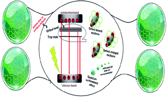
The significance and importance of carbon-based NPs and their promising applications were a strong inspiration for this work. An inexpensive and easy synthesis of carbon quantum dots (CQDs) has been carried out by using easily-available biomass such as wheat (Triticum), rice (Oryza sativa), pearl millet (Pennisetum glaucum) and sorghum. The as-prepared particles have better-controlled stability and luminescence activities as CQDs without using any external template. The well-defined emission properties of CQDs further encouraged the investigation of their role for sensing chromium (Cr3+) ions in aqueous media. Compared to the response to other metal ions, the fluorescence emission (photoluminescence) of CQDs shows significant enhancement in the presence of Cr3+ ions, with a limit of detection of 6 × 10⁻⁷ to 13 × 10−7 M and a linear range of 20 μM. The practicality of use and the average recovery of Cr3+ ions in the presence of different CQDs were also tested in water from different sources, such as tap water, buffer, distilled water and sewage water. The present route offers a simple, rapid, cost-effective and non-toxic means to determine metal ion impurities with improved selectivity and sensitivity.


1. Introduction

Currently, the designing of novel categories of photoreceptive and sensitive NPs (NPs) of semiconducting-, metallic- and lanthanide-based oxide materials has gained significant importance in the biomedical and materials sciences.1–3 The advanced fluorescence properties of these materials gives them applications in diagnostics, therapeutics, imaging, and the sensing of harmful ecological pollutants.4,5 Conversely, the extensive use of heavy metal components in these advanced materials poses a cytotoxic threat to nature and restricts their usage in clinical trials.6 Thus, from the perspective of human health and environmental security, there is an urgent need for alternative materials with extraordinary properties and low toxic side effects. In this respect, carbon-based NPs, primarily carbon quantum dots (CQDs), have been subject to comprehensive research. Their distinctive optical properties and their prospects for the development of efficient optoelectronic devices and sensors have widened their horizons in fluorescence sensing applications.7–9 The high photoluminescence (PL) properties of CQDs make them suitable candidates for bio-imaging applications.8 The adjustable emission properties of CQDs have promoted their role as efficient photo-sensitizers in different types of photo-catalytic applications.10 Their low toxicity, higher stability, appropriate biocompatibility, and excellent water solubility, because of the presence of carboxylic acid groups at their exterior surfaces, make CQDs a new class of environmentally friendly nanomaterials.11,12

Up to now, various synthetic approaches involving pyrolysis, electrochemical processes, laser ablation, oxidation processes, and microwave-assisted methods have been employed to prepare CQDs.13–15 For example, Lai et al.13b have carried out the solid-state synthesis of fluorescent CQDs by using ammonium citrate as a starting material. The systematic structural and optical analysis of CQDs with different concentrations of starting materials was undertaken by Ma et al.13c Cayuela et al. have used amine-derivatized carbon dots (a-CQDs) for detecting the presence of silver NPs in cosmetics.16 Wang et al. have used the application of heteroatom co-doping over CQDs for enhancing the PL activities of NPs. They have also developed easy-to-use test papers for the direct and rapid estimation of mercury (Hg2+) ions from real water samples.17 In a separate paper, Kiran and Misra have reported an effective method for the intracellular recognition of glucose via functionalization of CQDs with boronic acid.18 The as-developed sensor possesses a higher dynamic response towards the changing concentration of glucose. Recently, Martindale et al. have used CQDs as efficient photosensitizers in combination with molecular nickel for solar-light-driven hydrogen production in aqueous solution.19a It has also been found that CQDs can be efficiently produced by using available biomass and waste materials as starting materials. For example, different types of natural hydrocarbons, including oils, tea extracts, honey, milk, eggs, sugar and so on,19b,c have been employed for synthesizing a high yield of CQDs. However, different types of food waste, such as agricultural and vegetable wastes, were also employed for the synthesis of CQDs.19d All these methods avoided the utilization of harmful and toxic chemicals during the preparation of CQDs. But reports on the comparative effects of these methods on the optical and luminescence properties of the CQDs and on their environmental applications are to some extent limited. The use of edible seeds such as wheat and rice for the synthesis of CQDs has also been reported. Furthermore, on comparing the nutrient contents of these edible seeds with vegetables or their waste products, it has been found that the percentage concentrations of protein, crude lipid, carbohydrate and crude fibre contents are higher for edible seeds. It has also been shown in reports in the literature that the amount of carbohydrate formed by the fixation of carbon is greater in edible seeds when compared to vegetable and food products, and therefore, seeds act as a good source of starting material for the synthesis of CQDs.19e As a consequence, the use of these seeds could provide better alternatives for a starting material for preparing CQDs.

Therefore, by keeping the importance of biomass for preparing CQDs in mind, wheat (Triticum), rice (Oryza sativa), pearl millet (Pennisetum glaucum) and sorghum have been used for preparing CQDs. The obtained CQDs were denoted CQD1 (sorghum), CQD2 (millet), CQD3 (rice) and CQD4 (wheat). The main advantage of the synthesis is the use of water as a reaction medium for the conversion of biomass into CQDs. Furthermore, these starting materials are geographically some of the most widely used natural resources and this wide availability has never been recognised in the context of CQD production. The use of such renewable biomass as a starting material for preparing CQDs has significantly reduced the cost of the process. The as-prepared particles offer better control over the stability and luminescence activities of CQDs. The PL activity of the as-prepared particles was further employed to check the ratiometric sensing of chromium (Cr3+) ions. Depending on the fluorescence variations of the as-prepared particles, they can act as superior optical sensors for the effective determination of metal ions. The as-prepared particles were further utilized for estimating metal ion recoveries in real water samples. The present method is a simple, rapid, cost-effective and non-toxic way to determine metal ion impurities with superior selectivity and sensitivity.

2. Experimental section

2.1. Materials

Wheat (Triticum), rice (Oryza sativa), pearl millet (Pennisetum glaucum) and sorghum were purchased from a local market (Sector 14, Chandigarh, India) and used as a starting material for producing CQDs. The pH of the solution was maintained by using hydrochloric acid, sodium hydroxide, disodium hydrogen phosphate dodecahydrate (Na2HPO4·12H2O) and sodium dihydrogen phosphate dehydrate, purchased from Sigma-Aldrich, India. Stock solutions (25 μM) of various metal ions [aluminium (Al3+), cadmium (Cd2+), calcium (Ca2+), chromium (Cr3+), cobalt (Co3+), copper (Cu2+), europium (Eu3+) iron(II) (Fe2+), iron(III) (Fe3+), lead (Pb2+), lithium (Li+), manganese (Mn2+), mercury (Hg2+), potassium (K+), silver (Ag+), sodium (Na+), and zinc (Zn2+)] were obtained from their nitrate salts (Sigma-Aldrich). All the reagents were of high-quality analytical grade and used as received without any further purification. The corresponding aqueous solutions were prepared in doubly distilled water.

2.2. Synthesis of CQDs

The synthesis of CQDs was carried out by thermal calcination of 15 g of edible seeds, for example, wheat, rice, millet and sorghum seed, in a muffle furnace for two hours at 400 °C under a nitrogen atmosphere. After calcination, 11.23 g, 10.94 g, 11.51 g, and 11.3 g of material was obtained for CQD1 (sorghum), CQD2 (millet), CQD3 (rice) and CQD4 (wheat). The complete descriptive synthesis process is illustrated in Scheme 1. The black-coloured powders obtained were cooled down to room temperature and mechanically ground to a powder.
image file: c6ra15691f-s1.tif
Scheme 1 Complete descriptive illustration for the synthesis of CQDs.

From the total amount of powder obtained, 1 g was further dispersed in 50 mL of distilled water under stirring to form a carbonized solution of CQDs. The as-obtained solution was further centrifuged at 7000 rpm for 20 min to remove the larger particles from the solution. The supernatant obtained, containing the CQDs, was further separated out from the larger particles by filtering the solution using 0.22 μm filter membranes. The supernatant obtained was ultracentrifuged at 17[thin space (1/6-em)]000 rpm to obtain a solid extract of CQDs, which was used for analysis. The obtained solid (i.e., 0.35 g CQD1, 0.41 g CQD2, 0.39 g CQD3 and 0.28 g CQD4) was further lyophilised to remove all moisture. The elemental analysis of the obtained solid was carried out using CHN analysis (see Table S1, ESI). Primary studies on the biocompatibility of the as-prepared CQDs were made using the growth of a fungus in the presence of CQDs (Fig. S1, ESI). The fungus Echinodontium taxodii was used to test the activities of the CQDs. In the presence of NPs, the relative colony size of the fungus did not show any decrease, which provided evidence for the biocompatibility of the as-synthesized particles. Further studies confirmed the full biocompatibility of the as-prepared NPs and will be considered later in the paper.

2.3. Experimental instrumentation

The size and morphology of the CQDs were measured with a Hitachi H7500 electron microscope (100 kV). High resolution transmission electron microscopy (HRTEM) analysis was carried out using a Jeol JEM-2100F instrument. Particle size analysis (90° optics) and dynamic light scattering (DLS) analysis were carried out using a Malvern Instruments Zetasizer Nano S90 (Red badge; Model number ZEN 1690). X-ray photoelectron spectroscopic (XPS) analysis was carried out on Omicron NanoTechnology ESCA+. Ultraviolet-visible (UV-vis) absorbance and fluorescence analysis were performed on a Jasco UV-vis V550 and a Hitachi F7000 PL spectrophotometer, respectively. A PANalytical, D/Max 2500 X-ray diffractometer was used to estimate the crystallite size and the phases of the as-obtained CQDs using Cu-Kα radiation (λ = 1.5418 Å). Elemental analysis was carried out using an automatic PerkinElmer 2400 CHN/Analytischer Funktionstest vario EL II elemental analyzer. Thermal calcination of the biomass was carried out in an AICIL muffle furnace at 400 °C. The ultracentrifugation of the resultant solution was undertaken in a Remi R-24 centrifuge. The magnetic stirring of the solutions was carried out on a IKA C-MAG HS 7 stirrer. pH measurements were carried out using a Mettler Toledo digital pH meter. The fluorescence quantum yield of the as-prepared CQDs was estimated by employing the comparative mode, using rhodamine 6G as a reference in water (where ФF = 0.95).

3. Results and discussion

3.1. Structural characterization of CQDs

The light yellowish-brown coloured solutions of the CQDs were obtained from sorghum (CQD1), millet (CQD2), rice (CQD3) and wheat (CQD4), via simple calcination at 400 °C followed by pyrolysis for 2 h as described in Scheme 1. The as-used starting materials are principal cultivated crops which are universally used, easily accessible and an economical resource for producing highly luminescent CQDs, which makes this process unique. Fig. 1 shows the TEM images and the corresponding percentage counts from the DLS analysis for the as-obtained CQDs from the four different crops. The particles appeared to be spherical in shape with an average size of 5–10 nm in all cases. This method does not require any type of external template for the stabilization of NPs, which makes this process more economical and useful for practical applications.
image file: c6ra15691f-f1.tif
Fig. 1 TEM images and corresponding particle size distribution of (a) CQD1, (b) CQD2, (c) CQD3 and (d) CQD4.

The results were further verified by carrying out HRTEM analysis of the as-prepared NPs. Fig. 2 shows the HRTEM images of all four prepared samples. The particles appear to be spherical and well dispersed. The selected area electron diffraction (SAED) patterns show the diffused rings which indicate the poly-nanocrystalline nature of the CQDs. The lattice fringes are also visualized in the images. The calculated interplanar distances were found to be 0.2998 nm for CQD1, 0.312 nm for CQD2, 0.323 nm for CQD3 and 0.309 nm for CQD4. All these results were associated with the (002) planes of graphitized carbon. The results obtained were comparable with those found in previous reports in the literature.8d The respective X-ray diffraction (XRD) patterns for all the CQDs obtained show a single broad diffraction peak at 21° for the (002) plane (Fig. 3)20a–c with a JCPDS file number: 41-1487 (graphite). The crystalline size obtained was comparable with the results obtained from TEM and DLS analysis.


image file: c6ra15691f-f2.tif
Fig. 2 HRTEM images and the corresponding SAED patterns of (a) CQD1, (b) CQD2, (c) CQD3 and (d) CQD4.

image file: c6ra15691f-f3.tif
Fig. 3 XRD spectra of CQDs synthesised using sorghum (CQD1), millet (CQD2), rice (CQD3) and wheat (CQD4).

The structure and functional characterization of the CQDs were further carried out using XPS. The spectra obtained displayed characteristic dominant peaks at around 287.3, and 534.4 eV, which were associated with the binding energies of C1s and O1s, respectively (Fig. 4). The overall C1s and O1s were further deconvoluted to check the existence of the different bonds present in each type of CQD (Fig. S2, ESI). Fig. S2a shows the four types of deconvoluted peaks of C1s for the as-prepared CQDs. For CQD1, the peak positions were around 282.32, 285.14, 285.21 and 295.23 eV. These values were associated with the C1s states in C–C, C[double bond, length as m-dash]C, C–O, O–C[double bond, length as m-dash]O bonds in the as-formed NPs. However, the peaks appeared at 283.99, 284.47, 292.21 and 293.81 eV for CQD2, at 283.21, 285.93, 286.01 and 293.81 eV for CQD3 and at 283.48, 285.74, 286.69 and 294.82 eV for CQD4. In contrast, the O1s region showed two major deconvoluted peaks at 532.6 and 532.95 eV for CQD1, at 531.11 and 531.56 eV for CQD2, at 530.95 and 533.77 eV for CQD3, and at 532.16 and 532.55 eV for CQD4, which confirms the presence of C–OH, C–O–C and C[double bond, length as m-dash]O groups in the as-prepared NPs.21,22


image file: c6ra15691f-f4.tif
Fig. 4 XPS spectra of CQDs synthesised using sorghum (CQD1), millet (CQD2), rice (CQD3) and wheat (CQD4).

3.2. Optical and photoluminescence properties of CQDs

The absorption and emission spectra of the as-synthesized CQDs from four different sources are presented in Fig. 5. By interpreting the data, it has been found that the UV-vis absorption shoulder for CQD1 was found at 281.6 nm, whereas those for CQD2, CQD3 and CQD4 were observed at 259.8 nm, 261.5 nm and 274.2 nm, respectively (Fig. 5a). All these absorption peaks were associated with the corresponding n–π* and π–π* transition of C[double bond, length as m-dash]O bonds over the surface of the NPs.5 The bandgap calculations performed using Tauc plots were also carried out for CQDs in order to understand the effect of the four carbon sources (Fig. S3a, ESI). From the data, it was found that the band gap values were different for all the four samples (Fig. S3b, ESI). The results clearly demonstrated the sensitivity of the starting material for the size and optical properties of the CQDs. In order to further investigate the optical properties, the emission spectra of the CQDs were studied using fluorescence measurements (Fig. 5b).7 The corresponding emission spectra displayed characteristic peaks at 431 nm for CQD1, 347 and 421 nm for CQD2, 349 and 433 nm for CQD3 and 355 and 430 nm for CQD4, with a quantum yield of 17% for CQD1, 33% for CQD2, 30% for CQD3 and 16% for CQD4. The corresponding Stokes shift of the emission spectra for all the four types of CQDs with reference to the absorption spectra is around 149 nm for CQD1, 87 nm for CQD2, 88 nm for CQD3 and 81 nm for CQD4. The variation in Stokes shift for all the samples was related to the nature of the surface state on the NPs, which further affected the electronic transitions in the NPs.23 The tapering of the peaks was further associated with the enhanced degree of surface oxidation in CQDs.19c The size of the NPs also affected the PL intensity and Stokes shift in the CQDs. Such variations further affect the electronic structure of the relaxed, excited, and ground state of the CQDs. Such modification will further open up the uses of such materials for preparing fluorescence-sensitive sensors for different metal ions.24 Gaussian analysis25 of the emission spectra for all four of the samples was carried out to investigate the nature of the defects in the CQDs (Fig. 6). It was found that in all the samples the broad emission peak was divided into three peaks. The first peak was related to the defect-bound excitons (DBE1) and the second peak was correlated with the defect-charged excitons (DBE2) in CQDs.26–28 The third peak corresponded to the electron–hole recombination effect (DBE3) in CQDs (Scheme 2).25 The corresponding positioning, area under the curve and the full width at half maxima (FWHM) results for all the samples are given in Table S2 (ESI).
image file: c6ra15691f-f5.tif
Fig. 5 (a) UV-vis and (b) fluorescence spectra of the four different types of CQDs.

image file: c6ra15691f-f6.tif
Fig. 6 Gaussian analysis of the emission spectra of (a) CQD1, (b) CQD2, (c) CQD3 and (d) CQD4 at λex = 310 nm.

image file: c6ra15691f-s2.tif
Scheme 2 Schematic illustration showing the emission mechanism in CQDs.

The influence of the pH variations on the emission spectra for all the samples was also investigated (Fig. S4, ESI). On interpretation, it is found that with an increase in the pH value from 2 to 13, the PL intensity shows a significant enhancement of around 78.4% for CQD1, 79.5% for CQD2, 29.7% for CQD3 and 52.8% for CQD4. The corresponding increase for the pH from 2 to 7 is around 11.4% for CQD1, 47.8% for CQD2, 18.6% for CQD3 and 50.5% for CQD4. The location of the peak position does not show any variation. The changes clearly show that the variations are because of the deprotonation of available carboxylic groups29 on the surface of the CQDs. Such changes guide the formation of the delocalized π electron cloud, and thus increase the concentration of electrons over the surface of the CQDs.25 Furthermore, the changes in the pH also affect the dynamic equilibrium between the defect-bound excitons and defect-charged excitons.

The PL behaviour of the CQDs was further investigated as a function of increasing the excitation wavelengths ranging from 280 nm to 450 nm for all the four types of particles obtained (Fig. S5, ESI).23 From the graph, it is seen that emission intensity (λemiss) is increased with an increase in excitation wavelength (λex) from 280 to 310 nm. With further increases in the λex up to 450 nm, there is a significant decrease in the emission intensity. The corresponding values of the area under the curve and FWHM as function of λex are illustrated in Fig. S6 (ESI). The results were further verified via Gaussian analysis of emission spectra25 as a function of the excitation wavelength for all four samples, in order investigate the nature of the defect in the CQDs. It was found that DBE1, DBE2 and DBE3 showed significant variations with an increase in the excitation wavelength (λex) from 280 to 310 nm for all CQDs. In the case of CQD1 at λex = 280 nm, the FWHM for DBE1 was found to be 44 nm with respect to the 176.67 nm at λex = 310 nm. The area under the peak also showed a considerable increase of 84.7% in CQD1, and of 44.9%, 77.29% and 43.7% in CQD2, CQD3 and CQD4, respectively. The distinguishing variation was associated with the availability of the electron–hole pairs over the surface of the CQDs produced from different sources.25 Similarly, DBE2 and DBE3 peaks also showed linear variations with increase in λex from 280 to 310 nm. By further increasing the λex up to 450 nm, there was a drop in the FWHM for DBE1 from 176.76 nm at λex = 310 nm to 38.7 nm at λex = 450 nm. The variations were comparable with the other three types of CQDs produced from millet, rice and wheat. All these behavioural changes were associated with the resultant consequences of the different allocations of defect-bound excitons, emissive traps, available surface functional groups (evident from XPS), and reduced rotational arrangement of adjoining graphene sheets which further produces the vacant zig-zag sites and imperfections in the CQDs produced.25,20c Such comparative results open up a new way for utilizing these CQDs in fluorescence imaging applications. The influence of different applied voltages on the emission spectra of CQDs has also been investigated in order to understand the sensitivity of the particles (Fig. S7, ESI).30 The corresponding changes in the emission spectra were mainly explained by the activation and slow inactivation of the electrons in the excited state.31 It was found that a very high voltage of 900 V caused an enhancement in signal with an intensity of more than 10[thin space (1/6-em)]000 au, as compared to >500 au intensity for 400 V and 500 V. Such high intensity restricts the variations in the presence of metal ions. Therefore, 700 V voltage was appropriate for further studies. Such an effect further enhances the prospects of as-synthesized particles with improvements in sensitivity towards voltage variation and lesser tendencies towards photodynamic damage in NPs. The voltage-sensitive changes will be further utilized for optimizing the selection conditions for a given synthesis, so as to obtain an enhanced signal with reduced undesirable side effects.

3.3. Metal ion sensing activity of CQDs

The well-defined emission properties of CQDs encouraged the further investigation of their prospects in chemo-sensing applications for the identification of heavy and transition metal ions from aqueous media.32–34 These metal ions are considered as major constituents in various physiological activities of living things, and thus their selective and sensitive recognition is one of the promising areas of research. Furthermore, the pollution caused by excessive use of these metal ions further enhanced the curiosity of scientists to develop easy and affordable methods for the identification of metal ions from aqueous media. In this respect, fluorescence-based identification of metal ions using carbon NPs presented a new possibility for the selective and sensitive detection of metal ions.35,36 Additionally, the presence of a conjugated π electron system enhances the probability of CQDs to form complexes with a diverse range of metal ions. Thus, the fluorescence-based titration of CQDs synthesized from four different sources towards different metal ions (Ag+, Al3+, Ca2+, Cd2+, Co3+, Cr3+, Cu2+, Eu3+, Fe2+, Fe3+, Hg2+, K+, Li+, Mn2+, Na+, Pb2+, and Zn2+) provided as their nitrate salts (Fig. 7) at a concentration equal to 25 μM was carried out at λex = 310 nm and pH 7.0. In the presence of all the ions, the emission peak at λ = 430 nm showed a significant decrease in all cases. The variations were explained on the basis of the complexation of Cr3+ ions with the carboxylic and hydroxyl groups present over the surface of CQDs (as is evident from the spectra shown in Fig. 4). Additionally, out of all the ions, the presence Cr3+ ions showed a significant enhancement in the fluorescence intensity at λ = 352 nm for all the different types of CQDs under investigation. In all cases, this peak is mainly associated with the transition of electrons from the core of the CQDs. For CQD1, the enhancement was around 16.5%, whereas it was 41.8% for CQD2 and 25.2% for CQD3 and 33.5% for CQD4. The fluorescence intensity of the CQDs was mainly explained by introducing four phenomena, i.e., quantum confinement effect, presence of conjugated π electron domains in the carbon core, presence of different types of functional groups and crosslink-enhanced emission effect.36b After the interactions of Cr3+ ions with the surface groups, there was a probability of interaction of conjugated π electron domains in the carbon core with the metal ions and this causes the crosslink-enhanced emission effect.36b The detailed analysis of this aspect is still under investigation.
image file: c6ra15691f-f7.tif
Fig. 7 Emission spectra of (a) CQD1, (b) CQD2, (c) CQD3 and (d) CQD4 after the addition of different metal ions (25 μM) at λex = 310 nm and pH 7.0.

The corresponding fluorescence titrations were also performed in order to assess the sensitivity of CQDs to the Cr3+ ion. Fig. 8a shows the titration curves for CQD2, whereas the other titration curves are shown in Fig. S8 (ESI). The reliability of the process was tested by performing the titrations three times. The plot of ΔF versus [Cr3+] was also plotted to obtain calibration curves for CQDs with Cr3+ ions. Fig. 8b shows the linear dependency for all the types of fabricated particles as a function of different concentration ranges of Cr3+ ions (1–14 μM). The corresponding slopes, regression coefficients and standard deviations for all the CQDs are shown in Table 1.36 The limits detection (LODs) obtained were also calculated using 3σ IUPAC criteria (Table 1).


image file: c6ra15691f-f8.tif
Fig. 8 (a) Fluorescence emission spectra of CQD2 in the presence of various concentrations of Cr3+ ions and the corresponding plot of (b) ΔF as a function of Cr3+ ions in the presence of different CQDs.
Table 1 Summary of regression coefficients, slopes, standard deviations and limits of detection for different CQDs in the presence of Cr3+ ions
System Regression coefficient Slope Standard deviation LOD (M)
CQD1 0.9917 151.66 28.76 6.89 × 10⁻⁷
CQD2 0.9967 96.51 36.46 12.46 × 10⁻⁷
CQD3 0.9985 88.45 22.34 8.33 × 10⁻⁷
CQD4 0.9988 94.94 21.47 7.46 × 10⁻⁷


It was anticipated that the presence of Cr3+ in the surrounding the CQDs caused the variations in the fluorescence intensity because of the transference of charge. The reduced intensity at 423 nm in comparison to the emission band at 352 nm of CQD is in agreement with the internal charge transference from the π* of the CQD core.37 The intensity enhancement was further explained via the conformational changes because of the decrease in the energy of vibration and the rotational levels in the excited π* of the core in CQD. The results were further evaluated against the available results in the literature for Cr3+ ion sensing.38–41 For example, Liu et al. have used applications of hydroxyl-coated CQDs for the detection of Cr3+ ions in human body fluids.42 The sensors developed have shown a good linearity from 1 to 25 μM and a LOD of 60 nM. In a separate report, fluorescence turn-on emission of N-doped CQDs was chosen for detecting Cr3+ ions in aqueous solution by Wang and Meng.43 Liu et al. have employed the application of hydroxyl-coated CQDs prepared from carbon soot using a hydrothermal method for the sensing of metal ions.44 The sensitivity for Cr3+ detection is high in the method as developed, but the recovery experiments, as well as the interference studies, were not fully explained in this work.

Although various reports have shown a higher LOD, the sensitivity values were low in most of them. Furthermore, the synthetic processing of the sensors developed was quite complicated, time-consuming and costly. However, the CQDs synthesised in this current work possess higher sensitivity and a lower LOD for Cr3+ ions. The modification of the fluorescence behaviour of CQDs synthesized from sorghum (CQD1), millet (CQD2), rice (CQD3) and wheat (CQD4) has further enhanced the use of as-synthesized CQDs for Cr3+ ion sensing. The modulation of defect-bound excitons, defect-charged excitons and electron–hole recombination effect in four different types of synthesized CQDs resulted in an effective electron movement and the formation of an efficient sensor for Cr3+ ions. The prepared CQDs have also provided efficient materials for the selective detection of harmful pollutants. The prepared material has also shown good water solubility and has broadened the horizon for determining harmful pollutants in different water sources.

The efficiency of the as-prepared fluorescence sensors were also checked in the presence of co-existing ions. Briefly, the samples of the interfering cations were diluted to 100 μM and added to a solution which was a mixture of Cr3+ (20 μM) with different CQD samples. All the fluorescence analysis was carried out after proper mixing of the solutions. It was established that the extra addition of different ions produced negligible variations in the emission intensities at 352 nm. Furthermore, in the absence of the Cr3+ ion, the emission ratio (F/F0) was quite low, whereas by adding the Cr3+ ion, there was a significant increase in the intensities (Fig. 9). In addition, there was no change in the peak position. Therefore, the sensor developed is highly selective and efficient for Cr3+ ion sensing. The reliability and reproducibility of the current efforts were also scrutinized for all the CQDs developed. It was found that the particles obtained were stable, with a relative standard deviation (RSD) of 1.4% for CQD1, 1.37% for CQD2, 1.6% for CQD3 and 1.2% for CQD4.


image file: c6ra15691f-f9.tif
Fig. 9 Interference studies performed with other metal ions in a ratio of 3[thin space (1/6-em)]:[thin space (1/6-em)]1 to Cr3+ ions under the same conditions (λmax = 352 nm) for (a) CQD1, (b) CQD2, (c) CQD3 and (d) CQD4.

The realistic effectiveness of the prepared fluorescence sensor for real samples was also examined. Measurements of Cr3+ ion spiked samples were carried out in four different sources of water, i.e., tap water, drinking water, buffer solution and sewage samples. Briefly, four different concentrations of Cr3+ ions, i.e., 4.5 μg L−1, 7.5 μg L−1, 9.5 μg L−1 and 12.5 μg L−1 were added to the four different types of water samples in the presence of CQDs (Fig. S9, ESI). All the prepared samples displayed comparable variations when compared to the prepared samples under laboratory conditions. The fluorescence intensity changes in each case were estimated from the linear calibration curve obtained for each sample. The average recovery of the Cr3+ ion was then estimated from this curve and the corresponding standard deviations for different concentrations of the Cr3+ ion were calculated. All the prepared samples showed an average recovery of >87% with RSDs below 2%. Consequently, the prepared sensor is quite selective and appropriate for examining Cr3+ ions in real water samples.

4. Conclusions

The fluorescent CQDs were prepared using a biogenic green method with thermal calcination of easily available biomass such as wheat, rice, millet and sorghum seed. The as prepared CQDs does not require any external template for the stability and sensitivity. The as-synthesized particles also possessed a narrow size distribution in aqueous media. The structural and morphological studies have revealed that the prepared particles are spherical in shape, with an average size of 5–10 nm and with a quantum yield of 17% in CQD1, 33% in CQD2, 30% in CQD3 and 16% in CQD4. The well-defined emission properties of CQDs were also investigated as a function of pH, excitation wavelength and voltage variations. Gaussian analysis of the emission spectra has clearly shown the nature of the defect states in the CQDs. The emission properties have been further used to investigate their prospects in chemo-sensing applications for the identification of Cr3+ ions from aqueous media. The prepared particles have a higher LOD of around 6 × 10⁻⁷ to 13 × 10−7 M, with a linear range of 20 μM. The practical use of the as-synthesized particles was also carried out using real spiked samples. The particles developed have also been found to be usable in the presence of other metal ions. The current route offers a simple, rapid, cost-effective and harmless method to determine metal ion impurities with higher selectivity and sensitivity.

Acknowledgements

Savita Chaudhary is grateful to DST Inspire Faculty award [IFA-CH-17] and DST Purse Grant II for financial assistance. Sandeep Kumar is grateful to CSIR India for providing a senior research fellowship.

References

  1. S. Chaudhary, A. Umar and S. K. Mehta, J. Biomed. Nanotechnol., 2014, 10, 3004–3042 CrossRef CAS PubMed.
  2. B. Wang, A. Song, L. Feng, H. Ruan, H. Li, S. Dong and J. Hao, ACS Appl. Mater. Interfaces, 2015, 7, 6919–6925 CAS.
  3. (a) S. Chaudhary, S. Kumar and S. K. Mehta, J. Mater. Chem. C, 2015, 3, 8824–8833 RSC; (b) S. K. Mehta, S. Chaudhary, S. Kumar and S. Singh, J. Nanoparticle Res., 2010, 12, 1697–1709 CrossRef CAS.
  4. K. A. S. Fernando, S. Sahu, Y. Liu, W. K. Lewis, E. A. Guliants, A. Jafariyan, P. Wang, C. E. Bunker and Y. P. Sun, ACS Appl. Mater. Interfaces, 2015, 7, 8363–8376 CAS.
  5. W. Meng, X. Zhou, Z. Qiu, C. Liu, J. Chen, W. Yue, M. Wang and H. Bi, Carbon, 2016, 96, 532–540 CrossRef CAS.
  6. M. Smith and S. Nie, Acc. Chem. Res., 2010, 43, 190–200 CrossRef PubMed.
  7. (a) T. Han, T. Yan, Y. Li, W. Cao, X. Pang, Q. Huang and Q. Wei, Carbon, 2015, 91, 144–152 CrossRef CAS; (b) J. Ju, R. Zhang and W. Chen, Sens. Actuators, B, 2016, 228, 66–73 CrossRef CAS; (c) J. Ju and W. Chen, Biosens. Bioelectron., 2014, 58, 219–225 CrossRef CAS PubMed.
  8. (a) Y. P. Sun, B. Zhou, Y. Lin, W. Wang, K. A. S. Fernando and P. Pathak, J. Am. Chem. Soc., 2006, 128, 7756–7757 CrossRef CAS PubMed; (b) R. Zhang and W. Chen, Biosens. Bioelectron., 2014, 55, 83–90 CrossRef CAS PubMed; (c) X. Gao, Y. Lu, R. Zhang, S. He, J. Ju, M. Liu, L. Li and W. Chen, J. Mater. Chem. C, 2015, 3, 2302–2309 RSC; (d) X. Gao, C. Du, Z. Zhuang and W. Chen, J. Mater. Chem. C, 2016, 4, 6927–6945 RSC.
  9. (a) P. G. Luo, S. K. Sonkar, S. T. Yang, F. Yang, L. Yang, J. J. Broglie and Y. P. Sun, RSC Adv., 2014, 4, 10791–10807 RSC; (b) J. Ju, R. Zhang, S. He and W. Chen, RSC Adv., 2014, 4, 52583–52589 RSC; (c) J. Ju and W. Chen, Anal. Chem., 2015, 87, 1903–1910 CrossRef CAS PubMed.
  10. H. T. Li, X. D. He, Z. H. Kang, H. Huang, Y. Liu, J. J. Liu, S. Y. Lian, C. H. Tsang, X. B. Yang and S. T. Lee, Angew. Chem., Int. Ed., 2010, 49, 4430–4434 CrossRef CAS PubMed.
  11. M. M. Titirici, R. J. White, N. Brun, V. L. Budarin, D. S. Su, F. Del Monte, J. H. Clark and M. J. MacLachlan, Chem. Soc. Rev., 2015, 44, 250–290 RSC.
  12. K. K. R. Datta, V. V. Balasubramanian, K. Ariga, T. Mori and A. Vinu, Chem.–Eur. J., 2011, 17, 3390–3397 CrossRef CAS PubMed.
  13. (a) R. Ryoo, S. H. Joo and S. Jun, J. Phys. Chem. B, 1999, 103, 7743–7746 CrossRef CAS; (b) I. P. J. Lai, S. G. Harroun, S. Y. Chen, B. Unnikrishnan, Y. J. Li and C. C. Huang, Sens. Actuators, B, 2016, 228, 465–470 CrossRef CAS; (c) L. Ma, W. Xiang, H. Gao, J. Wang, Y. Ni and X. Liang, Dyes Pigm., 2016, 128, 1–7 CrossRef CAS.
  14. M. S. Shafeeyan, W. M. A. W. Daud, A. Houshmand and A. Shamiri, J. Anal. Appl. Pyrolysis, 2010, 89, 143–151 CrossRef CAS.
  15. S. Motojima and Q. Q. Chen, J. Appl. Phys., 1999, 85(7), 3919–3921 CrossRef CAS.
  16. A. Cayuela, M. L. Soriano and M. Valcárcel, Anal. Chim. Acta, 2015, 872, 70–76 CrossRef CAS PubMed.
  17. Y. Wang, S. H. Kim and L. Feng, Anal. Chim. Acta, 2015, 890, 134–142 CrossRef CAS PubMed.
  18. S. Kiran and R. D. K. Misra, J. Biomed. Mater. Res., Part A, 2015, 130, 2888–2897 CrossRef PubMed.
  19. (a) B. C. M. Martindale, G. A. M. Hutton, C. A. Caputo and E. Reisner, J. Am. Chem. Soc., 2015, 137, 6018–6025 CrossRef CAS PubMed; (b) R. A. Afre, T. Soga, T. Jimbo, M. Kumar, Y. Ando and M. Sharon, Microporous Mesoporous Mater., 2006, 96(1–3), 184–190 CrossRef CAS; (c) L. Wang and H. S. Zhou, Anal. Chem., 2014, 86, 8902–8905 CrossRef CAS PubMed; (d) R. Kumar, R. K. Singh and D. P. Singh, Renewable Sustainable Energy Rev., 2016, 58, 976–1006 CrossRef CAS; (e) A. K. Gupta and N. Kaur, Development in Crop Science, Carbohydrate reserves in plant, Synthesis and regulation, Elsevier, New York, 2000 Search PubMed.
  20. (a) H. Li, Z. Kang, Y. Liu and S. T. Lee, J. Mater. Chem., 2012, 22, 24230–24253 RSC; (b) Z. Z. Lou, H. Huang, M. Li, T. Shang and C. Chen, Materials, 2014, 7, 97–105 CrossRef; (c) Q. Zhao, L. J. Pan, H. Ma and T. Wang, Diamond Relat. Mater., 2012, 22, 33–36 CrossRef CAS.
  21. S. Li, L. Wang, C. C. Chusuei, V. M. Suarez, P. L. Blackwelder, M. Micic, J. Orbulescu and R. M. Leblanc, Chem. Mater., 2015, 27, 1764–1771 CrossRef CAS.
  22. W. J. Niu, D. Shan, R. H. Zhu, S. Y. Deng, S. Cosnier and X. J. Zhang, Carbon, 2016, 96, 1034–1042 CrossRef CAS.
  23. M. J. Krysmann, A. Kelarakis, P. Dallas and E. P. Giannelis, J. Am. Chem. Soc., 2012, 134, 747–750 CrossRef CAS PubMed.
  24. D. Y. Pan, J. C. Zhang, Z. Li, C. Wu, X. M. Yan and M. H. Wu, Chem. Commun., 2010, 46, 5116–5118 Search PubMed.
  25. Y. Hao, Z. Gan, X. Zhu, T. Li, X. Wu and P. K. Chu, J. Phys. Chem. C, 2015, 119, 2956–2962 CAS.
  26. T. Koyama, S. Shimizu, Y. Miyata, H. Shinohara and A. Nakamura, Phys. Rev. B: Condens. Matter Mater. Phys., 2013, 87, 165430 CrossRef.
  27. R. Matsunaga, K. Matsuda and Y. Kanemitsu, Phys. Rev. Lett., 2011, 106, 037404 CrossRef PubMed.
  28. S. Mouri, Y. Miyauchi, M. Iwamura and K. Matsuda, Phys. Rev. B: Condens. Matter Mater. Phys., 2013, 87, 045408 CrossRef.
  29. L. Liu, F. Feng, M. C. Paau, Q. Hu, Y. Liu, Z. Chen, Y. Bai, F. Guo and M. M. F. Choi, Talanta, 2015, 144, 390–397 CrossRef CAS PubMed.
  30. Y. Wang and A. Hu, J. Mater. Chem. C, 2014, 2, 6921–6939 RSC.
  31. F. Wang, Y. H. Chen, C. Y. Liu and D. G. Ma, Chem. Commun., 2011, 47, 3502 RSC.
  32. J. Hou, H. Li, L. Wang, P. Zhang, T. Zhou, H. Ding and L. Ding, Talanta, 2016, 146, 34–40 CrossRef CAS PubMed.
  33. J. Chena, Y. Li, K. Lv, W. Zhong, H. Wang, Z. Wu, P. Yi and J. Jiang, Sens. Actuators, B, 2016, 224, 298–306 CrossRef.
  34. L. Li, B. Yu and T. You, Biosens. Bioelectron., 2015, 73, 263–269 CrossRef PubMed.
  35. Y. Zou, F. Yan, L. Dai, Y. Luo, Y. Fu, N. Yang, J. Wun and L. Chen, Carbon, 2014, 77, 1148–1156 CrossRef CAS.
  36. (a) J. Zhao, Q. Hu, Y. Li and H. Liu, Chem. Eng. J., 2015, 264, 487–496 CrossRef CAS; (b) S. Zhu, Y. Song, X. Zhao, J. Shao, J. Zhang and B. Yang, Nano Res., 2015, 8, 321–339 CrossRef.
  37. P. G. Mahajan, D. P. Bhopate, G. B. Kolekar and S. R. Patil, Sens. Actuators, B, 2015, 220, 864–872 CrossRef CAS.
  38. R. P. Modi, V. N. Mehta and S. K. Kailas, Sens. Actuators, B, 2014, 195, 562–571 CrossRef CAS.
  39. B. Liu, H. Tan and Y. Chen, Anal. Chim. Acta, 2013, 761, 178–185 CrossRef CAS PubMed.
  40. N. Kaur, S. Kaur, R. Mehan, C. A. H. Aguilar, P. Thangarasu and N. Singh, Sens. Actuators, B, 2015, 206, 90–97 CrossRef CAS.
  41. (a) M. Salimi, B. Pourbahram, S. Mansouri-Majd and R. Hallaj, Electrochim. Acta, 2015, 156, 207–215 CrossRef; (b) Z. Yue, S. E. Bender, J. Wang and J. Economy, J. Hazard. Mater., 2009, 166, 74–78 CrossRef CAS PubMed; (c) F. Cai, X. Liu, S. Liu, H. Liu and Y. Huang, RSC Adv., 2014, 4, 52016–52022 RSC.
  42. L. Liu, Y. Li, L. Zhan, Y. Liu and C. Huang, Sci. China: Chem., 2011, 54, 1342–1347 CrossRef CAS.
  43. M. Wang and G. Meng, RSC Adv., 2016, 6, 72728–72732 RSC.
  44. L. Liu, Y. F. Li, L. Zhan and C. Z. Huang, Sci. China: Chem., 2011, 54, 1342–1347 CrossRef CAS.

Footnote

Electronic supplementary information (ESI) available. See DOI: 10.1039/c6ra15691f

This journal is © The Royal Society of Chemistry 2016
Click here to see how this site uses Cookies. View our privacy policy here.