Preparation of α-Fe2O3 hollow spheres, nanotubes, nanoplates and nanorings as highly efficient Cr(VI) adsorbents

Zhong Liua, Ruitao Yub, Yaping Donga, Wu Li*a and Wuzong Zhou*c
aQinghai Institute of Salt Lakes, Chinese Academy of Sciences, Xining 810008, China. E-mail: liwu2016@126.com
bNorthwest Plateau of Biology Institute, Chinese Academy of Sciences, Xining 810008, China
cEaStChem, School of Chemistry, University of St Andrews, St Andrews, KY16 9ST, UK. E-mail: wzhou@st-andrews.ac.uk

Received 12th June 2016 , Accepted 22nd August 2016

First published on 23rd August 2016


Abstract

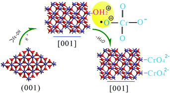
α-Fe2O3 nanoparticles with different morphologies, such as hollow spheres, nanotubes with a limited amount of the {0001} plane exposed, and nanoplates and nanorings with the {0001} plane predominantly exposed, have been synthesised by using NaH2PO4 and urea in a facile hydrothermal method. The mechanism of the morphology evolution from hollow sphere to nanoring has been investigated. It is proposed that the polymerisation of Fe3+/H2PO4 plays an important role in the formation of these morphologies. The adsorption of Cr(VI) from aqueous solution onto these α-Fe2O3 nanoparticles showed that the α-Fe2O3 with nanoring morphology has the highest removal efficiency, and the adsorption capacity reached 16.9 mg g−1. These results indicate that the adsorption mechanism of Cr(VI) onto hematite nanoparticles is a chemisorption process through doubly and triply coordinated hydroxyl groups on the outer surface of α-Fe2O3.


Introduction

The contamination of water bodies caused by heavy metals is a serious environmental problem because of their toxicity to many life forms.1 One of the common hazardous ions is Cr(VI) which usually occurs as highly soluble and toxic chromate anions (HCrO4 or Cr2O72−), both being suspected carcinogens and mutagens. Various kinds of methods have been carried out for Cr(VI) ion removal such as precipitation, reverse osmosis, solvent extraction and electrolysis, biological treatment, ion-exchange processes, and adsorption.2 Among them, the adsorption technique has advantages of high efficiency, cost effectiveness and being environment-friendly.3 During the past decade, many types of adsorbents have been studied for their Cr(VI) removal effectiveness, including activated carbons, metal oxides, polymeric adsorbents, and even certain types of bio-sorbents.4 Among them, hematite (α-Fe2O3) has attracted a great interest owing to its effective performance, natural abundance, low cost and environmental safety.5

Some studies have shown that α-Fe2O3 has a good capacity for Cr(VI) adsorption in aqueous solution. Cao et al. prepared flower-like α-Fe2O3 nanostructures that have a high capacity for Cr(VI) adsorption.6 Han et al. prepared α-Fe2O3 with a porous structure that can rapidly adsorb Cr(VI) ions in wastewater at room temperature.7 Liu et al. obtained porous α-Fe2O3 nanostructures with different morphologies exhibited excellent Cr(VI) removal capacity and a fast adsorption rate in a wide range of pH.8 Besides this, the adsorption ability and mechanism on different exposed planes of α-Fe2O3 have been partly calculated. Yin and Ellis revealed H2CrO4 adsorption to α-Fe2O3 {1[1 with combining macron]02} surface plane by strong H-bonding.9 Huang et al. systematically investigated Cr(VI) on the hematite nanorods, and concluded that Cr(VI) species were adsorbed on the {0001} and {11[2 with combining macron]0} planes in inner-sphere monodentate mononuclear and bidentate binuclear configurations.10 However, our knowledge of the morphology control of hematite crystals and the mechanism of Cr(VI) adsorption on the different surfaces of α-Fe2O3 is still very limited.

In the present work, with presence of NaH2PO4 and urea, the α-Fe2O3 nanoparticles with different morphologies, such as hollow spheres, nanotubes, nanoplates and nanorings, are successfully synthesised via a facile hydrothermal method. These α-Fe2O3 nanoparticles have good dispersion without using any substrates and show different exposed crystal planes. The microstructural studies of the specimens under different synthetic conditions lead to establishment of a new non-classical crystal growth route. The ability to remove Cr(VI) using these as-synthesised α-Fe2O3 is examined, and the possible mechanism of adsorption of Cr(VI) on the crystal surface is deduced.

Experimental

All chemicals were in analytical purity, and used without further purification. A typical experiment to prepare α-Fe2O3 is as follows: required amounts of NaH2PO4 and urea were added under stirring to 80 mL aqueous solution of 23 mmol L−1 of FeCl3. The solution was subsequently sealed in a 100 mL autoclave, and maintained at 220 °C for 24 h. The autoclave was then cooled down to room temperature gradually. The red precipitates were collected by centrifugation, and washed with deionised water and ethanol for three times, and finally dried in a desiccator at 60 °C for 12 h. The concentrations of NaH2PO4 and urea used in the synthesis are listed in Table 1. Powder X-ray diffraction (XRD) was performed on a PANalytical Empyrean diffractometer using Cu Kα radiation. The morphology of the samples were observed using scanning electron microscopy (SEM, LEO 1530VP). The microstructures and crystal structures of the particles were investigated using transmission electron microscopy (TEM) and high resolution TEM (HRTEM) on a JEOL JEM-2011FEF microscope. X-ray photoelectron spectroscopy (XPS) analysis was performed on a PHI 5300x multitechnique system with a Mg-Kα X-ray source (Perkin-Elmer Physical Electronics). The nitrogen adsorption–desorption isotherms were obtained using a JW-BK specific surface area instrument (Beijing, China) and the specific surface areas were calculated using the Brunauer–Emmett–Teller (BET) method.
Table 1 Compositions of the synthetic solutions
Sample number FeCl3 (mmol L−1) PO43− (mmol L−1) Urea (mmol L−1)
S1 23.00 0 0.50
S2 23.00 1.0 1.0
S3 23.00 2.50 2.50
S4 23.00 4.00 4.00


Batch adsorption studies were performed by mixing 0.12 g of the sample with 50 mL of Cr(VI) solution of various concentrations in a 150 mL conical flask with a shaking speed of 150 rpm. The adsorption experiments were carried out at the room temperature, and were conducted at the initial pH of the Cr(VI) solution without adjustment unless otherwise specified. To study the effect of pH, the pH values of the Cr(VI) solution were adjusted from 3 to 11 using 1.0 mol L−1 HCl and 1.0 mol L−1 NaOH solutions. The desorption process of Cr(VI) from the samples took place in 50 mL of 0.01 M NaOH to reach Cr(VI) desorption equilibrium in 24 h. The concentrations of Cr(VI) were determined using a GBC-908 Atomic Absorption Spectrometer (GBC Scientific Equipment Pty Ltd.). The removal efficiency (E%) and the equilibrium adsorption capacity (qe, mg g−1) were calculated using eqn (1) and (2), respectively:

 
image file: c6ra15245g-t1.tif(1)
 
image file: c6ra15245g-t2.tif(2)
where C0 is the initial concentration of Cr(VI) in the solution (mg L−1), Ct is the concentration of Cr(VI) at a given time (mg L−1), m is the mass of the adsorbent (g), and V is the total volume of the Cr(VI) solution (L).

Results and discussion

Initial characterisation of the samples was by XRD. XRD patterns of all the four samples (S1 to S4 as listed in Table 1), shown in Fig. 1, can be indexed to the hexagonal hematite structure (α-Fe2O3, JCPDS 33-0664) with unit cell parameters a = 0.5035 nm and c = 1.374 nm, indicating a good crystallinity of the samples. In the XRD patterns of samples S3 and S4, a few extra weak peaks are indexed to the unit cell of Fe4(PO4)3(OH)3, with a = 1.9554 nm, b = 0.7395 nm, c = 0.7439 nm, space group C2/c15, (PDF: 42-0429).
image file: c6ra15245g-f1.tif
Fig. 1 XRD patterns of the α-Fe2O3 nanoparticles with different morphologies, (a) hollow sphere of sample S1, (b) nanotube of sample S2, (c) hollow plate of sample S3, (d) nanoring of sample S4. The peaks in (d) are indexed to α-Fe2O3 (*) and Fe4(PO4)3(OH)3 (#), respectively.

The particle size and morphology were examined using SEM (Fig. 2). When the concentration of urea was 0.5 mmol L−1 without adding H2PO4 for sample S1, the particle morphology was hollow sphere and the diameter was in a range from 100 to 200 nm (Fig. 2(a)). As the concentrations of both H2PO4 and urea were 1.0 mmol L−1, the morphology of the α-Fe2O3 particles in sample S2 was nanotube with the length of about 300 nm and the diameter of about 80 nm (Fig. 2(b)). When the concentrations of H2PO4 and urea were increased to 2.5 mmol L−1, the produced α-Fe2O3 particles in sample S3 had a shape of hollow plate with about 150 nm in diameter and 50 nm in thickness. There is a hole in the face centre of each particle (Fig. 2(c)). As the concentrations of H2PO4 and urea were further increased to 4.0 mmol L−1, the morphology of the particles in sample S4 appeared as nanorings (Fig. 2(d)) with the similar radius to the hollow plates in sample S3. But the particles in sample S4 are much thinner than those in sample S3, and the holes in the face centre are bigger. Actually, the hollow plates and nanorings are closely related. The hollow plates must form first and the holes at the face centre are developed later. When the holes become large enough, the particles look like nanorings. Fig. S1 in ESI presents low magnification SEM images of these samples, showing the uniformity of the particle morphology and size.


image file: c6ra15245g-f2.tif
Fig. 2 SEM images of the hematite particles, (a) hollow spheres of S1, (b) nanotubes of S2, (c) nanoplates of S3, and (d) nanorings of S4.

The microstructures of these hematite particles were investigated by using HRTEM. Fig. 3(a) and (b) show the spherical particles of sample S1. The light image contrast in the centre indicates their hollow property. Also from the image contrast, the particle seems to be polycrystalline consisting of many nanocrystallites. On the other hand, the corresponding selected area electron diffraction (SAED) pattern in Fig. 3(b) from the whole particle shows a single crystal like diffraction pattern. A possible explanation is that all the nanocrystallites in the particle are self-orientated. Fig. 3(c) and (d) are HRTEM images from two selected top and bottom edge areas. The two d-spacings measured from each image are about 0.25 nm with an interplane angle of 60°, which can be indexed to the (11[2 with combining macron]0) and ([1 with combining macron]2[1 with combining macron]0) planes of the hematite structure. The marked planes in these two HRTEM images are parallel each other, supporting the self-orientated structure.


image file: c6ra15245g-f3.tif
Fig. 3 TEM analysis of the hollow spheres in S1. (a) Low magnification TEM image. (b) TEM image of a single hollow sphere with the corresponding SAED pattern. (c) and (d) HRTEM images recorded from the two marked areas in (b).

Fig. 4 shows TEM and HRTEM images of particles in sample S2. The mono-dispersed nanotubes have a shape of weaving shuttle with dimensions of 100 to 200 nm in diameter and 300 to 600 nm in length (Fig. 4(a)). The wall thickness of about 30 nm is almost uniform, therefore, the inner diameter as well as external diameter at both ends is smaller than that in the middle (Fig. 4(b)). This shape with non-uniform diameters is different from commonly seen nanotubes with a uniform diameter, such as carbon nanotubes, anodic TiO2 nanotubes,11 and crystalline titanate and niobate nanotubes.12,13 On the other hand, the HRTEM images from the wall of the nanotubes confirm the hematite crystal structure. The observed d-spacings on Fig. 4(c) and (d), 0.27 and 0.37 nm, can be indexed to the (01[1 with combining macron]4) and (0[1 with combining macron]12) with the interplane of 95.6°. Comparing the crystal orientations of the two HRTEM images, we also found that they are well orientated, indicating that the nanotubes can be regarded as porous single crystals. Although determination of crystal orientation along the long axis of the nanotubes is not simple, we are often unable to make sure whether the long axis is perpendicular to the electron beam.14 For an individual nanotube lying down on the carbon film, it is easy to approach to this position, where the observed orientation along the long axis on the projected image in Fig. 4(b) is parallel to the long axis of the nanotube. This direction can be derived from the marked planes of (01[1 with combining macron]4) and (0[1 with combining macron]12) to be [0001].


image file: c6ra15245g-f4.tif
Fig. 4 TEM investigation of the nanotubes in S2. (a) Low magnification TEM image. (b) High magnification TEM image of a single nanotube. (c) and (d) HRTEM images of the marked areas in (b).

Although the nanoplate morphology of the particles in sample S3 is different from the spherical particles in sample S1, the appearance of microstructures of these two samples on the TEM images are very similar. The HRTEM images of the nanoplates (Fig. 5(c) and (d)) also show self-orientated aggregation of nanocrystallites, and this orientation extends to the whole particle. The observed d-spacings of 0.25 nm can be indexed to {11[2 with combining macron]0} equivalent planes. That means the view direction parallel to the short axis of the nanoplate in Fig. 5(b) is along the [0001] zone axis of the hematite crystal structure.


image file: c6ra15245g-f5.tif
Fig. 5 TEM investigation of the nanoplates in sample S3. (a) Low magnification TEM image. (b) Higher magnification TEM image of a single plate particle. (c) and (d) HRTEM images obtained from the two marked areas in (b).

Fig. 6 shows TEM and HRTEM images of nanoring particles in sample S4. The particle size of ∼150 nm in diameter and ∼50 nm in thickness (Fig. 6(a)) is similar to the hollow plates in sample S3, but much smaller than the spheres in sample S1. It is obvious that the rings consist of many nanocrystallites. But these nanocrystallites connected each other and self-orientated to insure a certain mechanical strength. Viewing along the thickness direction, some d-spacings of 0.25 nm, corresponding to the {11[2 with combining macron]0} planes, can be observed on the HRTEM images (Fig. 6(c) and (d)). The view direction for the particle in Fig. 6(b) is along the [0001] zone axis, same as the nanoplates in Fig. 5.


image file: c6ra15245g-f6.tif
Fig. 6 TEM investigation of nanoring in sample S4. (a) Low magnification TEM image. (b) Higher magnification TEM image of a single nanoring particle. (c) and (d) HRTEM images obtained from the two marked areas in (b).

In order to identify the influence of NaH2PO4 on the morphology formation of hematite crystals, several syntheses with different amounts of NaH2PO4 and zero concentration of urea were performed while keeping other parameters constant (Fig. 7). Without the addition of NaH2PO4, relatively large round particles were produced (Fig. 7(a)). When 1.0 mmol L−1 NaH2PO4 was added to the synthetic solution, the produced particles were spindle-like with an average length of 200 nm and a mean diameter of 50 nm (Fig. 7(b)). With increasing the concentration of NaH2PO4 to 2.5 mmol L−1, the morphology of the particles changed to round disk (Fig. 7(c)). When the NaH2PO4 concentration was further increased to 4.0 mmol L−1, the disk shape was reproduced, but the thickness of these disks was significantly reduced (Fig. 7(d)). As the concentration of NaH2PO4 increased to 8 mmol L−1 (Fig. S2 in ESI), the α-Fe2O3 particles mainly keep the disk shape, but the size and the thickness are consequently reduced. XPS of the particles obtained in 4.0 mmol L−1 NaH2PO4 without urea (Fig. S3 in ESI) shows a peak at binding energy of 133.47 eV for P 2p together with peaks from Fe, O, and C. The above results imply that the particles are composite containing phosphate anions and NaH2PO4 plays a key role in determining the particle morphology. The four morphologies presented in Fig. 7 have a high degree of similarity with the morphologies in Fig. 2, and can be regarded as templates for the latter. On the other hand, the influence of urea on growth of hollow α-Fe2O3 crystals has been investigated in a similar synthetic system containing FeCl3, glucose, and urea.15 It was found that, even without addition of urea, hollow particles still formed. However, according to the observation in the present work, urea can certainly enhance the formation of hollow particles by gas-bubble-assisted effect.16


image file: c6ra15245g-f7.tif
Fig. 7 SEM images of the products prepared with different initial concentrations of NaH2PO4 without adding urea, (a) 0, (b) 1.0, (c) 2.5 and (d) 4.0 mmol L−1.

XPS was also carried out on some typical nanoring particles (Fig. 8). The binding energies are corrected for specimen charging by referencing the C 1s line to 284.8 eV. The spectrum reveals that the nanoring surface is mainly composed of Fe and O (Fig. 8(a)). However, there is a small peak at 399.47 eV for N 1s (Fig. 8(b)), indicating that there is a small amount of N on the surface of nanorings corresponding to urea deposition. At the same time, a peak at binding energy of 133.47 eV for P 2p is also observed, which could be attributed to the surface adsorbed phosphate anions (Fig. 8(c)).17


image file: c6ra15245g-f8.tif
Fig. 8 (a) XPS spectrum of α-Fe2O3 nanorings. (b) High-resolution spectrum of the N 1s and (c) P 2p region.

Based on the above experimental results, it can be concluded that the morphology evolution of α-Fe2O3 nanoparticles should be attributed to the co-effect of urea and H2PO4. Especially the H2PO4 concentration has a great influence on the morphology of the α-Fe2O3 nanoparticles. When 0.5 mmol L−1 of urea was used without adding H2PO4, a large number of primary particles precipitated from the solution. Due to their high surface energy, these primary particles were unstable and tended to aggregate into thermodynamically more stable large spheres. With urea decomposing into carbon dioxide and ammonia, a lot of bubbles were gradually generated and the pH value of the solution increased, promoting the nucleation process of α-Fe2O3 on the particle surface. As a result, sample S1 with hollow spheres was obtained.

In a solution with 1.0 mmol L−1 of H2PO4 and urea, the amount of H2PO4 was rather low compared with the initially formed α-Fe2O3 nuclei. The surface hydroxyl configuration on the {0001) facets of α-Fe2O3 were all neutral doubly coordinated, but on other normal low-index facets such as the {10[1 with combining macron]0}, {11[2 with combining macron]0}, {01[1 with combining macron]2} and {10[1 with combining macron]4} facets, triply-coordinated surface hydroxyl groups with positive charge exposed the crystal facets. Thus, the adsorption capacities and affinities for phosphate to α-Fe2O3 were much lower for the {0001} facets,18,19 and then the [0001] direction would grow faster than the other facets, and the spindle-like α-Fe2O3 was obtained (Fig. 7(b)).

The situation was totally different as the concentration of H2PO4 increased to 2.5 mmol L−1, where nanodisks appeared instead of the spindle particles (Fig. 7(c)), and the {0001} facets were the main exposed crystal facets. In the reaction system, two kinds of ions (Fe3+ and H2PO4) could be adsorbed or coordinated with the surface hydroxyls. The adsorption of Fe3+ ions favoured the growth of α-Fe2O3, but the coordination of H2PO4 restrained the growth process. An increase in the concentration of H2PO4 ions would induce the intense protonation of the surface hydroxyl groups, and the adsorption of H2PO4 on the {0001} facets was enhanced, but the adsorption of Fe3+ ions was weakened due to the effect of electrostatic attraction. All the special doubly coordinated hydroxyl groups on the {0001} facets did not facilitate the adsorption of H2PO4 ions. As a result, nanodisks with the exposed {0001} facets were obtained due to a slower growth of the {0001} facets in comparison with the other facets (Fig. 7(d)). Finally, the exceed H2PO4 had a dissolution effect on the α-Fe2O3 surface follow the eqn (3):18

 
Fe2O3 + 2xH3PO4 + 6H+ = 2[Fe(H2PO4)x]3−x + 3H2O (3)

This dissolution process mainly occurred on the {0001} facets due to the fact that the {0001} facets had the highest concentration of exposed Fe3+ ions and the second among the normal low-index facets20,21 with single effect of H2PO4, this dissolution process will happen if the time of hydrothermal reaction reached 48 h.18,21 According to Zhang et al., the thickness of the α-Fe2O3 barrels can be controlled to some extent by adjusting the amount of urea with its gas-bubble-assisted effect.15,22 The hollow structure and rough surface can also form with this effect. With the help of this effect, the dissolution process along the [0001] direction could be achieved in 24 h, leading to the holes formation in samples S2–S4. The intensity of the peaks (511) and (600) of Fe4(PO4)3(OH)3 obviously increased in the XRD patterns of samples S3 and S4 (Fig. 1), which could be regarded as another evidence for the dissolution process. The formation routes of the different morphologies of α-Fe2O3 are illustrated in Fig. 9.


image file: c6ra15245g-f9.tif
Fig. 9 Sketch of the morphology evolution of α-Fe2O3 nanoparticles under the influence of H2PO4 ions and urea in 24 h.

Fig. 10 shows the Cr(VI) removal efficiency by 2.4 g L−1 as-prepared α-Fe2O3 with two initial Cr(VI) concentrations (25 mg L−1 and 50 mg L−1). It is observed that the Cr(VI) adsorption can reach to equilibrium within the first half an hour. Therefore, Cr(VI) can easily access to the surface of the well-dispersed α-Fe2O3 nanoparticles. At the concentration of 25 mg L−1 (Fig. 10(a)), the Cr(VI) removal efficiency is up to 97.4% by nanorings in sample S4. The hollow spheres in sample S1 have the lowest removal efficiency (90%). The removal efficiency is decreased when the concentration Cr(VI) increases to 50 mg L−1 (Fig. 10(b)). Although the Cr(VI) removal efficiency by nanorings decreases to 81.2% in the latter case, the adsorption capacity can still reach to 16.9 mg g−1, that is higher than other α-Fe2O3 nanoparticles.5 In comparison with previous reports (Table S1 in ESI), the nanorings (sample S4) have very high level capacity of the Cr(VI) removal (16.9 mg g−1) among the non-porous α-Fe2O3 materials.


image file: c6ra15245g-f10.tif
Fig. 10 Adsorption kinetic of different Cr(VI) concentrations on the as-prepared α-Fe2O3; (a) 25 mg L−1, (b) 50 mg L−1 (adsorbent dosage: 2.4 g L−1, no pH adjustment, shaking speed: 150 rpm, T: 25 °C).

The difference in Cr(VI) removal efficiency among the four samples prepared in the present work is mainly attributed to the different surface areas and exposed surface planes. Because the adsorption occurs on the particle surface, a higher surface area will have a higher adsorption capacity. The surface areas of these α-Fe2O3 particles are 31.1 m2 g−1 (sample S1), 34.2 m2 g−1 (sample S2), 38.5 m2 g−1 (sample S3), and 36.7 m2 g−1 (sample S4). The difference is not significant. On the other hand, the Cr(VI) removal efficiency also depends on the type of exposed crystallographic planes. The more the {0001} planes exposed, the high the adsorption capacity.

Furthermore, pH value of the solution has also an influence on removal of Cr(VI). In this study, this effect of the four different α-Fe2O3 samples was investigated by changing the initial pH from 3 to 11 and the results are shown in Fig. 11. The equilibrium adsorption capacities are reduced with pH increased. As the pH value increases to 11, the adsorption capacity is decreased to 14.7 mg g−1. This phenomenon can be understood as the low pH value in the solution would leave higher concentration of positively charged hydroxyl groups on the α-Fe2O3 surface, which has a larger power to attract the CrO42− anions.


image file: c6ra15245g-f11.tif
Fig. 11 Effect of initial pH on the removal of Cr(VI). (Adsorbent dosage: 2.4 g L−1, Cr(VI) concentration: 50 mg L−1, shaking speed: 120 rpm, contact time: 150 min, T: 25 °C.)

Desorption is another important process reflecting the economical and enhancement values of the adsorption. In this study, the removal efficiencies of the regenerated α-Fe2O3 nanoparticles are shown in Fig. 12. The results indicate that the adsorption capability decrease gradually in first three cycles and then decrease with increase of cycles at a relatively slower rate. The nanorings in sample S4 has high removal ability without loss of much efficiency after 5 cycles. The results may be related to the higher proportion of outer surface of the nanorings in sample S4 than samples S2 and S3. The regeneration process needs more outer surface area, pore and channel for the desorption of Cr(VI). Nevertheless, all the four samples of α-Fe2O3 can be reused for removal of Cr(VI) and can make the overall process cost effective.


image file: c6ra15245g-f12.tif
Fig. 12 5th consecutive recycled Cr(VI) removal efficiency of as-prepared samples S1–S4.

From the adsorption results in the systems with different pH values and different morphologies of α-Fe2O3, the adsorption mechanism may be deduced to be chemical adsorption at the doubly and triply coordinated hydroxyl groups on the α-Fe2O3 surface. Because these two types of hydroxyl groups are in neutral and positive overall charging states, they can adsorb CrO42− ions by electrostatic attraction. Different planes in α-Fe2O3 have different densities of these hydroxyl groups. Among the low index planes, the (0001) plane has the highest density of the doubly coordinated hydroxyl groups (13.7 nm−2) in comparison with the (10[1 with combining macron]0) and (11[2 with combining macron]0) planes (2.9 nm−2, 5.0 nm−2).23 Besides this, the (0001) plane does not have singly coordinated hydroxyl with a negative charge. Consequently, the nanoplates in sample S3 and nanorings in S4 with more (0001) planes exposed have high adsorption capacities than the nanospheres in S1 and nanotubes in S2. Because of the open structure of nanorings have a higher surface area than the closed structure of hollow nanoplate, sample S4 removes Cr(VI) more efficiently than S3. The pH value can usually influence the electronic structures of the surface hydroxyl groups. Under an acidic condition, the coordinated hydroxyl groups easily form FeOH2+, which predominate over the surface.24 Therefore, α-Fe2O3 has a high adsorbent capacity at low pH values. The important role of the surface hydroxyl groups in the formation of some novel morphologies, e.g. snowflake-like α-Fe2O3 and 8-branch Cu2O, have also been reported recently.25,26 Consequently, the adsorption mechanism of Cr(VI) species on α-Fe2O3 studied in the present work can be proposed as depicted in Fig. 13.


image file: c6ra15245g-f13.tif
Fig. 13 Possible mechanism of Cr(VI) adsorption on α-Fe2O3.

Conclusion

The α-Fe2O3 nanoparticle with different morphologies, such as hollow spheres, nanotubes, nanoplates and nanorings, were synthesised by using NaH2PO4 and urea via a facile hydrothermal method. These α-Fe2O3 nanoparticles have good dispersion without solid substrates. The adsorption of Cr(VI) results showed that the α-Fe2O3 with nanoring-like morphology has the highest removal efficiency and adsorption capacity. The adsorption mechanism of Cr(VI) onto hematite nanoparticles is chemisorption process through outer surface of doubly and triply coordinated hydroxyl groups.

Acknowledgements

This work was financially supported by the National Nature Science Foundation of China (No. 51302280, 51574286), Natural Science Foundation in Qinghai province (No. 2014-ZJ-936Q). CAS “Light of West China” Program and Youth Innovation Promotion Association (2016377), CAS.

Notes and references

  1. Y. Mu, Z. H. Ai, L. Z. Zhang and F. H. Song, ACS Appl. Mater. Interfaces, 2015, 7, 1997–2005 CAS.
  2. J. P. Beukes, P. G. van Zyl and M. Ras, J. South. Afr. Inst. Min. Metall., 2012, 112, 347–352 CAS.
  3. X. Li, R. Zhao, B. L. Sun, X. F. Lu, C. C. Zhang, Z. J. Wang and C. Wang, RSC Adv., 2014, 4, 42376–42382 RSC.
  4. C. E. Barrera-Diaz, V. Lugo-Lugo and B. Bilyeu, J. Hazard. Mater., 2012, 223, 1–12 CrossRef PubMed.
  5. H. I. Adegoke, F. AmooAdekola, O. S. Fatoki and B. J. Ximba, Korean J. Chem. Eng., 2014, 31, 142–154 CrossRef CAS.
  6. C. Y. Cao, J. Qu, W. S. Yan, J. F. Zhu, Z. Y. Wu and W. G. Song, Langmuir, 2012, 28, 4573–4579 CrossRef CAS PubMed.
  7. C. L. Han, J. S. Xie, C. H. Deng and D. F. Zhao, J. Chil. Chem. Soc., 2012, 57, 1372–1374 CrossRef CAS.
  8. E. T. Liu, H. P. Zhao, H. Li, G. F. Li, Y. L. Liu and R. Chen, New J. Chem., 2014, 38, 2911–2916 RSC.
  9. S. X. Yin and D. E. Ellis, Surf. Sci., 2009, 603, 736–746 CrossRef CAS.
  10. X. P. Huang, X. J. Hou, F. H. Song, J. C. Zhao and L. Z. Zhang, Environ. Sci. Technol., 2016, 50, 1964–1972 CrossRef CAS PubMed.
  11. Z. X. Su and W. Z. Zhou, J. Mater. Chem., 2011, 21, 357–362 RSC.
  12. Q. Chen, W. Z. Zhou, G. H. Du and L. M. Peng, Adv. Mater., 2002, 14, 1208–1211 CrossRef CAS.
  13. G. H. Du, Q. Chen, Y. Yu, S. Zhang, W. Z. Zhou and L. M. Peng, J. Mater. Chem., 2004, 14, 1437–1442 RSC.
  14. W. Z. Zhou and H. F. Greer, Eur. J. Inorg. Chem., 2016, 941–950 CrossRef CAS.
  15. X. H. Zhang, Y. Z. Chen, H. Liu, Y. Wei and W. Wei, CrystEngComm, 2013, 15, 6184–6190 RSC.
  16. X. Liu, Y. F. Li, W. W. Zhu and P. F. Fu, CrystEngComm, 2013, 15, 4937–4947 RSC.
  17. Z. Liu, B. L. Lv, Y. Xu and D. Wu, J. Mater. Chem. A, 2013, 1, 3040–3046 CAS.
  18. C.-J. Jia, L.-D. Sun, Z.-G. Yan, L.-P. You, F. Luo, X.-D. Han, Y.-C. Pang, Z. Zhang and C.-H. Yan, Angew. Chem., Int. Ed., 2005, 44, 4328–4333 CrossRef CAS PubMed.
  19. C. J. Jia, L. D. Sun, F. Luo, X. D. Han, L. J. Heyderman, Z. G. Yan, C. H. Yan, K. Zheng, Z. Zhang, M. Takano, N. Hayashi, M. Eltschka, M. Klaui, U. Rudiger, T. Kasama, L. Cervera-Gontard, R. E. Dunin-Borkowski, G. Tzvetkov and J. Raabe, J. Am. Chem. Soc., 2008, 130, 16968–16977 CrossRef CAS PubMed.
  20. B. Lv, Z. Liu, H. Tian, Y. Xu, D. Wu and Y. Sun, Adv. Funct. Mater., 2010, 20, 3987–3996 CrossRef CAS.
  21. B. L. Lv, Y. Xu, D. Wu and Y. H. Sun, CrystEngComm, 2011, 13, 7293–7298 RSC.
  22. W. Cheng, K. B. Tang, Y. X. Qi, J. Sheng and Z. P. Liu, J. Mater. Chem., 2010, 20, 1799–1805 RSC.
  23. R. M. Cornell and U. Schwertman, The Iron Oxides: Structure, Properties, Reaction, Occurrences and Use, WILEY-VCH GmbH&Co.KGaA, 2nd edn, 2003 Search PubMed.
  24. Z. H. Ruan, J. H. Wu, J. F. Huang, Z. T. Lin, Y. F. Li, Y. L. Liu, P. Y. Cao, Y. P. Fang, J. Xie and G. B. Jiang, J. Mater. Chem. A, 2015, 3, 4595–4603 CAS.
  25. Z. Liu, C.-Y. Chiang, W. Li and W. Z. Zhou, Chem. Commun., 2015, 51, 9350–9353 RSC.
  26. K. Self and W. Z. Zhou, Cryst. Growth Des., 2016 DOI:10.1021/acs.cgd.6b00883.

Footnote

Electronic supplementary information (ESI) available: More SEM images, XPS, Cr(VI)-adsorption capacities. See DOI: 10.1039/c6ra15245g

This journal is © The Royal Society of Chemistry 2016
Click here to see how this site uses Cookies. View our privacy policy here.