Enhanced photocatalytic performance and morphology evolvement of PbWO4 dendritic nanostructures through Eu3+ doping

Dan Yuea, Dong Chenb, Wei Luc, Mengnan Wangb, Xiaoli Zhangb, Zhenling Wang*b and Guodong Qian*a
aState Key Laboratory of Silicon Materials, Cyrus Tang Center for Sensor Materials and Applications, School of Materials Science and Engineering, Zhejiang University, Hangzhou 310027, China. E-mail: gdqian@zju.edu.cn
bThe Key Laboratory of Rare Earth Functional Materials and Applications, Zhoukou Normal University, Zhoukou 466001, P. R. China. E-mail: zlwang2007@hotmail.com
cUniversity Research Facility in Materials Characterization and Device Fabrication, The Hong Kong Polytechnic University, Hong Kong, P. R. China

Received 9th June 2016 , Accepted 22nd August 2016

First published on 22nd August 2016


Abstract

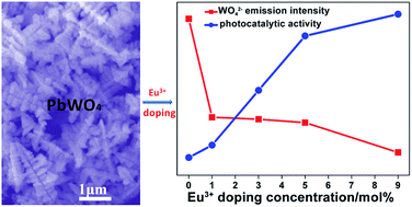
Eu3+ doped PbWO4 dendritic nanostructures have been prepared by a facile hydrothermal technique and characterized by XRD, SEM, TEM, XPS and PL spectroscopy. The results indicate that the changing of the Eu3+ doping concentration in a reasonable range can adjust the morphology, and has no obvious effect on the phase structure of the obtained materials. Moreover, the Eu3+ doping can lead to the enhancement of photocatalytic activity of PbWO4. Compared with pure PbWO4 dendritic nanostructures, the PbWO4:x%Eu3+ samples exhibit improved photocatalytic activity in the decomposition of Rhodamine B (RhB). The enhanced performance of the photocatalyst might be ascribed to efficient separation of electron and hole pairs after doping Eu3+ into the PbWO4 lattice.


Introduction

Photocatalysis is a green technology for environmental remediation, especially for degrading organic dyes or environmental pollutants. In recent years, the research into various nanocrystalline photocatalytic materials has mainly focused on TiO2. Undoubtedly, TiO2 is known as one of the most excellent photocatalysts. However, it has a wide band gap of 3.2 eV, which causes it to be so only in response to UV light.1–3 Hence, exploiting new photocatalysts is more anticipated, among which tungstate is considered to be a promising candidate. Tungstate crystals have been investigated as photocatalysts because of their high average refractive index and low radiation damage, etc.4–8 For example, Bi2WO6 has high photocatalytic activities for degradation of rhodamine B (RhB) under visible-light irradiation2 and a WO3/CdWO4 photocatalyst exhibited enhanced photocatalytic efficiency for the degradation of different organic dyes under visible light irradiation.9 Reduced graphene oxide (RGO) hybridized CdWO4 shows much higher photocatalytic activity than pure CdWO4 for methylene blue degradation, because the RGO with a two-dimensional π–π conjugation structure can attract the photo-generated electrons and thus help to reduce the recombination rate of photo-generated charges.10

As an important member of the tungstate family, lead tungstate (PbWO4) has been attracting increasing attention due to its optical applications in high-energy physics and scintillating crystal, etc.11–16 To date, the controlled synthesis of PbWO4 nano/microstructures has been investigated, and various morphologies such as nanowires, hollow spheres, hollow nanospindles, polyhedral have been reported by solid-state reaction method,17 solvothermal route,18 wet chemical route.19 PbWO4 microcrystals with various morphologies (helixlike, chainlike, rodlike and crosslike structures) have been successfully synthesized by changing the pH value, temperature, and the addition of surfactant into the reaction solution.20,21 Beside various morphologies, PbWO4 is a popular multi-functional material possessing luminescence, photocatalysis and so on. Such as, PbWO4 microspheres exhibited photocatalytic activity in dye degradation, the pH value and different hydrothermal temperature play key role in controlling the PbWO4 morphology and photocatalytic activity.15 The photocatalytic activity of the erythrocyte-like Y-doped PbWO4 mesocrystals is further investigated in terms of the degradation of the acid orange II under UV irradiation.22 However, it has been observed that the application of PbWO4 in photocatalysis is usually restricted due to the high recombination rate of photoexcited electron–hole pairs which plays important role in photocatalytic reaction. In the course of developing photocatalysts, some techniques have been reported and adopted to improve the performance of photocatalysts, such as doping,1,23 coupling plasmon resonance,24 size and crystal facet of semiconductors and controlling morphology.9,25 Among them, the doping is a simple and effective method that has been proved more effective for improving photocatalytic efficiency. It was also found that the doping could not only alter the phase structure and morphology, but also affect the property of the obtained samples. Recently, rare earth ions (RE3+) have proved to be the most promising doping ions because of the apparently enhancement of photogenerated electron transfer rate in the interfacial.26 Due to the transitions of RE 4f electrons, the doping of Eu3+ could make the optical absorption of substrate material be increased, and separated the photo-generated electron–hole (e–h+) pair as well.27 Eu3+ is one of interesting luminescent rare earth ions due to its unique optical properties and its applications in a number of optical and biotechnological areas.

In this work, the aim was to design an enhanced PbWO4 photocatalyst through the Eu3+ ion doping and investigate the effect of the Eu3+ ion doping concentration on the structure, luminescence properties, and their relationship with the photocatalytic properties. The Eu3+ ion doped PbWO4 dendritic nanostructures were obtained by a simple hydrothermal method. The rhodamine B (RhB) was employed to appraise the photocatalytic activities of the PbWO4:Eu3+ sample. The effect of Eu3+ ion doping concentration on the luminescent properties, morphology and photocatalytic efficiency was also investigated in detail.

Results and discussion

Fig. 1a shows the XRD patterns of Eu3+-doped PbWO4 with different doping concentrations. All of the diffraction peaks of each sample can be identified as tetragonal structures with the same space groups of I41/a, and are in agreement with the standard data of bulk PbWO4 (JCPDS card no. 19-0708). No peaks corresponding to any other phases or impurities were detected, indicating the high purity of these samples. However, when increasing the Eu3+ doping concentration from 0% to 9%, the (112) diffraction peaks of PbWO4:x%Eu3+ in the range of 2θ = 26.5–28.5° are shifted slightly to the lower 2θ value, as shown in Fig. 1b. The ionic radii of Pb2+ is 1.19 Å, which is larger than that of Eu3+ ion (0.95 Å). Hence, it is reasonable to observe the peak shift towards lower 2θ value, and to some extent the results might imply that Pb2+ sites in PbWO4 are partially occupied by the dopant Eu3+ ions. In PbWO4:x%Eu3+ system, the imbalance of the electrical charges and the difference in the ionic radii between the host Pb2+ and the dopant Eu3+ ions lead to changes in the ion-to-ion distance and spatial arrangement. It means that Eu3+ ions enter the crystal lattice of PbWO4 or introduce a high disorder into the lattice, which might be the reason for the different effects on the morphology and property of the obtained samples.
image file: c6ra15045d-f1.tif
Fig. 1 (a) XRD patterns of PbWO4:x%Eu3+ at different concentrations (x = 0, 1, 3, 5, and 9) and the standard data for bulk PbWO4 (JCPDS card no. 19-0708); (b) diffraction peak positions of PbWO4:x%Eu3+ in the range of 2θ = 26.5° to 28.5°.

It should be denoted that there are some impurity peaks in the XRD patterns when the Eu3+ doping concentration was further increased to 12% or above, which probably results from the formation of other tungstates or tungstate composites. The effect of Eu3+ doping with higher concentration on phase structure, composition, morphology, optical absorption property and photocatalytic activity of the obtained tungstates will be systematically investigated in future. Hence, in the present work, the morphology, optical absorption and photocatalytic performance of PbWO4 with Eu3+ doping concentration of 0–9% were discussed and compared.

Fig. 2a shows SEM image of the as-prepared PbWO4. It indicates that the sample consists of dendritic nanostructures which have many symmetric arms extending radially from the center. Further observations show that each arm is perpendicular to the center trunk, and those arms are in good symmetry and have the characteristic of self-similarity. Fig. 2b shows the morphology of PbWO4 dendritic structures acquired in STEM-HAADF (high angle annular dark field) topology mode. The EDS elemental mappings (Fig. 2c–e) show that Pb, W and O elements are evenly distributed into the PbWO4 dendritic structures.


image file: c6ra15045d-f2.tif
Fig. 2 (a) SEM and (b) STEM-HAADF images of PbWO4 dendritic structures; and EDS elemental mapping for (c) Pb, (d) W and (e) O of the dendritic crystal shown in (b).

It can be clearly seen that the obtained sample is composed of PbWO4 dendritic structures, as shown in Fig. 3. The SAED pattern (Fig. 3b, inset) can be indexed to the (002), (112) and (110) planes of the PbWO4 dendritic structures with the tetragonal phase structure, which is in agreement with the XRD result. The HRTEM image (Fig. 3c) of the top of the branch marked as an arrow in Fig. 3b displays single crystalline nature. The values of the interplanar spacings of PbWO4 dendritic structures are 0.302, 0.193 and 0.325 nm, which are identical to the (002), (110) and (112) facet distances of the PbWO4 dendritic structures, respectively.


image file: c6ra15045d-f3.tif
Fig. 3 (a and b) TEM images of PbWO4 dendritic structures (the inset is the SAED pattern recorded from the branched part), and (c) HRTEM image taken from the branch marked with arrow shown in (b).

The morphology of PbWO4 was affected by doping of Eu3+ ions. Fig. 4 shows the SEM images of PbWO4:x%Eu3+ with different doping concentrations. With increasing Eu3+ doping concentrations from 1% to 9%, the morphology change of PbWO4 is very interesting. It can be seen that both PbWO4:1%Eu3+ (Fig. 4a) and PbWO4:3%Eu3+ (Fig. 4b) are composed of dendritic nanostructures with one trunk (long axis) and four branches (short axes), and the size of the latter is larger than that of the former. The trunk lengths of PbWO4:1%Eu3+ and PbWO4:3%Eu3+ are 9.5 μm and 15 μm respectively, while the lengths of branches for these two samples are 2.5 μm and 5 μm respectively. When gradually increasing doping concentrations to 5%, the sample is also composed of dendritic nanostructures with the trunk length of 12 μm (Fig. 4c), except that the four branches of the dendritic nanostructure became short and disappeared partially. When the doping concentration is increased to 9%, the four branches of the dendritic nanostructure disappeared completely, and the sample is composed of trunk-like structure with the length of 14 μm, as shown in Fig. 4d. It was found that the doping concentration of rare earth ions played a key role in the morphology control of PbWO4. The morphology of PbWO4:9%Eu3+ is very special. From the XRD pattern of PbWO4:9%Eu3+ as shown in Fig. 1, it can be seen that the intensity of the plane diffraction peaks (200) at 2θ = 32.7°, (312) at 2θ = 55.3° is much stronger than those of the other samples. It indicates that the Eu3+ doping has little effect on the phase structure, but has evidently effect on the crystal growth direction and final morphology formation of PbWO4, especially at high doping concentration of Eu3+ ions. The possible reason is partly attributed to the strong effect of the dopant ion on the crystal growth rate through surface charge modification upon increasing doping concentration. A schematic showing the morphology evolution process of PbWO4:x%Eu3+ with different doping concentrations is illustrated in Fig. 4e.


image file: c6ra15045d-f4.tif
Fig. 4 SEM images of PbWO4:x%Eu3+ with different doping concentrations: (a) 1%, (b) 3%, (c) 5%, and (d) 9%; (e) schematic illustration of the morphology evolution process of PbWO4:x%Eu3+ with the increase of Eu3+ doping concentration.

The existence of Eu3+ and the chemical state of each atom were investigated by X-ray photoelectron spectroscopy (XPS). The elements of Pb, W, O and Eu can be detected in the survey spectrum (Fig. 5a). Fig. 5b–d show the high-resolution XPS spectra of Pb 4f, W 4f and O 1s, respectively. The values of binding energy of Pb 4f5/2, Pb 4f7/2, W 4f5/2, W 4f7/2 and O 1s in PbWO4:5%Eu3+ are 143.2, 138.3, 36.8, 34.7 and 530.1 eV, respectively. The XPS spectrum of Eu 3d was shown in Fig. 5d. The characteristic peak of Eu 3d region was displayed at 1136 eV, which is due to the binding energy of Eu 3d5/2. The XPS results indicate that Eu doped PbWO4 has been successfully obtained. The PbWO4:5%Eu3+ was further characterized by EDS, and Fig. S1a shows the STEM-HAADF image of PbWO4:5%Eu3+ dendritic structures. From EDS elemental mappings and EDX spectrum (Fig. S1b–f, ESI), it can be seen that the sample are composed of Pb, W, O and Eu elements and these elements are evenly distributed into the PbWO4 dendritic structures.


image file: c6ra15045d-f5.tif
Fig. 5 XPS analysis of PbWO4:5%Eu3+: (a) survey, (b) Pb 4f, (c) W 4f, (d) O 1s, (e) Eu 3d.

The optical absorption performance relevant to the band gap of a semiconductor is considered to be a key factor in determining its photocatalytic activity. Fig. 6 shows the UV-vis diffuse reflectance spectra (DRS) of the PbWO4:x%Eu3+ with different doping concentrations. According to the spectra, all the sample show intense absorption in UV (200–400) light region. However, PbWO4:x%Eu3+ (x = 1, 3, 5, 9) samples display the visible-light response. Clearly, upon increasing Eu3+ doping concentrations from 1% to 9%, the absorption intensity of sample at 520 nm increases gradually, and their corresponding band gaps are 2.42 eV approximately.


image file: c6ra15045d-f6.tif
Fig. 6 UV-vis DRS of PbWO4:x%Eu3+ with different doping concentrations.

Based on the UV-vis DRS analysis, PbWO4 have visible response through Eu3+ doping. Fig. 7 displays the photocatalytic degradation of RhB with the PbWO4:x%Eu3+ under UV light irradiation. As a contrast, the curves showing the degradation of RhB by pure PbWO4 and the blank experiment (without addition of any catalysts) were also presented. It can be seen from Fig. 7 that there is only little impact of RhB concentration in the blank experiment after 4 h, indicating that the photolysis of the RhB solution is negligible in this experimental conditions. It was found that the photodegradation rate of RhB by PbWO4, PbWO4:1%Eu3+, PbWO4:3%Eu3+, PbWO4:5%Eu3+ and PbWO4:9%Eu3+ is 41.13%, 45.44%, 64.43%, 83.33%, and 90.85%, respectively. The photocatalytic activity of PbWO4:9%Eu3+ is close to that of Y-doped PbWO4 mesocrystals, and PbWO4 microspheres15,22 The pseudo-first-order kinetic curves of RhB photodegradation were plotted to quantitatively compare the degradation rate. The efficiency of RhB photodegradation by the photocatalysts was determined quantitatively using the following model: ln(C/C0) = −kt, where C0 and C are the concentrations of dye at time 0 and t, respectively, and k is the apparent rate constant (h−1). The experiment data show that the k values of PbWO4, PbWO4:1%Eu3+, PbWO4:3%Eu3+, PbWO4:5%Eu3+ and PbWO4:9%Eu3+ are 0.1586, 0.1733, 0.3082, 0.5175, and 0.6897 h−1 respectively. Namely, the photocatalytic activity of PbWO4:9%Eu3+ is 4.35 times than that of the pure PbWO4. As mentioned above, the photodegradation rate of RhB by PbWO4 with higher Eu3+ doping concentration was not given and compared because of the presence of impurity phase or other tungstate composites.


image file: c6ra15045d-f7.tif
Fig. 7 Photodegradation curves of RhB over PbWO4:x%Eu3+ (x = 0, 1, 3, 5, 9) samples.

To further investigate the mechanism of photocatalytic process over the PbWO4:9%Eu3+ dendritic catalyst, a series of scavengers were developed to scavenge the relevant species and their concentration is about 2.0 mM. The results of the trapping experiments were shown in Fig. S2. The quenchers used in this reaction system were potassium iodide (KI) for h+ and ˙OH, sodium oxalate (Na2C2O4) for h+, benzoquinone (BQ) for ˙O2 and isopropanol (IPA) for ˙OH respectively.28–32 It should be seen that IPA had little impact on the RhB solution, indicating that ˙OH on the surface of the PbWO4:9%Eu3+ dendritic catalyst did not play a major role in the photodegradation system. However, the degradation efficiency decreased obviously after the addition of KI, BQ or Na2C2O4. These results indicated that h+ and ˙O2 were produced and acted as dominating active species in the photodegradation process. In addition, Fig. S3 show that the removal rate of TOC with PbWO4:x%Eu3+ (x = 0, 1, 3, 5, 9) samples on the degradation of RhB. After the degradation of RhB for 4 h, the removal rate of TOC with PbWO4:x%Eu3+ (x = 0, 1, 3, 5, 9) reached 35.6%, 41.7%, 62.1%, 70.6% and 78.1%, respectively. Due to the degradation curve data were measured after the photocatalyst separation step by centrifugation, the removal rate of TOC was lower than photocatalytic degradation ratio (Fig. 7). The decrease in the carbon content indicates the degradation of the RhB dye into nontoxic compounds. It indicate that PbWO4:x%Eu3+ sample have good photocatalytic activity.

It is well known that electron–hole separation is one of the key factors limiting the efficiency of the photocatalytic process.33 With the Eu3+ ions doped into PbWO4, and the excited electrons were trapped by the Eu3+, which effectively suppresses the electron–hole recombination. Furthermore, the XRD results suggested that Eu3+ with low concentration is incorporated into the lattice of PbWO4 and occupied the sites of Pb2+, creating abundant oxygen vacancies and surface defects for electron trapping and pollutant adsorption, also accelerating the separation of electron–hole pairs and RhB photodegradation. Hence, it can greatly promote the separation of electrons and holes, and the photooxidation ability of holes (h+) will be enhanced. Meanwhile, the electrons are easier to react with oxygen molecules to form another active species superoxide radical anion (˙O2) for the degradation of RhB. All of the above processes can effectively improve the performance of the photocatalyst. And the result of the trapping experiments was also in agreement with the supposed photocatalytic mechanism.

The efficiency of charge carrier trapping and transfer and better understanding the fate of electron–hole pair in semiconductor particles could be investigated using PL emission spectrum.34 Excited by the UV light of 280 nm, the emission spectra of PbWO4 with different doping concentrations of Eu3+ were obtained, as shown in Fig. 8. It can be seen that pure PbWO4 exhibited a blue-green emission band in the range of 400–680 nm with the emission peak centered at 485 nm. With increasing Eu3+ doping concentrations from 1% to 9%, the emission spectra not only contain the intrinsic blue-green emission of WO42− groups but also include 5D07FJ (J = 1, 2, 3) emission lines of Eu3+ ions. The doping of Eu3+ into PbWO4 decreased the PL emission intensity of WO42− groups gradually.


image file: c6ra15045d-f8.tif
Fig. 8 PL emission spectra of PbWO4:x%Eu3+ with different doping concentrations (x = 0, 1, 3, 5, 9).

According to the above result, there exist an opposite relationship between the photocatalytic activity and PL intensity of WO42− groups for PbWO4:x%Eu3+ photocatalysts. The higher PL intensity of WO42− groups, the higher recombination rate of photo-generated charge carriers which bring about lower photocatalytic efficiency. In other words, a lower PL intensity shows a lower recombination rate of photo-generated electron–hole pairs, so more photo-generated electrons and holes can participate in the oxidation and reduction reactions, resulting in improvement of the photocatalytic performance.35 This investigation strongly provides the evidence that the WO42− groups in tungstate photocatalysts mainly play a key role in the process of photocatalysis and establishes the relationship between photocatalytic activity and PL intensity through Eu3+ as doping ion and fluorescent probe.

Experimental

Preparation of PbWO4:x%Eu3+

All chemicals were analytical grade reagents and used directly without further purification. Pb(NO3)2 (99.99%) and Na2WO4 (≥99.0%) were obtained from Sinopharm Chemical Reagent Co., Ltd. Eu(NO3)3 solutions were prepared by dissolving the corresponding oxides in diluted nitric acid. PbWO4:x%Eu3+ (x = 0, 1, 3, 5, 9) dendritic nanostructures were prepared by hydrothermal technique. Typically, 10.0 mL of 0.2 mol L−1 Na2WO4 solution was added to 10.0 mL of 0.2 mol L−1 Pb(NO3)2 aqueous solution and the mixture was continuously stirred for 15 min. The obtained suspension was then transferred into a Teflon bottle held in a stainless steel autoclave, which was sealed and hydrothermally treated at 160 °C for 24 h. After the autoclave was cooled to room temperature naturally, the precipitates were separated by centrifugation, washed with ethanol and distilled water twice, respectively, and dried at 80 °C for 24 h to obtain the undoped sample. Additionally, a series of Eu3+-doped PbWO4 samples (1%, 3%, 5% and 9%) were prepared. The synthesis procedure was similar to that mentioned above except that 10.0 mL of 0.2 mol L−1 Na2WO4 was added to the mixed solutions of Pb(NO3)2 and Eu(NO3)3 with a desired molar ratio, and the other conditions were kept unchanged.

Characterization

Phase structure of the obtained samples was characterized by a Bruker D8 Advance X-ray diffractometer (XRD) with Cu-Kα radiation (λ = 0.15406 nm). The accelerating voltage and emission current were 40 kV and 40 mA, respectively. The TEM image and selected area electron diffraction (SAED) pattern were obtained on a JEOL-2010 transmission electron microscope at an accelerating voltage of 200 kV. Photoluminescence (PL) spectrum was recorded using an FLS920P Edinburgh Analytical Instrument apparatus equipped with a 450 W xenon lamp as the excitation source. The diffuse reflection spectrum (DRS) was obtained using a UV-visible spectrophotometer with BaSO4 as a reflectance standard. X-ray photoelectron spectroscopy (XPS) analysis was performed on a Thermo Scientific Escalab 250Xi instrument with Mg Kα radiation (1253.6 eV) at a scan step of 0.1 eV. Total organic carbon (TOC) analyses were conducted on a multi N/C 2100 (Analytik Jena AG, Germany) TOC analyzer.

Photocatalytic activity measurements

Photocatalytic activities of the as-prepared samples were evaluated by the degradation of rhodamine B in aqueous solution under simulated UV light (a 500 W mercury lamp) in XPA photochemical reactor. The detailed process was as follows: the PbWO4:x%Eu3+ (x = 0, 1, 3, 5, 9) powders (0.05 g) were dispersed in an aqueous RhB solution (5 mg L−1, 50 mL) and then transferred into a 80 mL cylindrical quartz reactor with constant magnetic stirring equipped with a water circulation facility. To reach an adsorption–desorption equilibrium before irradiation, the suspensions were first stirred for 30 min in dark condition. Then the light was switched on, and the solutions were kept stirring. At given time intervals, 5 mL of the suspension was sampled and centrifuged to remove the photocatalyst from the aqueous solution so as to obtain the supernatant for the analysis of RhB. The concentrations of RhB were monitored with a UV-vis spectrometer in terms of the absorbance at 553 nm during the photo-degradation process.

Conclusions

In summary, this work demonstrated a simple hydrothermal method of synthesizing undoped and Eu3+ doped PbWO4 dendritic nanostructures. It has been found that the different doping concentrations of Eu3+ ions in the reaction system play a key role in controlling the morphology and photoluminescent property of the obtained samples. Eu3+ doped PbWO4 had higher photocatalytic activity than pure PbWO4 in the photocatalytic degradation of RhB. The enhanced photocatalytic performance of PbWO4:x%Eu3+ was probably attributed to the increased electron–hole separation and surface defects.

Acknowledgements

This work is financially supported by the National Natural Science Foundation of China (no. 51572303), Program for Innovative Research Team (in Science and Technology) in University of Henan Province (14IRTSTHN009), and Innovation Scientists and Technicians Troop Construction Projects of Henan Province (2013259).

Notes and references

  1. R. Asahi, T. Morikawa, T. Ohwaki, K. Aoki and Y. Taga, Science, 2001, 293, 269–271 CrossRef CAS PubMed .
  2. N. Tian, Y. Zhang, H. Huang, Y. He and Y. Guo, J. Phys. Chem. C, 2014, 118, 15640–15648 CAS .
  3. F. Deng, Y. Liu, X. Luo, D. Chen, S. Wu and S. Luo, Sep. Purif. Technol., 2013, 120, 156–161 CrossRef CAS .
  4. D. Yue, Q. Li, W. Lu, Q. Wang, M. Wang, C. Li, L. Jin, Y. Shi, Z. Wang and J. Hao, J. Mater. Chem. C, 2015, 3, 2865–2871 RSC .
  5. Y. Xiong, B. Wang, H. Zhuang, X. Jiang, G. Ma, Y. Yi, W. Hu and Y. Zhou, RSC Adv., 2014, 4, 36738–36741 RSC .
  6. D. Ye, D. Li, W. Chen, Y. Shao, G. Xiao, M. Sun and X. Fu, Res. Chem. Intermed., 2009, 35, 675–683 CrossRef CAS .
  7. Y. Tian, G. Hua, W. Xu, N. Li, M. Fang and L. Zhang, J. Alloys Compd., 2011, 509, 724–730 CrossRef CAS .
  8. D. Wang, G. Xue, Y. Zhen, F. Fu and D. Li, J. Mater. Chem., 2012, 22, 4751–4758 RSC .
  9. I. Aslam, C. Cao, M. Tanveer, M. H. Farooq, W. S. Khan, M. Tahir, F. Idrees and S. Khalid, RSC Adv., 2015, 5, 6019–6026 RSC .
  10. J. Xu, M. Chen and Z. Wang, Dalton Trans., 2014, 43, 3537–3544 RSC .
  11. J. Geng, J. J. Zhu and H. Y. Chen, Cryst. Growth Des., 2006, 6, 321–326 CAS .
  12. W. Wang, S. Lee, H. Piao, D. Choi and Y. Son, CrystEngComm, 2015, 17, 6548–6554 RSC .
  13. C. Yu, B. Yu, H. He, W. Fan, L. Zhu and W. Zhou, Chin. J. Catal., 2015, 36, 2178–2185 CrossRef CAS .
  14. X. Wang, B. Liu and Y. Yang, Opt. Laser Technol., 2014, 58, 84–88 CrossRef CAS .
  15. C. Yu, F. Cao, X. Li, G. Li, Y. Xie, J. C. Yu, Q. Shu, Q. Fan and J. Chen, Chem. Eng. J., 2013, 219, 86–95 CrossRef CAS .
  16. Y. Zheng, G. Chen, Y. Yu, J. Sun, Y. Zhou and F. He, RSC Adv., 2015, 5, 46897–46903 RSC .
  17. F. Dong, Y. Huang, S. Zou, J. Liu and S. C. Lee, J. Phys. Chem. C, 2011, 115, 241–247 CAS .
  18. E. Auffray, M. Korjik and S. Zazubovich, J. Lumin., 2015, 168, 256–260 CrossRef CAS .
  19. D. Chen and J. Ye, Adv. Funct. Mater., 2008, 18, 1922–1928 CrossRef CAS .
  20. B. Liu, S. H. Yu, L. J. Li, Q. Zhang, F. Zhang and K. Jiang, Angew. Chem., Int. Ed., 2004, 43, 4745–4750 CrossRef CAS PubMed .
  21. H. Huang, X. Han, X. Li, S. Wang, P. K. Chu and Y. Zhang, ACS Appl. Mater. Interfaces, 2015, 7, 482–492 CAS .
  22. Y. Xiong, B. Wang, W. Hu and L. Dai, Mater. Charact., 2015, 105, 24–29 CrossRef CAS .
  23. Y. Zhou, G. Chen, Y. Yu, L. Zhao, Q. Yu and Q. He, Catal. Sci. Technol., 2016, 6, 1033–1041 CAS .
  24. S. Linic, P. Christopher and D. B. Ingram, Nat. Mater., 2011, 10, 911–921 CrossRef CAS PubMed .
  25. M. Farbod and M. Kajbafvala, Powder Technol., 2013, 239, 434–440 CrossRef CAS .
  26. S. Huang, Z. Lou, Z. Qi, N. Zhu and H. Yuan, Appl. Catal., B, 2015, 168, 313–321 CrossRef .
  27. Y. Liang, N. Guo, L. Li, R. Li, G. Ji and S. Gan, RSC Adv., 2015, 5, 59887–59894 RSC .
  28. J. Bandara and J. Kiwi, New J. Chem., 1999, 23, 717–724 RSC .
  29. L. S. Zhang, K. H. Wong, H. Y. Yip, C. Hu, J. C. Yu, C. Y. Chan and P. K. Wong, Environ. Sci. Technol., 2010, 44, 1392–1398 CrossRef CAS PubMed .
  30. P. Ju, Y. Wang, Y. Sun and D. Zhang, Dalton Trans., 2016, 45, 4588–4602 RSC .
  31. J. Liu, G. Zhang, J. C. Yu and Y. Guo, Dalton Trans., 2013, 42, 5092–5099 RSC .
  32. W. J. Wang, L. Z. Zhang, T. C. An, G. Y. Li, H. Y. Yip and P. K. Wong, Appl. Catal., B, 2011, 108–109, 108–116 CrossRef CAS .
  33. J. Reszczynska, T. Grzyb, J. W. Sobczak, W. Lisowski, M. Gazda, B. Ohtani and A. Zaleska, Appl. Catal., B, 2015, 163, 40–49 CrossRef CAS .
  34. H. Meng, X. Li, X. Zhang, Y. Liu, Y. Xu, Y. Han and J. Xu, Dalton Trans., 2015, 44, 19948–19955 RSC .
  35. X. Bai, L. Wang and Y. Zhu, ACS Catal., 2012, 2, 2769–2778 CrossRef CAS .

Footnote

Electronic supplementary information (ESI) available. See DOI: 10.1039/c6ra15045d

This journal is © The Royal Society of Chemistry 2016