Inhibitory effects of enzyme-treated dried sardine extract on IgE-mediated degranulation of RBL-2H3 cells and a murine model of Japanese cedar pollinosis

Seiji Awanea, Kosuke Nishiab, Masaya Nakamotoa, Katsuhiro Osajimac, Tomokazu Suemitsuc and Takuya Sugahara*abd
aFaculty of Agriculture, Ehime University, Ehime, Japan. E-mail: mars95@agr.ehime-u.ac.jp
bFood and Health Sciences Research Center, Ehime University, Ehime, Japan
cSenmi Ekisu Co., Ltd., Ehime, Japan
dSouth Ehime Fisheries Research Center, Ehime University, Ehime, Japan

Received 9th June 2016 , Accepted 1st September 2016

First published on 1st September 2016


Abstract

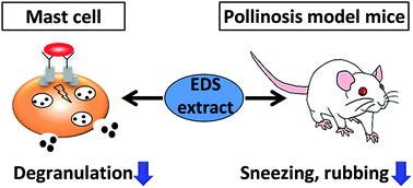
The effects of small dried sardine on IgE-mediated allergic responses are herein reported. Small dried sardine extract digested with an acidic protease from Aspergillus niger suppressed degranulation of rat basophilic leukemia RBL-2H3 cells in a dose-dependent manner without cytotoxicity. Size-exclusion chromatography analysis and a dialysis experiment indicated that the molecular weight of bioactive components contained in enzyme-treated dried sardine extract (EDS extract) ranges from 4800 to 14[thin space (1/6-em)]000. Immunoblot analysis revealed that EDS extract does not affect the signaling pathway via the spleen tyrosine kinase Syk. Although degranulation of RBL-2H3 cells induced by either antigen or a calcium ionophore A23187 was suppressed by EDS extract, the elevation in intracellular Ca2+ concentration was not affected. Immunofluorescence staining revealed that EDS extract significantly suppresses microtubule formation in RBL-2H3 cells during degranulation induced by antigen. In addition, oral administration of EDS extract significantly suppressed passive cutaneous anaphylaxis reaction in mice and an allergic symptom in Cry j1- and Cry j2-induced pollinosis model mice. In conclusion, small dried sardine treated with a protease has potential as a health-promoting foodstuff with an anti-allergic effect.


Introduction

Allergic disorders, such as anaphylaxis, hay fever, asthma, and atopic dermatitis, are an increasing problem all over the world. Now, about 25% of people have allergic symptoms in developed countries.1 Allergic symptoms are divided into four categories based on the difference in their mechanisms to generate immune responses.2 Type I allergic symptoms, such as asthma, atopic dermatitis, and pollinosis, are most common allergic reactions. Especially, Japanese cedar pollinosis is becoming a serious problem in Japan, which is defined as a seasonal allergic rhinitis caused by pollens of Japanese cedar (Cryptomeria japonica). Patients with Japanese cedar pollinosis experience sneezing, a runny nose, a stuffy nose, and itchy eyes every spring. The number of patients with Japanese cedar pollinosis is estimated to be 27% of the population in Japan. Hence, Japanese cedar pollinosis is now called the national affliction in Japan.3 Recently, many kinds of symptomatic treatments have been widely studied and attempted to relieve the symptoms of Japanese cedar pollinosis.

Type I allergic reactions are provoked by cross-linkage of an antigen to immunoglobulin E (IgE) bound to the high-affinity IgE receptor (FcεRI) on the surface of mast cells and basophils. Aggregation of antigen-IgE-bound FcεRI leads to mast cell degranulation and secretion of inflammatory cytokines through activating intracellular signaling processes.4 The initial signaling event is activation of Src family non-receptor tyrosine kinases Lyn and Fyn. Activated Lyn induces phosphorylation of another kinase Syk, which leads to Ca2+ mobilization. As a result, chemical mediators, such as histamine and eicosanoids, are released from intracellular granules, and inflammatory cytokines are secreted, which induces contraction of smooth muscle, vasodilation, and increased vascular permeability.5 On the other hand, the activated Fyn induces phosphorylation of an adapter protein Gab2, thereby causing degranulation by microtubule-dependent translocation of granules to the plasma membrane.6 Thus, mast cells play a crucial role in type I allergic reactions, and prevention of mast cell degranulation is of great importance for the relief of allergic symptoms.

The anti-allergic treatment with anti-histamine, synthetic steroids, and other drugs is not perfect, because a long-term administration of these drugs sometimes causes side effects. Therefore, an alternative way is required to relieve allergic symptoms without such drugs. Anti-allergic foods have recently attracted much attention as substitutes for drugs. Various food components have been reported to suppress degranulation.7–10 As a result of screening with various foodstuffs, we found small dried sardine as an anti-allergic food material.

Small dried sardine is essential for Japanese cuisine “Washoku”, which has been registered by UNESCO as an intangible cultural heritage. Small dried sardine is used for making the traditional broth “Dashi”, a fish stock used for Washoku. In addition, sardine contains abundant nutrients possessing health benefits such as n-3 polyunsaturated fatty acids, vitamin D, and calcium. Effects of n-3 polyunsaturated fatty acids, such as docosahexaenoic acid and eicosapentaenoic acid, on chronic inflammatory diseases including asthma, rheumatoid, arthritis, and inflammatory bowel disease have been reported.11–13 Proteins contained in sardine have been reported to improve diabetes with fructose-induced metabolic syndrome in rats.14 A hypertension-suppressive effect of peptides derived from sardine has also been reported.15

Although various functionalities of ingredients contained in sardine have been reported, the anti-allergic effect of sardine has not yet been reported. In the present study, the anti-allergic effect of small dried sardine was examined using rat basophilic leukemia cell line, RBL-2H3 cells, which have been commonly used for screening of substances inhibiting mast cell degranulation in vitro16 and using murine models of passive cutaneous anaphylaxis (PCA) and pollinosis in vivo. Our data suggest that small dried sardine would contribute to attenuation of allergic symptoms. The findings demonstrated in the present study would be of significance in providing a new dimension to the functionality of small dried sardine.

Experimental

Reagents

Dulbecco's modified Eagle's medium (DMEM), fetal bovine serum (FBS), penicillin, streptomycin, bovine serum albumin (BSA), mouse anti-dinitrophenol (DNP) monoclonal IgE, DNP–human serum albumin (HSA) conjugate, wortmannin, a calcium ionophore A23187, Triton X-100, Evans blue, and mineral oil were purchased from Sigma-Aldrich (St. Louis, MO, USA). Goat anti-actin antibody and horseradish peroxidase (HRP)-labeled anti-goat IgG antibody were purchased from Santa Cruz Biotechnology (Santa Cruz, CA, USA). Alexa Fluor 488-labeled anti-α-tubulin mouse monoclonal antibody, HRP-labeled anti-rabbit IgG antibody, and rabbit antibodies against phosphoinositide 3-kinase (PI3K) p85, phosphorylated PI3K p85/p55, Syk, phosphorylated Syk, Lyn, phosphorylated Lyn, phospholipase C (PLC)γ1, phosphorylated PLCγ1, PLCγ2, and phosphorylated PLCγ2 were purchased from Cell Signaling Technology (Danvers, MA, USA). Purified pollen allergens (Cry j1 and Cry j2) were purchased from Hayashibara (Okayama, Japan). All other chemicals were purchased from Wako Pure Chemical Industries (Osaka, Japan) or Nacalai Tesque (Kyoto, Japan) unless otherwise noted.

Cells and cell culture

RBL-2H3 cells were obtained from American Type Culture Collection (Rockville, MD, USA) and cultured in DMEM supplemented with 100 U mL−1 of penicillin, 100 μg mL−1 of streptomycin, and 10% FBS at 37 °C under humidified 5% CO2.

Animals

Female BALB/c mice were purchased from Japan SLC (Shizuoka, Japan) and kept in an animal room under 12 h light/dark cycle at a temperature of 24 ± 1 °C. Animals received standard chow and water ad libitum. All animal experiments described in this study were carried out in accordance with the protocol approved by the Laboratory Animal Care Committee of Ehime University. Mice were maintained in accordance with the Guidelines for the Care and Use of Laboratory Animals of Ehime University.

Sample preparation

Enzyme-digested small dried sardine extract (EDS extract) was kindly provided by Senmi Ekisu Co., Ltd. (Ehime, Japan). Small dried sardine powder was suspended in 3.7-fold volume of water, and the pH was adjusted to 3.3 to 4.3. The suspension was next digested with 0.3% (w/v) of an acidic protease from Aspergillus niger (Nagase Chemtex; Osaka, Japan) at 40–50 °C for 15–20 h. After neutralizing the suspension (pH = 5.2–5.6), the enzyme was deactivated by heating at 97–99 °C for 30 min. The suspension was then centrifuged, and the supernatant was used as the EDS extract. Protein concentrations of EDS extract were determined using a DC protein assay kit (Bio-Rad Laboratories; Hercules, CA, USA) with BSA as a standard.

To estimate the molecular size of bioactive components, EDS extract was dialyzed using a dialysis membrane with molecular weight cut-off (MWCO) of 500 (Spectrum Laboratories; Rancho Dominguez, CA, USA) or MWCO of 14[thin space (1/6-em)]000 (Wako Pure Chemical Industries) against 10 mmol L−1 sodium phosphate buffer (NaPB; pH 7.4) for 24 h at 4 °C. To evaluate the heat stability, EDS extract was heated at 100 °C for 15 min. These treated samples were used for the degranulation assay described below.

Degranulation assay

Antigen-induced degranulation assay. The assay was performed as previously described17 with some modifications.18 The measurement of released β-hexosaminidase from cells has been used as an indicator of mast cell degranulation.19 RBL-2H3 cells suspended in DMEM containing 100 U mL−1 of penicillin, 100 μg mL−1 of streptomycin, and 10% FBS were seeded into a 96-well culture plate (BD Falcon; Franklin Lakes, NJ, USA) at 4.0 × 10⁴ cell per well and cultured for 12 h at 37 °C under humidified 5% CO2. The cells were then treated with anti-DNP monoclonal IgE at 50 ng mL−1 and incubated for 2 h at 37 °C. After washing the cells with modified Tyrode's (MT) buffer (20 mmol L−1 HEPES, 135 mmol L−1 NaCl, 5 mmol L−1 KCl, 1.8 mmol L−1 CaCl2, 1 mmol L−1 MgCl2, 5.6 mmol L−1 glucose, and 0.05% BSA, pH 7.4) twice, anti-DNP IgE-sensitized cells were treated with 120 μL of MT buffer containing various concentrations of EDS extract, 0.5 μmol L−1 wortmannin, or 10 mmol L−1 NaPB (vehicle) and incubated for 10 min at 37 °C. Ten μL of DNP–HSA diluted in MT buffer at 0.625 μg mL−1 was then added to each well and incubated for 30 min at 37 °C. After incubation, the supernatant was collected from each well, and the cells were sonicated in 130 μL of MT buffer containing 0.1% Triton X-100 for 5 s on ice. Both supernatant and cell lysate were transferred into a new 96-well microplate at 50 μL per well and incubated for 5 min at 37 °C. One hundred μL of 3.3 mmol L−1 4-nitrophenyl-2-acetamido-2-deoxy-β-D-glucopyranoside (Wako Pure Chemical Industries) dissolved in 0.1 mol L−1 citrate buffer (pH 4.5) was then added to each well and incubated for 25 min at 37 °C. The enzyme reaction was terminated by adding 100 μL of 2 mol L−1 glycine buffer (pH 10.4), and the absorbance was measured at 405 nm using a Model 550 microplate reader (Bio-Rad Laboratories). β-Hexosaminidase release rate (%) was calculated as follows:
image file: c6ra14967g-t1.tif
where “A” is the absorbance of each well.
A23187-induced degranulation assay. RBL-2H3 cells were inoculated and treated with various concentrations of EDS extract as described above, except sensitization with anti-DNP IgE. Ten μL of A23187 diluted in MT buffer at 3 μmol L−1 was added to each well, and the cells were incubated for 30 min at 37 °C. The β-hexosaminidase release rate was then measured as described above.

Cell viability

Cytotoxicity of EDS extract to RBL-2H3 cells was examined using a WST-8 assay kit (Kishida Chemical; Osaka, Japan) according to the manufacturer's instruction. Anti-DNP IgE-sensitized cells were treated with various concentrations of EDS extract and stimulated with DNP–HSA as described above. After the cells were washed with PBS once, 100 μL of 10% FBS–DMEM containing 10% WST-8 solution was added to each well of the culture plate and incubated for 30 min at 37 °C. The absorbance was then measured at 450 nm using a microplate reader.

Size-exclusion chromatography analysis

Size exclusion chromatography analysis was performed on a LaChrom Elite HPLC system (Hitachi, Tokyo, Japan) with a TSK-gel G4000SWXL column (7.8 × 300 mm, Tosoh, Tokyo, Japan) as previously described18 with some modifications. The column was preequilibrated with phosphate-buffered saline (PBS, pH 7.4), and the column temperature was set at 15 °C. One milliliter of EDS extract (1.5 mg mL−1) was applied to the column and eluted with PBS with a flow rate at 1.0 mL min−1. A gel filtration molecular weight markers kit (Sigma-Aldrich) was used to acquire a calibration curve.

Immunoblot analysis

RBL-2H3 cells were seeded into a 24-well culture plate (BD Falcon) at 2.5 × 10⁵ cells per well and cultured for 12 h at 37 °C under humidified 5% CO2. The cells were then treated with anti-DNP monoclonal IgE at 50 ng mL−1 and incubated for 2 h at 37 °C. After washing the cells with MT buffer twice, anti-DNP IgE-sensitized cells were treated with 490 μL of MT buffer containing EDS extract (1.5 mg mL−1) or 10 mmol L−1 NaPB (vehicle) and incubated for 10 min at 37 °C. The cells were then stimulated with DNP–HSA at 50 ng mL−1 and further incubated for 10 min. After removing the added reagents, cells were lysed and immunoblotting was performed with various antibodies as previously described.20

Measurement of intracellular Ca2+ concentration ([Ca2+]i)

[Ca2+]i was measured using a Calcium Kit Fluo 3 (Dojindo Laboratories; Kumamoto, Japan) according to the manufacturer's instruction. RBL-2H3 cells were seeded into a white 96-well culture plate (Nunc; Roskilde, Denmark) and treated with anti-DNP IgE as described above. The IgE-sensitized RBL-2H3 cells were then washed with PBS twice and incubated with 100 μL of Fluo-3 AM for 1 h at 37 °C. After washing the cells with PBS, the cells were treated with 120 μL of MT buffer containing EDS extract (1.3 mg mL−1) or 10 mmol L−1 NaPB (vehicle) and incubated for 10 min at 37 °C. The cells were then stimulated by adding 10 μL of MT buffer containing DNP–HSA at 0.625 μg mL−1 or 3 μmol L−1 A23187, and the fluorescent intensity was immediately monitored with an excitation wavelength of 490 nm and an emission wavelength of 530 nm using a SH-8000Lab microplate reader (Corona Electric; Ibaraki, Japan).

Immunofluorescence microscopy

RBL-2H3 cells were seeded into 35 mm culture dishes (BD Falcon) at 5.0 × 10⁵ cells per dish and cultured for 12 h at 37 °C under humidified 5% CO2. The cells were treated with anti-DNP monoclonal IgE at 50 ng mL−1 and incubated for 2 h at 37 °C. After washing the cells with MT buffer twice, anti-DNP IgE-sensitized cells were treated with 2.45 mL of MT buffer containing EDS extract (1.5 mg mL−1) or 10 mmol L−1 NaPB (vehicle) and incubated for 10 min at 37 °C. The cells were then stimulated with 50 ng mL−1 of DNP–HSA and further incubated for 30 min at 37 °C. After removing the added reagents, the cells were washed with PBS and fixed with methanol containing 1 mmol L−1 EDTA for 3 min at −20 °C. After removing fixative, the cells were rinsed with PBS for 5 min three times. The cells were then permeabilized with PBS containing 0.1% Triton X-100 for 5 min three times and blocked with 5% BSA–PBS containing 0.3% Triton X-100 for 1 h. To visualize tubulin, the cells were stained with Alexa Fluor 488-conjugated anti-α-tubulin antibody diluted in 1% BSA–PBS containing 0.3% Triton X-100 for 12 h at 4 °C. After removing the added reagents, the cells were washed with PBS. The cells were then treated with an anti-fading agent SlowFade (Molecular Probes; Eugene, OR, USA) to prevent fading and observed under a Fluoview FV1000 confocal microscope (Olympus; Tokyo, Japan). Images were analyzed with FV10-ASW2.1 software (Olympus).

IgE-mediated PCA in mice

The assay was performed according to the method of Knoops et al.21 with some modifications. After acclimating to their housing environment for 1 week, 7 week-old female BALB/c mice were intradermally injected with 10 μL of PBS containing 0.1 μg of anti-DNP IgE and with PBS alone into the left and right ears, respectively. After 24 h, 0.2 mL of PBS containing 0.2 mg of DNP–HSA and 0.5% Evans blue were injected into their tail vein. One hour before DNP–HSA injection, the mice were orally administered with 20 μL of 10 mmol L−1 NaPB for the control group and of EDS extract for the EDS extract-administered group (6 mg protein per kg body weight). The mice were then euthanized 30 min after DNP–HSA injection, and their ears were excised. The extravagated dye was then extracted from each ear with 0.5 mL of formamide for 16 h at 70 °C, and the absorbance was measured at 620 nm using an Ultrospec 3000 spectrophotometer (Amersham Pharmacia Biotech; Uppsala, Sweden).

A mouse model of Japanese cedar pollinosis

A mouse model of Japanese cedar pollinosis was developed according to Nomiya et al.22 by treating mice with Japanese cedar pollen allergen as scheduled in Fig. 1. Following adaptation period for 1 week, 6 week-old female BALB/c mice were randomly divided into 3 groups as follows; control group (7 mice), EDS extract-administered group (7 mice), and intact group (7 mice). The mice in control and EDS extract-administered groups were intranasally treated with 2.0 μg of purified pollen allergens (Cry j1 and Cry j2) dissolved in 10 μL of PBS using a glass microsyringe (Hamilton; Reno, NV, USA) on days 0, 7, 14, and 21, while the mice in intact group were with PBS alone. The mice in control and EDS extract-administered groups were then intranasally challenged with 0.4 μg of purified pollen allergens (Cry j1 and Cry j2) in 10 μL of PBS for 6 consecutive days from day 28 to day 33, while the mice in intact group were given 10 μL of PBS alone. The mice in EDS extract-administered group were orally administered with 20 μL of EDS extract (6 mg protein per mL) for 7 consecutive days from day 28 to day 34, while the mice in intact and control groups were with 20 μL of 10 mmol L−1 NaPB (vehicle). On day 34, all mice, including intact group, were treated with 0.4 μg of Cry j1 and Cry j2. Nasal rubbing frequency was counted for 30 min immediately after the final challenge. Blood was collected on day 35, and serum IgE and IgG1 levels were measured by in-house-developed ELISA as described in Nishimoto et al.23
image file: c6ra14967g-f1.tif
Fig. 1 Experimental design used to investigate the effect of EDS extract on pollinosis induction using purified pollen allergen (Cry j1 and Cry j2) in BALB/c mice.

Statistical analysis

Data obtained were expressed as mean ± standard deviation (SD). Dunnett's test, Tukey–Kramer test, or Student's t test was used to assess the statistical significance of the difference. p-Values <0.05 or <0.01 were considered statistically significant.

Results

Effect of EDS extract on degranulation of RBL-2H3 cells

An effect of EDS extract on degranulation of RBL-2H3 cells was examined. EDS extract was added at various concentrations to the culture medium of anti-DNP IgE-sensitized RBL-2H3 cells, and degranulation was induced with DNP–HSA as antigen. As shown in Fig. 2A, degranulation was suppressed by treating cells with EDS extract in a dose-dependent manner as well as with wortmannin used as a positive control. Statistically significant differences against control in the β-hexosaminidase release rate were observed at equal to or higher than 720 μg mL−1 of protein concentration, suggesting that EDS extract has a degranulation-suppressive activity on RBL-2H3 cells stimulated by antigen. In addition, EDS extract was found to exhibit no cytotoxicity at any concentrations tested as shown in Fig. 2B. From these results, EDS extract was used around 1.5 mg mL−1 of protein concentration for further experiments, which showed the highest degranulation-suppressive activity without cytotoxicity.
image file: c6ra14967g-f2.tif
Fig. 2 Effects of EDS extract on degranulation and cell viability of RBL-2H3 cells. (A) After anti-DNP IgE-sensitized RBL-2H3 cells were treated with EDS extract at indicated concentrations, with 0.5 μmol L−1 wortmannin, or with 10 mmol L−1 NaPB as control, degranulation was induced with DNP–HSA. Released β-hexosaminidase was used as a marker of degranulation. Data are represented as mean ± SD (n = 3). *p < 0.05 against control by Dunnett's test. (B) Viability of RBL-2H3 cells was measured using a WST-8 assay kit after treating anti-DNP IgE-sensitized RBL-2H3 cells with EDS extract at indicated concentrations or with 10 mmol L−1 NaPB as control, followed by stimulation with DNP–HSA. Data are represented as mean ± SD (n = 3). No statistical significance was found against control by Dunnett's test.

Characteristics of bioactive components in EDS extract

EDS extract was heated at 100 °C for 15 min to investigate the molecular characteristic of bioactive components in EDS extract and used for the antigen-induced degranulation assay. The result showed that the degranulation-suppressive activity of EDS extract was not affected by heating (Fig. 3A), indicating that the bioactive components in EDS extract are stable to heat.
image file: c6ra14967g-f3.tif
Fig. 3 Effects of dialysis and heat treatment of EDS extract on degranulation of RBL-2H3 cells. (A) EDS extract (white bar; 1.5 mg mL−1) or 10 mmol L−1 NaPB (gray bar) used as control was heated at 100 °C for 15 min. After anti-DNP IgE-sensitized RBL-2H3 cells were treated with heated/non-heated EDS extract or with NaPB, degranulation was induced with DNP–HSA. Relative β-hexosaminidase release to control is shown. Data are represented as mean ± SD (n = 3). **p < 0.01 against control by Student's t test. (B) EDS extract was dialyzed using a dialysis membrane of MWCO of 500 (closed circle) or of 14[thin space (1/6-em)]000 (open circle) against 10 mmol L−1 NaPB. After anti-DNP IgE-sensitized RBL-2H3 cells were treated with dialyzed EDS extract at indicated concentrations or with NaPB (gray circle) as control, degranulation was induced with DNP–HSA. Released β-hexosaminidase was used as a marker of degranulation. Data are represented as mean ± SD (n = 3). *p < 0.05 or **p < 0.01 against control by Dunnett's test.

EDS extract was next dialyzed using a dialysis membrane with MWCO of 500 or 14[thin space (1/6-em)]000 to estimate the molecular size of bioactive components in EDS extract and used for the antigen-induced degranulation assay. The result showed that EDS extract dialyzed with a 14[thin space (1/6-em)]000 MWCO membrane lost its degranulation-suppressive activity, whereas EDS extract dialyzed with a 500 MWCO membrane retained the degranulation-suppressive activity on RBL-2H3 cells (Fig. 3B), indicating that the molecular weight of the bioactive components in EDS extract would be roughly between 500 and 14[thin space (1/6-em)]000 based on the dialysis experiments. EDS extract dialyzed with a 500 MWCO membrane was thus used for further experiments.

In addition, EDS extract was analyzed and fractionated by size exclusion chromatography. When the elution profile was monitored by the absorbance at 214 nm, components of the sample were continuously eluted, and no obvious peaks were observed (Fig. 4A). We thus observed the absorbance at 280 nm, and a profile different from that monitored at 214 nm was obtained (Fig. 4B). The absorbance monitored at 240 nm is shown in Fig. 4C. Based on these profiles, EDS extract was roughly fractionated into six fractions and subjected to the antigen-induced degranulation assay. As shown in Fig. 4D, the result showed that the major bioactive component contained in EDS extract existed in the fraction IV. According to a calibration curve (Fig. 4E) obtained with gel filtration molecular weight markers, the molecular weight of the major bioactive component in the fraction IV was estimated to range from 4800 to 14[thin space (1/6-em)]000 from the size exclusion chromatography. Interestingly, the fraction IV contained substances strongly absorbed at the wavelength of 240 nm (Fig. 4F).


image file: c6ra14967g-f4.tif
Fig. 4 Analysis and fractionation of EDS extract by size exclusion chromatography. A chromatogram of EDS extract (1.5 mg protein) was monitored at 214 nm (A), 280 nm (B), and 240 nm (C), and six fractions of EDS extract were obtained. (D) Anti-DNP IgE-sensitized RBL-2H3 cells were treated with each fraction at the same protein concentration, and degranulation was induced with DNP–HSA. NaPB was used as control. Released β-hexosaminidase was used as a marker of degranulation. Data are represented as mean ± SD (n = 3). *p < 0.05 or **p < 0.01 against control by Dunnett's test. (E) A calibration curve of the size exclusion chromatography was obtained with gel filtration molecular weight markers. (F) Absorbance spectra at the retention time of 15.51 min is shown.

Effect of EDS extract on signaling pathways involved in antigen-induced degranulation

The effect of EDS extract on signaling pathways involved in the antigen-induced degranulation was examined by immunoblot analysis. As shown in Fig. 5, EDS extract did not affect phosphorylation of any of Lyn, Syk, PI3K, or PLCγ1/2, indicating that EDS extract suppresses the antigen-induced degranulation of RBL-2H3 cells through signaling pathways other than the Lyn-dependent pathway.
image file: c6ra14967g-f5.tif
Fig. 5 Effect of EDS extract on signaling pathways involved in degranulation of RBL-2H3 cells stimulated by antigen. After anti-DNP IgE-sensitized RBL-2H3 cells were treated with EDS (1.5 mg mL−1) or with 10 mmol L−1 NaPB as control, degranulation was induced with DNP–HSA. After 10 min, each cell lysate was prepared and immunoblotting was performed. A representative blot from two independent experiments is shown. p-Lyn, p-Syk, p-PLCγ, and p-PI3K indicate phosphorylated Lyn, phosphorylated Syk, phosphorylated phospholipase Cγ, and phosphorylated PI3K, respectively.

Effect of EDS extract on [Ca2+]i

Ca2+ is one of major second messengers in the intracellular signaling, and the elevation in [Ca2+]i is a critical process in degranulation. Thus, the effect of EDS extract on [Ca2+]i in RBL-2H3 cells was examined. As shown in Fig. 6A, the antigen-induced elevation in [Ca2+]i was not suppressed by EDS extract. In addition, the effect of EDS extract on degranulation induced by a calcium ionophore A23187, which carries Ca2+ into cytosol across the plasma membrane, was examined. EDS extract suppressed A23187-induced degranulation (Fig. 6B), whereas it did not inhibit the A23187-induced elevation in [Ca2+]i (Fig. 6C). These results suggest that EDS extract would be involved in an intracellular event after the elevation in [Ca2+]i.
image file: c6ra14967g-f6.tif
Fig. 6 Effects of EDS extract on [Ca2+]i and calcium ionophore-induced degranulation of RBL-2H3 cells. (A) After anti-DNP IgE-sensitized RBL-2H3 cells were incubated with Fluo-3 AM, the cells were treated with EDS extract (1.3 mg mL−1) or with 10 mmol L−1 NaPB as control. Fluorescence intensity was measured immediately after inducing degranulation with DNP–HSA. Data are represented as mean (n = 3). Gray circle, NaPB-treated cells stimulated with antigen; open circle, EDS extract-treated cells stimulated with DNP–HSA; closed circle, NaPB-treated cells not stimulated with DNP–HSA. (B) After RBL-2H3 cells were treated with various concentrations of EDS extract or with NaPB as control, degranulation was induced with a calcium ionophore A23187. Released β-hexosaminidase was used as a marker of degranulation. Data are represented as mean ± SD (n = 3). *p < 0.05 or **p < 0.01 against control by Dunnett's test. (C) After RBL-2H3 cells were incubated with Fluo-3 AM, the cells were treated with EDS extract (1.3 mg mL−1) or with NaPB as control. Fluorescence intensity was measured immediately after inducing degranulation with A23187. Data are represented as mean (n = 3). Gray circle, NaPB-treated cells stimulated with A23187; open circle, EDS extract-treated cells stimulated with A23187; closed circle, NaPB-treated cells not stimulated with A23187.

Effect of EDS extract on microtubule formation

Microtubule formation is a crucial process for degranulation, which is involved in the movement of intracellular granules and the fusion of cell membrane with granule membrane. Microtubule formation is not triggered by increased [Ca2+]i.24 Thus, the effect of EDS extract on microtubule formation was observed by fluorescence microscopy. As shown in Fig. 7, microtubules spread out to the plasma membrane after antigen stimulation to build tracks for granule transport. On the other hand, the microtubule formation in RBL-2H3 cells stimulated with antigen was inhibited in the presence of EDS extract. These results suggest that EDS extract suppresses degranulation by affecting the microtubule formation occurring after the elevation in [Ca2+]i.
image file: c6ra14967g-f7.tif
Fig. 7 Effects of EDS extract on microtubule formation. After anti-DNP IgE-sensitized RBL-2H3 cells were treated with EDS (1.5 mg mL−1) or with 10 mmol L−1 NaPB as control, degranulation was induced with DNP–HSA as antigen. After 30 min, cells were fixed with methanol, permeabilized with PBS containing 0.1% Triton X-100, and blocked with 5% BSA–PBS. After staining with Alexa Fluor 488-conjugated anti-α-tubulin antibody, the cells were observed under a confocal microscope.

Effect of EDS extract in PCA model mice

The anti-allergic activity of EDS extract was evaluated in vivo using PCA model mice. Anti-DNP IgE-injected mice were orally administered with 10 mmol L−1 NaPB for control group or with EDS extract for EDS extract-administered group, and PCA reaction was induced with DNP–HSA. As shown in Fig. 8, the PCA reaction was significantly suppressed by oral administration of EDS extract (p < 0.01 against control group), indicating that EDS could exhibit the inhibitory effect on degranulation in vivo.
image file: c6ra14967g-f8.tif
Fig. 8 Effect of EDS extract on IgE-mediated PCA model mice. Six-week-old female BALB/c mice were intravenously injected with DNP–HSA and 0.5% Evans blue 24 h after intradermal injection of anti-DNP IgE and PBS alone into left and right ears of mice, respectively. EDS extract (6 mg protein per kg body weight) was orally administrated 1 h before DNP–HSA injection. Evans blue was extracted from each ear and absorbance of the dye was measured at 620 nm. Absorbance of the dye extracted from left ear was subtracted with that from right ear. Data are represented as mean ± SD (n = 7). **p < 0.01 against control by Student's t test.

Effect of EDS extract in a mouse model of pollinosis induced with Cry j1 and Cry j2

Finally, we investigated the effect of EDS extract on IgE-mediated allergic reactions in a mouse model of Cry j1 and Cry j2-induced pollinosis. Cry j1 and Cry j2 are the major allergens of Japanese cedar (C. japonica) pollen. Mice were sensitized and challenged with pollen allergens (Cry j1 and Cry j2) as scheduled in Fig. 1. The mice in EDS extract-administered group were orally administered with EDS extract for 7 consecutive days, while the mice in intact and control groups were with 10 mmol L−1 NaPB. Oral administration of EDS extract did not affect the body weight of mice (data not shown). On day 34, all mice were treated with pollen allergens, and sneezing and rubbing frequencies were counted, which are clinical signs of hypersensitivity in pollinosis model mice.25,26 As shown in Fig. 9A and B, the sneezing frequency significantly decreased in EDS extract-administrated group compared with that in control group (p < 0.05), although there was no difference in the rubbing frequency. These results suggest that EDS extract has a potential to attenuate the symptom of cedar pollinosis in vivo. Biochemical analysis was also performed by measuring total serum IgE and IgG1 levels in mice. As shown in Fig. 9C and D, there was no difference in serum IgE and IgG1 among any groups, indicating that the administration of EDS extract does not suppress the production of either class of antibodies.
image file: c6ra14967g-f9.tif
Fig. 9 Effect of EDS extract on a mouse model of pollinosis. Six-week-old female BALB/c mice in control and EDS extract-administered groups were intranasally treated with 2.0 μg of Cry j1 and Cry j2 on days 0, 7, 14, and 21, while intact group was with PBS alone. Mice in control and EDS extract-administered groups were then intranasally challenged with 0.4 μg of Cry j1 and Cry j2 for 6 consecutive days on days 28 to 33, while intact group was given PBS alone. EDS extract-administered group was given 20 μL of EDS extract (6.0 mg protein per mL) once a day from days 28 to 34, while intact and control groups were 20 μL of 10 mmol L−1 NaPB. On day 34, all mice were treated with 0.4 μg of Cry j1 and Cry j2. (A) Nasal rubbing frequency was counted for 30 min after the final challenge on day 34. Data are represented as mean ± SD (n = 7). No statistical significance was found against control by Tukey–Kramer test. (B) Sneezing frequency was counted for 30 min after the final challenge on day 34. Data are represented as mean ± SD (n = 7). *p < 0.05 against control by Tukey–Kramer test. NS indicates no statistical significance. (C) Serum IgE levels in each mouse on day 35. Data are represented as mean ± SD (n = 7). No statistical significance was found against control by Tukey–Kramer test. (D) Serum IgG1 levels in each mouse on day 35. Data are represented as mean ± SD (n = 7). No statistical significance was found against control by Tukey–Kramer test.

Discussion

In this study, EDS extract was revealed to suppress the antigen-induced degranulation of RBL-2H3 cells (Fig. 2A and B) without cytotoxicity. Bioactive components in EDS extract were suggested to be heat-stable (Fig. 3A). From the dialysis experiments, the molecular weight of the bioactive components in EDS extract was suggested to be between approximately 500 and 14[thin space (1/6-em)]000 (Fig. 3B). According to the size exclusion chromatography and the antigen-induced degranulation assay using the fractions, the molecular weight of the bioactive components in EDS extract was suggested to range from 4800 to 14[thin space (1/6-em)]000 (Fig. 4). These data, taken together, suggested that the estimated molecular weight of the bioactive substance in EDS extract would be between 4800 and 14[thin space (1/6-em)]000. Interestingly, the fraction IV had the maximum absorption at the wavelength of 241 nm. Thus, the bioactive components in EDS extract may be the substance responsible for the absorption at 241 nm.

Because n-3 polyunsaturated fatty acids are insoluble in water and low-molecular-weight substances like vitamin D were assumed to be removed by dialysis, bioactive components in EDS extract would not be those compounds but peptides. We attempted to identify the bioactive component in the fraction IV after size exclusion chromatography; however, the sample is still a mixture of various substances when the chromatography was monitored at 214 nm as shown in Fig. 4A, and no obvious peaks were observed. Thus, further purification of the sample is required for identification of the bioactive component using LC-MS/MS.

The mechanism underlying the inhibition of degranulation by EDS extract was investigated. The signaling pathway involved in degranulation is initiated by phosphorylation of Src family non-receptor tyrosine kinases Lyn and Fyn after cross-linkage of antigen to FcεRI via IgE. The activated Lyn phosphorylates Syk, a tyrosine kinase, which leads to activation of several downstream signaling molecules such as PI3K, Akt, and PLCγ, subsequently inducing Ca2+ mobilization.5,27 Fyn phosphorylates an adapter protein Gab2, leading to microtubule-dependent translocation of granules to the plasma membrane.28 As shown in Fig. 5A, EDS extract did not suppress the activation of the Lyn signaling pathway. Moreover, the elevation in [Ca2+]i was not suppressed by EDS extract (Fig. 6A and C). These results indicate that EDS extract suppresses an intracellular event after elevation in [Ca2+]i induced by the antigen–antibody interaction.

Microtubule formation has been reported to occur after elevation in [Ca2+]i, which is crucial for degranulation process.24 EDS extract was found to suppress degranulation by inhibiting microtubule formation (Fig. 7). From these results, EDS extract is likely to affect the Fyn signaling pathway and thereby inhibit degranulation of RBL-2H3 cells through preventing microtubule formation, because the Fyn signal pathway leads to microtubule formation as mentioned above.

We further investigated whether the outcomes of in vitro studies are involved in those of in vivo studies. The anti-allergic effect of EDS extract was examined using murine models of passive cutaneous anaphylaxis and pollinosis. We revealed that oral administration of EDS extract significantly inhibits the PCA reaction (Fig. 8) and reduces the sneezing frequency compared with the control group (Fig. 9B). Type I allergic reactions including Japanese cedar pollinosis are induced by a couple of Ig classes, which are vital biomarkers of allergy.29 Thus, serum IgE and IgG1 levels in mice orally administered with EDS extract were measured. We found that the IgE amount increased by inducing pollinosis and slightly decreased by oral administration of EDS extract, although there was no statistical significance among groups (Fig. 9C), indicating that EDS extract does not suppress IgE production. On the other hand, IgG1 levels were not changed by inducing pollinosis or by oral administration of EDS extract (Fig. 9D). The result indicated that IgG1 might not be responsible for allergic reactions of cedar pollinosis. These in vivo data seemed to reflect an impact of EDS extract on the degranulation of RBL-2H3 cells as was noted in vitro as described above.

Conclusions

EDS extract suppressed degranulation of RBL-2H3 cells. The bioactive components in EDS extract were expected to be heat-stable with the molecular weight ranging from 4800 to 14[thin space (1/6-em)]000. The inhibitory effect of EDS extract was shown to result from the suppressed microtubule formation. In addition, oral administration of EDS extract significantly suppressed an allergic reaction in PCA model mice and sneezing frequency in a mouse model of pollinosis. Taken together, these findings suggest that EDS extract has an anti-allergy effect that controls mast cell degranulation and would be valuable as a functional food factor.

Acknowledgements

Animal experiments and fluorescence microscopy were accomplished at the Advanced Research Support Center (ADRES), Ehime University.

References

  1. S. J. Galli, M. Tsai and A. M. Piliponsky, Nature, 2008, 454, 445–454 CrossRef CAS PubMed.
  2. P. G. H. Gell and R. R. A. Coombs, in Clinical Aspects of Immunology, Blackwell, 1963, pp. 317–337 Search PubMed.
  3. T. Yamada, H. Saito and S. Fujieda, J. Allergy Clin. Immunol., 2014, 133, 632–639 CrossRef PubMed.
  4. K. Amin, Respir. Med., 2012, 106, 9–14 CrossRef PubMed.
  5. S. Kraft and J.-P. Kinet, Nat. Rev. Immunol., 2007, 7, 365–378 CrossRef CAS PubMed.
  6. J. Rivera and A. M. Gilfillan, J. Allergy Clin. Immunol., 2006, 117, 1214–1225 CrossRef CAS PubMed.
  7. M. Suzuki, K. Yoshino, M. Maeda-Yamamoto, T. Miyase and M. Sano, J. Agric. Food Chem., 2000, 48, 5649–5653 CrossRef CAS PubMed.
  8. A. Singh, S. Holvoet and A. Mercenier, Clin. Exp. Allergy, 2011, 41, 1346–1359 CrossRef PubMed.
  9. S. Onishi, K. Nishi, S. Yasunaga, A. Muranaka, K. Maeyama, A. Kadota and T. Sugahara, J. Funct. Foods, 2014, 6, 606–614 CrossRef CAS.
  10. K. Nishi, M. Teranishi, S. Yasunaga, M. Iitsuka, S. Matsumoto and T. Sugahara, Int. Dairy J., 2014, 39, 89–95 CrossRef CAS.
  11. P. C. Calder, Br. J. Clin. Pharmacol., 2013, 75, 645–662 CAS.
  12. C. M. Yates, P. C. Calder and G. E. Rainger, Pharmacol. Ther., 2014, 141, 272–282 CrossRef CAS PubMed.
  13. J. Miyata and M. Arita, Allergol. Int., 2015, 64, 27–34 CrossRef CAS PubMed.
  14. Z. Madani, K. Louchami, A. Sener, W. J. Malaisse and D. Ait Yahia, Int. J. Mol. Med., 2012, 29, 311–318 CAS.
  15. L. Otani, T. Ninomiya, M. Murakami, K. Osajima, H. Kato and T. Murakami, Biosci., Biotechnol., Biochem., 2009, 73, 2203–2209 CrossRef CAS PubMed.
  16. E. Passante and N. Frankish, Inflammation Res., 2009, 58, 737–745 CrossRef CAS PubMed.
  17. J. Watanabe, H. Shinmoto and T. Tsushida, Biosci., Biotechnol., Biochem., 2005, 69, 1–6 CrossRef CAS PubMed.
  18. M. Kondo, K. Nishi and T. Sugahara, J. Funct. Foods, 2015, 14, 659–669 CrossRef CAS.
  19. A. J. MacDonald, D. M. Haig, H. Bazin, A. C. McGuigan, R. Moqbel and H. R. Miller, Immunology, 1989, 67, 414–418 CAS.
  20. M. Ishida, K. Nishi, H. Watanabe and T. Sugahara, Food Chem., 2013, 136, 322–327 CrossRef CAS PubMed.
  21. L. Knoops, J. Louahed, J. Van Snick and J. C. Renauld, J. Immunol., 2005, 175, 335–341 CrossRef CAS.
  22. R. Nomiya, M. Okano, T. Fujiwara, M. Maeda, Y. Kimura, K. Kino, M. Yokoyama, H. Hirai, K. Nagata, T. Hara, K. Nishizaki and M. Nakamura, J. Immunol., 2008, 180, 5680–5688 CrossRef CAS.
  23. S. Nishimoto, K. Kanda, M. Yamawaki, M. Okabe, K. Akiyama, Y. Kakinuma and T. Sugahara, J. Toxicol. Sci., 2009, 34, 459–468 CrossRef CAS PubMed.
  24. K. Nishida, S. Yamasaki, Y. Ito, K. Kabu, K. Hattori, T. Tezuka, H. Nishizumi, D. Kitamura, R. Goitsuka, R. S. Geha, T. Yamamoto, T. Yagi and T. Hirano, J. Cell Biol., 2005, 170, 115–126 CrossRef CAS PubMed.
  25. M. Tsunematsu, T. Yamaji, D. Kozutsumi, R. Murakami, S. Kimura and K. Kino, Life Sci., 2007, 80, 1388–1394 CrossRef CAS PubMed.
  26. M. Tsunematsu, T. Yamaji, D. Kozutsumi, R. Murakami, H. Nagai and K. Kino, Biomed. Res., 2008, 29, 119–123 CrossRef CAS PubMed.
  27. D. D. Metcalfe, R. D. Peavy and A. M. Gilfillan, J. Allergy Clin. Immunol., 2009, 124, 639–646 CrossRef CAS PubMed.
  28. P. Dráber, V. Sulimenko and E. Dráberová, Front. Immunol., 2012, 3, 34–51 Search PubMed.
  29. T. A. E. Platts-Mills, Am. J. Respir. Crit. Care Med., 2001, 164, S1–S5 CrossRef CAS PubMed.

This journal is © The Royal Society of Chemistry 2016
Click here to see how this site uses Cookies. View our privacy policy here.