Borage oil supplementation decreases lipopolysaccharide-induced inflammation and skeletal muscle wasting in mice

Pei-Yin Chena, Chin-San Liubc, Li-Yun Lind, Yi-Chin Linef, Hai-Lun Sungh, Chien-Chun Lief, Haw-Wen Cheni, Tsu-Shing Wangj, John Wang*k and Kai-Li Liu*ef
aDepartment of Recreation and Holistic Wellness, Mingdao University, Changhua, 52345, Taiwan
bDepartment of Neurology and Vascular and Genomic Center, Changhua Christian Hospital, Changhua, 500 Taiwan
cGraduate Institute of Integrated Medicine, College of Chinese Medicine, China Medical University, Taichung, 404, Taiwan
dDepartment of Food Science and Technology, Hung Kuang University, No. 1018, Sec. 6, Taiwan Boulevard, Shalu District, Taichung City 43302, Taiwan
eDepartment of Nutrition, Chung Shan Medical University, No. 110, Sec. 1, Chien-Kuo N. Rd., Taichung 40203, Taiwan. E-mail: kaililiu@csmu.eud.tw; Fax: +886 4 23248175; Tel: +886 4 24730022 ext. 12136
fDepartment of Nutrition, Chung Shan Medical University Hospital, Taichung, Taiwan
gSchool of Medicine, Chung Shan Medical University, Taichung, Taiwan
hDivision of Allergy, Asthma and Rheumatology, Department of Pediatrics, Chung Shan Medical University Hospital, Taichung, Taiwan
iDepartment of Nutrition, China Medical University, No. 91, Hsueh-Shih Road, Taichung, 40402, Taiwan
jDepartment of Biomedical Sciences, Chung Shan Medical University, Taichung, Taiwan
kDepartment of Nutrition, Master Program of Biomedical Nutrition, Hung Kuang University, No. 1018, Sec. 6, Taiwan Boulevard, Shalu District, Taichung City 43302, Taiwan. E-mail: shengw@seed.net.tw; Tel: +886 4 26318652

Received 31st May 2016 , Accepted 11th October 2016

First published on 14th October 2016


Abstract

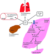
Because in vitro data have shown gamma-linolenic acid (GLA) to be protective in LPS-induced macrophage inflammation and myotube atrophy, we explored the therapeutic value of borage oil (BO), a GLA rich oil, in LPS-induced inflammation and muscle wasting in C57BL/6JNarl mice. Supplementation with BO was more potent than supplementation with soybean oil (SO) in decreasing LPS-induced expression of pro-inflammatory cytokines and glutathione in serum and tissues. Notably, GLA did not reverse LPS-induced inflammatory cytokine expression in C2C12 myotubes transfected with a constitutively active mutant IκB kinase-β plasmid, which suggested the importance of the inhibition of nuclear factor-κB (NF-κB) activation by GLA. Moreover, BO prevented LPS-induced skeletal muscle weight loss as well as molecule expression of ubiquitin-proteasome pathway and the autophagy-lysosomal pathway which played a key role in skeletal muscle protein degradation. BO but not SO reduced the LPS-induced increase in toll-like receptor 4 (TLR4) expression and activation of mitogen-activated protein kinases (MAPKs) and NF-κB in gastrocnemius muscle. In summary, supplementation with BO is more effective than supplementation with SO in preventing LPS-induced inflammation and muscle wasting. Blockade of the TLR4/MAPKs/NF-κB pathway is crucial in the action of BO on LPS-induced inflammation and wasting in skeletal muscle.


Introduction

Sepsis, a systemic inflammatory response to bacterial infections, is a serious health problem with high mortality in hospitalized patients.1 Lipopolysaccharide (LPS), derived from the cell wall of Gram-negative bacteria, can generate the overproduction of pro-inflammatory mediators, which play an important role in the development of not only multiple organ injury but also muscle wasting in sepsis.2–4 Wasting of skeletal muscle, the most abundant tissue in the body leads to weakness, fatigue, and eventually death.5,6 Data have shown that increased breakdown of muscle protein rather than depressed protein synthesis is the major factor contributing to sepsis-induced muscle wasting.5,6 Upregulation of ubiquitin-proteasome pathway (UPP) and the autophagy-lysosome pathway (ALP) are mainly responsible for the skeletal muscle proteolysis during severe catabolic diseases including sepsis.3,4

In response to various atrophic conditions, myofibrillar proteins such as myosin heavy chain (MyHC) conjugated with multiple ubiquitin (Ub) proteins can be degraded within the proteasome complex. Increased expression of muscle-specific E3 Ub ligases, atrogin-1/muscle wasting F-box (MAFbx), and muscle-specific ring finger protein 1 (MuRF1) is the rate-limiting step for muscle protein degradation by the UPP.7 Overexpression of MAFbx causes C2C12 myotube atrophy, whereas mice deficient in either MAFbx or MuRF1 are resistant to muscle denervation atrophy.8 Sepsis induced by either LPS administration or cecal ligation and puncture results in significantly increased expression of MAFbx and MuRF1 in skeletal muscle.9 Phenolic compounds such as curcumin, apigenin, and lutolin can decrease LPS-induced MAFbx expression and wasting in skeletal muscle and C2C12 myotubes.10,11

Constitutive activation of the ALP at basal levels is crucial for cellular homeostasis by which autophagosomes deliver cytosolic proteins and organelles to lysosomes for degradation and recycling.12 Conjugation of the autophagy-related gene microtubule-associated protein 1 light chain 3A (LC3A) with phosphatidylethanolamine to form LC3B is a key process in mature autophagosome formation12 and is increased in LPS-induced atrophic myotubes and skeletal muscle.3,4 Without a change in UPP activity, the autophagy inhibitor 3-methyladenine abolishes LPS-induced mouse skeletal muscle loss by decreasing the LC3B level and autophagosome formation.3 The above findings suggest that regulation and control of the activity of the UPP and the ALP may be a useful therapeutic strategy for reducing LPS-induced skeletal muscle wasting.

In mammalian cells, member proteins of the nuclear factor-κB (NF-κB) family, including p65 (RelA), p50/p105 (NF-κB1), p52/p100 (NF-κB2), RelB, and c-Rel, form a dimeric complex, mostly composed of p50 and p65 subunits. In unstimulated cells, NF-κB is bound to the inhibitory protein IκB in cytoplasm. Upon activation, IκB is phosphorylated by the IκB kinase (IKK) complex, which results in degradation of IκB and then nuclear translocation and activation of NF-κB-mediated transcriptional response to regulate various cellular events including the inflammation.13 Binding of LPS to toll-like receptor 4 (TLR4) activates the mitogen-activated protein kinases (MAPKs), which in turn increases the transcriptional activity of NF-κB to induce the production of pro-inflammatory mediators, including tumor necrosis factor-α (TNF-α), interleukin (IL)-6, and IL-1, associated with tissue damage and muscle protein degradation during sepsis.14–16 Moreover, there is evidence that NF-κB activation is associated with LPS-induced expression of MuRF1 and molecules related to autophagosome formation in mouse skeletal muscle.4,17 In light of the above findings, inhibiting NF-κB activation may be a therapeutic target in the management of inflammation and muscle wasting during sepsis.

Gamma-linolenic acid (GLA), an 18-carbon polyunsaturated fatty acid, is usually not present in the human diet unless fungal oils and plant oils of borage, evening primrose, black currant, and hemp are included as a diet supplement. Although GLA can be generated from linoleic acid (LA) by the action of Δ-6-desaturase, various pathophysiological states and lifestyle factors diminished the Δ-6-desaturase activity. Supplementation with GLA or GLA-rich oil has been shown to have health benefits in asthma, ulcerative colitis, rheumatoid arthritis, atopic dermatitis, cardiovascular disease, and cancer.18,19 Our previous results demonstrated that GLA ameliorated LPS-induced inflammation as well as UPP- and ALP related molecule expression in RAW264.7 macrophages and C2C12 myotubes, respectively.20,21 Borage oil from Borago officinalis seeds generally contains 22–24% GLA, which is relatively high compared with most other plant oils.22 Data from our lab have shown that supplementation with borage oil diminished inflammatory cytokine expression in the skeletal muscle of high-fat diet/streptozotocin-induced diabetic mice.23 Increasing awareness of the therapeutic value of GLA led us to investigate the effect and possible mechanisms of borage oil, a GLA rich oil, on LPS-induced inflammation and skeletal muscle wasting in mice.

Materials and methods

Materials

The C2C12 murine skeletal muscle cell line (BCRC number 60083) was purchased from the Bioresource Collection and Research Centre (Hsinchu, Taiwan). Dulbecco's modified Eagle's medium (DMEM), medium supplements for cell culture, OPTI media and Lipofectamine 2000, and Tri-Reagent were obtained from Invitrogen Corporation (Carlsbad, CA). Fetal bovine serum (FBS) and horse serum for cell culture were purchased from HyClone (Logan, UT). LA and GLA were from NuChek Prep, Inc. (Elysian, MN). LPS, borage oil, and soybean oil were got from Sigma Chemical Co. (St. Louis, MO). Antibodies against MuRF1, MAFbx, p50, p65, forkhead type transcription factor 1 (FoxO1) and glyceraldehyde 3-phosphate dehydrogenase (GAPDH), as well as total and phosphorylated IκB-α and IKKα/β, were from Santa Cruz Biotechnology (Santa Cruz, CA). Antibodies against LC3 and MF20 were from MBL (San Diego, CA) and eBioscience (San Diego, CA), respectively. Antibodies against total and phosphorylated MAPKs and AKT as well as poly-ADP ribose polymerase (PARP) were from Cell Signaling Technology Inc. (Beverly, MA). Reagents for synthesizing complementary DNA were from Promega Corp. (Madison, WI). TaqMan primers and probes of IL-6, TNF-α and β-actin as well as TaqMan Universal PCR Master Mix were from Applied Biosystems (Foster City, CA). Primers of MuRF1 and MAFbx for real-time quantitative polymerase chain reaction (PCR) were designed by use of the Universal Probe Library Assay Design Center (Roche Diagnostics, Indianapolis, IN). The oligonucleotide primers of MuRF1 and MAFbx were synthesized by MDBio Inc. (Taipei, Taiwan). All other chemicals and reagents were analytical grade and were obtained commercially.

Fatty acid analysis by gas chromatography

The fatty acid profiles of soybean oil and borage oil were determined as fatty acid methyl esters by gas chromatography. Four parallel samples were prepared from each oil and 1 N NaOH was added. The mixture was vortexed for 30 s and heated at 85 °C for 15 min. After cooling to the room temperature, 14% BF3–methanol solution was added to the mixture, vortexed for 30 s, and then boiled for 15 min. After cooling to the room temperature, NaCl saturated solution was added and mix well and the upper organic layers were dried with anhydrous sodium sulfate. Separation of the fatty acid methyl esters were performed by gas chromatography (Agilent 6890 Gas Chromatograph, Agilent Technologies, Inc, Wilmington, DE) with flame ionisation detector, equipped with a 60 m × 0.25 mm i.d. DB-1 fused-silica capillary column. The flow rate of carrier gas, helium, was 1.0 mL min−1, and the injector and detector temperatures were maintained at 250 °C and 300 °C, respectively. The oven temperature was programmed to start at 220 °C for 5 min, then heated to 240 °C at a rate of 6 °C min−1, then increased to 240 °C at a rate of 2 °C min−1 and held for 3 min. Retention times of fatty acid methyl esters were compared with retention times of authentic standards in order to identify fatty acids. Fatty acid profiles of soybean oil and borage oil were reported as % of total fatty acids.

Experimental protocol

C57BL/6JNarl male mice aged 5 weeks were purchased from the National Laboratory Animal Center (Taipei, Taiwan) and were housed under standard laboratory conditions (22 ± 2 °C and 60–80% relative humidity, 12 h light–dark cycle) with free access to food and water. After one-week acclimation, the mice were randomly divided into four groups (n = 10), avoiding any intergroup difference in mean initial body weights (Table 1). Two group were given water only, and two group were supplemented with either soybean oil, or borage oil (150 μL per mouse by intragastric gavage, every other day) for 4 weeks. Body weight and food intake were measured once and twice per week, respectively. After four weeks of supplementation, mice were injected intraperitoneally with a single dose of saline (control) or LPS (1 mg kg−1) after food was removed for 6 h. After 18 h LPS treatment, body weight was measured as well as blood was taken from a facial vein and the mice were then sacrificed by cervical dislocation. Liver, lung as well as skeletal muscle of gastrocnemius (GA), soleus and extensor digitorum longus (EDL) were then removed, weighed and stored at −80 °C for analysis. Muscle from hind legs were embedded in paraffin and sectioned for hematoxylin and eosin staining as well as immunohistochemistry. Experimental protocols were approved in advance by the Institutional Animal Care and Use Committee of Chung Shan Medical University (protocol# 1292) and were accordance with the guidelines of the Taiwan Society for Laboratory Animal Sciences for the care and use of laboratory animals.
Table 1 Effects of soybean oil and borage oil on body weight, food intake, and muscle weights in LPS-treated micea
Treatment N LPS LSO LBO
a Data are the mean ± SD of 10 mice per group. Values in the same row with different superscript letters are significantly different (p < 0.05). N, normal control mice; LPS, LPS treated mice consuming the chow diet; LSO, LPS treated mice supplemented with soybean oil; LBO, LPS treated mice supplemented with borage oil; GA muscle, gastrocnemius muscle; EDL muscle, extensor digitorum longus muscle.
Initial body weight (g) 16.8 ± 0.5a 17.3 ± 0.4a 17.9 ± 0.2a 16.9 ± 0.7a
Body weight before LPS injection (g) 22.9 ± 0.3a 22.9 ± 0.1a 22.8 ± 0.4a 23.8 ± 0.4a
Body weight after 18 h LPS injection (g) 22.9 ± 0.2a 21.7 ± 0.1b 21.8 ± 0.4b 23.0 ± 0.4a
Body weight loss after 18 h LPS injection (%) 0.0 ± 0.0c 4.4 ± 0.0a 4.5 ± 0.1a 4.1 ± 0.1b
Food intake (g per day) 2.8 ± 0.4a 2.7 ± 0.5a 2.8 ± 0.4a 2.7 ± 0.0a
GA muscle (mg) 268.4 ± 7.7ab 240.1 ± 1.5b 251.0 ± 2.1b 303.6 ± 20.8a
Soleus muscle (mg) 16.0 ± 0.7a 12.7 ± 0.4b 14.2 ± 0.2a 14.9 ± 0.7a
EDL muscle (mg) 19.5 ± 0.3a 16.1 ± 1.0b 17.8 ± 0.3ab 19.6 ± 0.2a


IL-6 and TNF-α measurements

The IL-6 and TNF-α levels in plasma were measured by plasma IL-6 and TNF-α ELISA kit (eBioscience, San Diego, CA) according to the manufacturer's instruction.

Glutathione (GSH) determination

The homogenates of GA muscle and red blood cells as well as C2C12 myotubes were pretreated with 100 mM iodoacetic acid in phosphate-buffered saline (PBS) for 5 min to prevent glutathione autoxidation during lysis. The 10% perchloric acid containing 2.5 mM 1,10-phenanthroline was added for 30 min at room temperature and the mixture was then centrifuged at 6000 × g for 10 min. The acid-soluble GSH in the cellular supernatants was measured by high-performance liquid chromatography.24 Protein contents were analyzed according to the method of Lowry et al.25

RNA isolation and real-time reverse-transcriptase PCR (real-time RT-PCR)

Total RNA was extracted from liver, lung, GA muscle and C2C12 myotubes by using Tri-Reagent as described by the manufacturer. RNA extracts were suspended in RNase-free water and were frozen at −80 °C until analyzed. RNA was reverse transcribed with M-MMLV reverse transcriptase for synthesis of complementary DNA. Complementary DNA was amplified with TaqMan Universal PCR Master Mix, primers and probes, and the reactions were measured in the StepOne System (Applied Biosystems). The sequences of the real-time quantitative PCR primers were as follows: IL-6 (Mm00446190_ml), TNF-α (Mm00443258_ml), IL-1β (Mm 00434228_m1), β-actin (Mm01205647_gl), mMAFbx 5′-GGTGGCACTGGTTTAGAGGA-3′ (forward) and 5′-ATCGGCTCTTCCGTTGAAA-3′ (reverse) using probe 31, mMuRF1 5′-GTGTACGGCCTGCAGAGG-3′ (forward) and 5′-CTTCGTGTTCCTTGCACATC-3′ (reverse) using probe 31. Relative expression compared with the internal control β-actin was determined by using the 2−ΔΔCt method.26

Total protein extraction and western blotting

Total protein extracts were prepared from homogenized GA muscle in PBS. Protein contents were quantified by the modified Lowry assay.25

Equal amounts of proteins were denatured and separated on SDS–polyacrylamide gels and were then transferred to polyvinylidene difluoride membranes (New Life Science Product, Inc., Boston, MA). The blots were incubated sequentially with primary antibodies and horseradish peroxidase-conjugated secondary antibodies (Bio-Rad, Hercules, CA). Immunoreactive protein bands were developed by the enhanced chemiluminescence kit, were visualized by use of a luminescent image analyzer (LAS-1000 plus, Fuji Photo Film Company, Japan), and were quantified by use of an AlphaImager 2200 (Alpha Innotech Corp., San Leandro, CA).

Nuclear protein isolation

GA muscle samples were homogenized in ice-cold buffer containing 10 mM HEPES, 10 mM KCl, 1 mM MgCl2, 0.1 mM EDTA, 0.5 mM dithiothreitol, 0.5 mM PMSF, 4 μg mL−1 leupeptin, 20 μg mL−1 aprotinin at 4 °C for 15 min and were then centrifuged at 500 × g for 1 min to remove unbroken tissue. The supernatant were added 1% NP-40 at 4 °C for 10 min and were then centrifuged at 5000 × g for 2 min. The pellets were resuspended in 50 μL hypotonic extraction buffer (10 mM HEPES, 410 mM KCl, 1 mM MgCl2, 0.1 mM EDTA, 0.5 mM dithiothreitol, 0.5 mM PMSF, 4 μg mL−1 leupeptin, 20 μg mL−1 aprotinin, and 25% glycerol) and were shaken constantly for 30 min at 4 °C. The samples were then centrifuged at 12[thin space (1/6-em)]000 × g for 15 min. The supernatant fluid containing nuclear proteins was used to analyze p65 protein expression.

Hematoxylin and eosin staining

Muscle from hind legs were fixed in 10% formalin solution for 24 h at room temperature. The muscle samples were processed in a fully-enclosed tissue processor (Leica ASP300; Leica Microsystems (SEA) Pte Ltd, Singapore). The muscle tissues were dehydrated in series by alcohol, cleared in xylene, and impregnated with liquid paraffin wax at 56 °C. Tissue blocks were sectioned to 5 μm thickness and were stained by hematoxylin–eosin.

Measurement of myofiber diameter

Hematoxylin–eosin staining of muscle tissue was photographed at 400× magnification, respectively. The diameters were measured in the 50 largest myofibers in each of 5 random fields by using an AlphaImager 2200 (Alpha Innotech Corporation, San Leandro, CA). The diameter was obtained from the mean of three independent measurements per myofiber. Then the 10 largest diameters from each field were selected and the mean diameter was calculated. Data were expressed as the percentage of the LPS treatment alone.

Immunohistochemistry

Immunohistochemistry staining was performed on paraffin-embedded sections with an automatic immunostaining device and UltraView detection kit (Ventana XT Medical Systems, Tucson, AZ). Antigen retrieval was performed automatically by the device according to the manufacturer's instructions. Expression of the NF-κB p50 and FoxO1 in muscle tissue sections was analyzed by immunostaining with a polyclonal antibodies to NF-κB p50 specifically against the nuclear localization sequence (1[thin space (1/6-em)]:[thin space (1/6-em)]200) and a monoclonal antibodies to FoxO1 specifically against the nuclear localization sequence (1[thin space (1/6-em)]:[thin space (1/6-em)]150). Human breast cancer tissue and human tonsil tissue were used as positive control stainings of p50 and FoxO1 expression, respectively. All tissue sections were visualized with diaminobenzidine and were counterstained with hematoxylin. The nuclear NF-κB p50 and FoxO1 were measured by counting the percentage of positive nuclei in histological sections immunostained with antibodies of anti-p50 and anti-FoxO1. An investigator blinded to the experimental groups examined the immunostained sections by using an upright fluorescence microscope in the Instrument Center of Chung Shan Medical University, which is supported by the Ministry of Science and Technology, Ministry of Education, and Chung Shan Medical University. The total number of cells in the fixed domain and the number of cells with nuclear staining for the NF-κB p50 and FoxO1 were counted under five ×400 fields in the fixed domain. The number of cells with nuclear staining for the NF-κB p50 and FoxO1 were expressed as percentages of the total number of cells counted.

Cell cultures and treatments

The mouse C2C12 myoblasts were cultured in DMEM supplemented with penicillin (100 units per mL), streptomycin (100 μg mL−1), glutamine (2 mM), and 10% FBS at 37 °C in a humidified atmosphere of 5% CO2. When cells reached 80% to 90% confluence, the medium was switched to the differentiation medium containing DMEM and 2% horse serum, and the myoblasts were fused into myotubes after 6 days of incubation, as described previously.21,23 Differentiated C2C12 myotubes were incubated in DMEM supplemented with 5% horse serum in the presence of 100 μM GLA or LA for 12 h followed by incubation with or without 100 ng mL−1 LPS for various periods of time as indicated.

Plasmids and transient transfection

IKK-2 WT (plasmid #11103) and IKK2-S177E S181E (IKK-2 SE, plasmid #11105) expression plasmids were purchased from Addgene (Cambridge, MA). pSV-β-galactosidase control vector and the pNF-κB-Luc reporter plasmid were from Promega Co. and Stratagene Inc. (La Jolla, CA), respectively. At 50% to 60% confluence, the C2C12 myoblasts were used for transfection with Lipofectamine 2000 reagent. The transfected myoblasts were cultured in differentiation medium for 6 days and were then treated as indicated in the figure legend.

Reporter gene assay

NF-κB transcriptional activity was determined by activity of the reporter enzyme luciferase by use of the Luciferase Assay System (Promega Co.). Luciferase activity was corrected on the basis of β-galactosidase activity by using the β-galactosidase enzyme assay system with reporter lysis buffer (Promega Co.).

Statistical analysis

Data are expressed as means ± SD and were evaluated for statistical significance by one-way ANOVA and Tukey's multiple-range test by use of Statistical Analysis System (Cary, NC). A value of p < 0.05 was considered to be statistically significant.

Results

Borage oil supplementation reduces body weight loss, pro-inflammatory cytokine production, and GSH depletion in LPS-challenged mice

The total content of saturated fatty acids in soybean oil and borage oil was similar (15.4% and 17.9%, respectively). The amount of oleic acid and linoleic acid in soybean oil was higher than that in borage oil. Notably, considerable amount of GLA was observed in borage oil (23.0 ± 0.53%), whereas in soybean oil this fatty acid was not detectable (Table 2).
Table 2 Fatty acid compositions (%) of soybean oil and borage oila
Fatty acid Soybean oil Borage oil
a Values are means ± SD expressed as the percentage of total fatty acids identified. ND not detected in measurable quantities.
Palmitic acid (16[thin space (1/6-em)]:[thin space (1/6-em)]0) 10.70 ± 0.00 11.4 ± 0.25
Stearic acid (18[thin space (1/6-em)]:[thin space (1/6-em)]0) 4.7 ± 0.01 6.5 ± 0.26
Oleic acid (18[thin space (1/6-em)]:[thin space (1/6-em)]1) 30.0 ± 1.19 17.1 ± 0.32
Linoleic acid (18[thin space (1/6-em)]:[thin space (1/6-em)]2) 54.5 ± 1.22 37.8 ± 1.72
Gamma linolenic acid (18[thin space (1/6-em)]:[thin space (1/6-em)]3) ND 23.0 ± 0.53
Gadoleic acid (20[thin space (1/6-em)]:[thin space (1/6-em)]1) 0.2 ± 0.01 4.2 ± 0.25


There was no significant difference in the initial body weight of the mice in the experimental groups. Supplementation with soybean oil and borage oil did not modify body weight or food intake during 28 days of feeding. Body weight was significantly decreased 18 h after LPS injection, and supplementation with borage oil but not soybean oil prevented the LPS-induced body weight loss (p < 0.05, Table 1).

Because abnormally elevated pro-inflammatory cytokine production is likely involved in organ dysfunction in sepsis,2 we examined the effect of borage oil supplementation on pro-inflammatory cytokine production in serum, lung, liver, and GA muscle of LPS-treated mice. As shown in Tables 3 and 4, the concentration of IL-6 and TNF-α in serum as well as mRNA expression of IL-1β, IL-6, and TNF-α in the lung, liver, and GA muscle of LPS-treated mice were markedly higher than those of PBS control-treated mice (p < 0.05). Supplementation with borage oil was more potent than supplementation with soybean oil against LPS-induced pro-inflammatory cytokine production in serum and the aforementioned organs (p < 0.05).

Table 3 Effects of soybean oil and borage oil on plasma cytokine concentration and GSH level in red blood cells and GA muscle of LPS-treated miceb
Treatmenta N LPS LSO LBO
a Plasma IL-6 and TNF-α ELISA kit were used to measure plasma concentration of IL-6 and TNF-α. HPLC system was used to measure GSH content in red blood cells and GA muscle.b Data are the mean ± SD of 10 mice per group and are expressed as the concentration (pg mL−1) or as the percentage of the amount in mice treated with LPS alone and consuming the chow diet. Values in the same row with different superscript letters are significantly different (p < 0.05).
Plasma
IL-6 (pg mL−1) 0.1 ± 0.0d 46.5 ± 11.6a 23.5 ± 2.2b 13.6 ± 3.1c
TNF-α (pg mL−1) 4.2 ± 0.7d 79.9 ± 19.2a 57.5 ± 3.6b 37.4 ± 7.6c
[thin space (1/6-em)]
Red blood cells
GSH (% of LPS) 276.4 ± 30.8a 100.0 ± 0.0b 258.4 ± 23.9a 247.8 ± 14.7a
[thin space (1/6-em)]
GA muscle
GSH (% of LPS) 293.5 ± 34.9a 100.0 ± 0.0c 134.0 ± 7.7c 181.3 ± 26.5b


Table 4 Effects of soybean oil and borage oil on pro-inflammatory cytokine expression in lung, liver, and GA muscle of LPS-treated C57BL/6 miceb
Treatmenta N LPS LSO LBO
a Real-time RT-PCR was used to measure IL-1β, IL-6, and TNF-α mRNA expression in lung, liver, and GA muscle.b Data are the mean ± SD of 10 mice per group and are expressed as the percentage of expression in mice treated with LPS alone and consuming the chow diet after adjustment for β-actin. Values in the same row with different superscript letters are significantly different (p < 0.05).
Lung
IL-1β 24.9 ± 8.6c 100.0 ± 0.0a 120.0 ± 31.4a 69.2 ± 16.7b
IL-6 22.7 ± 4.4d 100.0 ± 0.0b 124.8 ± 14.3a 53.9 ± 18.4c
TNF-α 48.8 ± 10.6b 100.0 ± 0.0a 93.5 ± 14.2a 30.8 ± 12.3c
[thin space (1/6-em)]
Liver
IL-1β 10.5 ± 6.3d 100.0 ± 0.0b 363.5 ± 56.3a 60.4 ± 11.2c
IL-6 42.2 ± 14.7c 100.0 ± 0.0b 258.5 ± 11.2a 31.5 ± 12.8c
TNF-α 33.5 ± 18.4c 100.0 ± 0.0b 260.9 ± 15.7a 45.3 ± 14.0c
[thin space (1/6-em)]
GA muscle
IL-1β 6.8 ± 4.0c 100.0 ± 0.0a 36.4 ± 3.9b 30.7 ± 10.0b
IL-6 7.5 ± 1.6d 100.0 ± 0.0a 55.3 ± 15.7b 26.9 ± 7.7c
TNF-α 30.3 ± 9.8d 100.0 ± 0.0a 69.9 ± 14.3b 31.9 ± 6.9c


GSH, the master antioxidant, protects cells against oxidative stress which is involved in the development of sepsis.27 After 18 h of LPS treatment, GSH levels in serum and GA muscle were decreased and this reduction in GSH levels was significantly lessened by supplementation with borage oil and soybean oil (Table 3, p < 0.05).

Borage oil supplementation prevents muscle wasting in LPS-challenged mice

Isolated GA, soleus, and EDL muscle was collected and weighted immediately after harvest. The weights of GA, soleus, and EDL muscle in LPS treated mice were significantly lower than those of PBS control-treated mice. Supplementation with borage oil more effectively improved the weight loss of GA, soleus, and EDL muscle in LPS treated mice than supplementation with soybean oil (p < 0.05, Table 1). Consequently, the hematoxylin and eosin staining of skeletal muscle showed that LPS treatment caused the fiber diameter reduction and muscle fiber atrophy. Supplementation with borage oil blunted the effect of LPS on shrinkage of skeletal muscle. Moreover, supplementation with borage oil and soybean oil obstructed the LPS-induced the decrease of MyHC protein expression in the order of borage oil > soybean oil (p < 0.05, Fig. 1).
image file: c6ra14163c-f1.tif
Fig. 1 Effect of soybean oil and borage oil supplementation on LPS-induced muscle wasting in the GA muscle of C57BL/6 mice. (A) Graphs represented the H&E staining of the GA longitudinal sections (400× magnification, scale bar = 100 μm). Diameters of myofibers was then determined as described in the Materials and methods. (B) A representative western blot gel shows the protein levels of MyHC in GA muscle and GAPDH was used as the internal housekeeping protein control. Data are the mean ± SD of 10 mice per group and are expressed as the percentage of expression in mice treated with LPS alone and consuming the chow diet. Values not sharing the same letter are significantly different (p < 0.05). N, normal control mice; LPS, LPS treated mice consuming the chow diet; LSO, LPS treated mice supplemented with soybean oil; LBO, LPS treated mice supplemented with borage oil.

UPP and ALP are involved in the increased proteolysis in muscle of LPS-treated mice.3,4 In agreement with previous data, the expression of MuRF1, MAFbx, and LC3B in GA muscle was enhanced by LPS treatment compared with PBS treatment. Supplementation with borage oil was more potent than supplementation with soybean oil in diminishing the LPS-induced expression of the UPP- and ALP-related molecules (P < 0.05, Fig. 2).


image file: c6ra14163c-f2.tif
Fig. 2 Effect of soybean oil and borage oil supplementation on LPS-induced expression of MuRF1, MAFbx, and LC3B in GA muscle of C57BL/6 mice. (A) Real-time RT-PCR was used to measure MuRF1 and MAFbx mRNA expression in GA muscle. (B) A representative western blot gel shows the protein levels of MuRF1, MAFbx, and LC3B in GA muscle. Data are the mean ± SD of 10 mice per group and are expressed as the percentage of expression in mice treated with LPS alone and consuming the chow diet after adjustment for β-actin (for mRNA) and GAPDH (for protein). Values not sharing the same letter are significantly different (p < 0.05).

Borage oil supplementation inhibits TLR4 expression as well as activation of AKT, MAPKs and NF-κB in GA muscle of LPS-challenged mice

Our previous data have demonstrated that the inhibitory effect of GLA on LPS-induced UPP- and ALP-related molecule expression in C2C12 myotubes is related to the blockage of TLR4 expression as well as MAPKs and NF-κB activation rather than modulation of AKT/FoxO pathway.21 In the present in vivo study, LPS treatment raised AKT phosphorylation where as the nuclear FoxO1 expression remained unchanged in GA muscle. Borage oil supplementation significantly decreased LPS-induced AKT activation and no effect on nuclear FoxO1 expression in GA muscle (p < 0.05, Fig. 3). Notably, LPS treatment augments the TLR4 expression, MAPKs phosphorylation and NF-κB activation as evidenced by increases in IKK-β and IκB-α phosphorylation, IκB-α degradation as well as nuclear p65 and p50 expression in GA muscle (p < 0.05, Fig. 4 and 5). Borage oil but not soybean oil supplementation hindered the effect of LPS on the increase in TLR4 expression as well as MAPKs and NF-κB activation (p < 0.05, Fig. 4 and 5).
image file: c6ra14163c-f3.tif
Fig. 3 Effect of soybean oil and borage oil supplementation on AKT activation and nuclear FoxO1 expression in GA muscle of LPS treated C57BL/6 mice. (A) A representative western blot gel shows total and phosphorylated AKT in the cytosolic factions of GA muscle. Data are the mean ± SD of 10 mice per group and are expressed as the percentage of expression in mice treated with LPS alone and consuming the chow diet after adjustment for total AKT protein. (B) In GA muscle, the immunohistochemical staining for nuclear FoxO1 expression was visualized with diaminobenzidine and was counterstained with hematoxylin. Human tonsil tissue was used for the positive control staining (PS). The number of cells with nuclear staining of FoxO1 were expressed as percentages of total number of cells counted. Values not sharing the same letter are significantly different (p < 0.05).

image file: c6ra14163c-f4.tif
Fig. 4 Effect of soybean oil and borage oil supplementation on LPS-induced an increase in TLR4 expression and MAPKs activation in GA muscle of C57BL/6 mice. A representative western blot gel shows the protein content of TLR4 as well as total and phosphorylated MAPKs, in the cytosolic factions of GA muscle. Data are the mean ± SD of 10 mice per group and are expressed as the percentage of expression in mice treated with LPS alone and consuming the chow diet after adjustment for GAPDH (for TLR4) and total protein (for MAPKs). Values not sharing the same letter are significantly different (p < 0.05).

image file: c6ra14163c-f5.tif
Fig. 5 Effect of soybean oil and borage oil supplementation on LPS-induced NF-κB activation in GA muscle of C57BL/6 mice. (A) A representative western blot gel shows the protein content of total and phosphorylated IKKβ and IκB-α in the cytosolic fractions, and (B) p65 expression in nuclear fractions. (C) Nuclear p50 subunit of NF-κB in muscle tissue determined by immunohistochemical staining in which human breast cancer tissue was used for the positive control stainings (PS). Data are expressed as the percentage of expression in mice treated with LPS alone and consuming the chow diet except nuclear p50 immunohistochemical staining. The number of cells with nuclear staining for the p50 subunit of NF-κB was expressed as a percentage of the total number of cells counted. Values not sharing the same letter are significantly different (p < 0.05).

GLA and LA ameliorate LPS-induced GSH depletion in C2C12 myotubes

It is well established that intracellular GSH depletion leads to the overproduction of pro-inflammatory cytokine and cellular damage.27 In agreement with the in vivo data, LPS treatment caused a substantial decrease in GSH in C2C12 myotubes when compared with the control. Addition of GLA and LA, however, significantly abolished the LPS-induced GSH exhaustion (p < 0.05, Fig. 6).
image file: c6ra14163c-f6.tif
Fig. 6 Effect of LA and GLA on GSH expression in LPS treated C2C12 myotubes. C2C12 myotubes were pre-treated with 100 μM LA and GLA for 12 h and then were treated with either vehicle control or 100 ng mL−1 LPS for 24 h. HPLC and modified Lowry method were used to measure the acid-soluble glutathione and protein contents, respectively. Data are the mean ± SD of three separate experiments and are expressed as the percentage of the culture treated with LPS alone. Values not sharing the same letter are significantly different (p < 0.05).

Modulating NF-κB transcriptional activity is important in the inhibitory effect of GLA and LA on LPS-induced pro-inflammatory cytokine expression

In order to explore the mechanism of anti-inflammatory action of borage oil, we performed transfection experiments with a constrictively active form of IKK-β (IKK-2 SE) or wild-type IKK-β (IKK-2 WT), which induces the activation of NF-κB transcriptional activity independent or dependent LPS stimulation, respectively, in C2C12 myotubes. Transfection of C2C12 myotubes with IKK-2 WT dramatically induced NF-κB transcriptional activity and pro-inflammatory cytokine expression after LPS treatment and these increases was significantly inhibited by pre-treatment with GLA and LA (p < 0.05, Fig. 7). Notably, when we used a IKK-2 SE plasmid, which blocked the ability of GLA and LA to inhibit LPS-induced NF-κB transcriptional activity, protection against the LPS-induced increase in pro-inflammatory cytokine expression by GLA and LA was vanished (Fig. 7).
image file: c6ra14163c-f7.tif
Fig. 7 Effect of LA and GLA on LPS-induced NF-κB reporter assay and pro-inflammatory cytokine expression in C2C12 myotubes transfected with constitutively active mutant IKKβ. C2C12 myoblasts were transiently transfected with IKK-2 WT and IKK-2 SE with or without pNF-κB-Luc reporter gene for 24 h and were then cultured in differentiation media for 6 days. (A) Cells were pre-treated with 100 μM LA or GLA for 12 h and incubated with either vehicle control or 100 ng mL−1 LPS for the next 4 h to assay the levels of luciferase and β-galactosidase activities. (B) IL-1β, IL-6 and TNF-α mRNA expression. Data are the mean ± SD of three separate experiments and are expressed as the percentage of the culture treated with LPS after adjustment for β-galactosidase activity (for luciferase activity) and β-actin (for mRNA expression). Within treatments with the same plasmid transfection, values not sharing the same letter are significantly different (p < 0.05).

Discussion

It is well established that over-production of pro-inflammatory mediators is a major factor leading to multiple organ injury and skeletal muscle wasting in LPS-induced sepsis.2–4 In addition, a reduced GSH level in red blood cells is associated with increased mortality in septic patients.28 Dietary GLA-rich borage oil and eicosapentaenoic acid-rich fish oil as compared with LA-rich corn oil can diminish lung injury and pro-inflammatory eicosanoid synthesis in endotoxic rats.29 In humans, compared with corn oil feeding, patients fed diets containing fish oil and borage oil have less septic shock and delayed progression of sepsis-related organ dysfunction.30 The present study demonstrated for the first time that supplementation with GLA-rich borage oil was more potent than LA-rich soybean oil in preventing LPS-induced skeletal muscle wasting as well as expression of muscle-specific E3 Ub ligases and LC3B in mouse GA muscle. Moreover, mice supplemented with borage oil showed a decrease in LPS-induced pro-inflammatory cytokine expression in serum, liver, lung, and GA muscle. In addition, borage oil supplementation reversed the down-regulation by LPS of GSH levels in serum and GA muscle. Of note is that supplementation with borage oil ameliorated LPS-induced multiple organ inflammation and muscle wasting which maybe causally involved in the therapeutic effect of borage oil on LPS-induced body weight loss.

TLR4, known as LPS pattern recognition receptor, is an important mediator of host inflammatory response to infection in which activation of MAPKs and NF-κB signaling pathways are involved.31 TLR4 expression and p38 MAPK activation are reported to be increased in LPS-treated C2C12 myotubes. Moreover, LPS-induced muscle proteolysis as well as the expression of muscle-specific E3 Ub ligases and LC3B are impeded by TLR4 knockout or p38 MAPK inhibitor.3 Therefore, the inhibitory effect of borage oil on LPS-induced TLR4 expression and MAPK activation may be a mechanism of the therapeutic action of borage oil in modulating LPS-induced muscle wasting.

In various clinical settings of muscle wasting, expression of muscle-specific Ub ligases and ALP-related molecules is induced by FoxO 1/3, which is phosphorylated and inactivated by the PI3K/AKT signaling pathway.32,33 However, because LPS induces an increase in AKT activation and FoxO 1/3 phosphorylation, LPS-induced myotube atrophy is independent of the AKT/FoxO signaling pathway.3 In agreement with these in vitro data, our data showed that LPS-induced muscle wasting was not through the AKT/FoxO signaling pathway as evidenced by the finding that LPS treatment increased AKT activation and did not influence nuclear FoxO1 expression in GA muscle. Previous data have shown that LPS-induced sepsis and pulmonary inflammation augment NF-κB activation as well as the expression of MuRF1 and ALP-related gene expression in mouse skeletal muscle.4,17 LPS-induced muscle wasting as well as MuRF1 expression and LC3B formation are weakened in MISR transgenic mice with muscle-specific inhibition of NF-κB activation.4,17 Previous data from C2C12 myotubes showed that modulation of NF-κB activity is important in the inhibitory effects of GLA on LPS-induced expression of UPP- and ALP-related molecules as well as myotube atrophy.21 Additionally, the present study demonstrated when the ability to inhibit NF-κB activation was impeded by transfection with a constitutively active mutant IKK-β (IKK-2 SE) plasmid, the GLA and LA could no longer inhibit the LPS-induced pro-inflammatory cytokine expression in C2C12 myotubes. Importantly, our findings revealed that LPS treatment increased NF-κB activation in mouse GA muscle and that this increase in activation was significantly decreased by borage oil supplementation. Taken together, our data suggest that the inhibitory effect on NF-κB activation may be important in the healthy benefits of borage oil against LPS-induced inflammation and wasting in mouse GA muscle.

A trend toward the use of natural products to modulate inflammatory disorders because of less side effects and cytotoxicity. Data from our lab and others suggest that eicosapentaenoic acid and docosahexaenoic acid, usually derived from fish oil, have anti-inflammatory properties in macrophages, adipocytes and myotubes.34–36 Moreover, eicosapentaenoic acid administration reduces atrophy of myotubes and skeletal muscle induced by cancer, starvation, hyperthermia, and sepsis through a down-regulation of UPP activity.37–40 In addition to eicosapentaenoic acid and docosahexaenoic acid, our data showed that supplementation with GLA-rich borage oil decreased LPS-induced multiple organ inflammation and muscle wasting. The therapeutic value of borage oil supplementation against LPS action was more potent than that of soybean oil supplementation. Modulating TLR4 expression as well as MAPKs and NF-κB activation may be an underlying mechanism by which borage oil diminishes LPS-induced expression of pro-inflammatory cytokines as well as UPP- and ALP-related molecules in GA muscle. In addition to sepsis, skeletal muscle wasting is a major feature of other inflammatory-related catabolic diseases such as cancer cachexia5 and the therapeutic efficacy of supplementation with borage oil for modulating muscle wasting is worthy of future study.

Conflict of interest

No conflicts of interest are declared.

Acknowledgements

This study was supported by the grant NSC 102-2320-B-040-001 from the Ministry of Science and Technology.

References

  1. C. Alberti, C. Brun-Buisson, S. V. Goodman, D. Guidici, J. Granton, R. Moreno, M. Smithies, O. Thomas, A. Artigas, J. R. Le Gall and G. European Sepsis, Am. J. Respir. Crit. Care Med., 2003, 168, 77–84 CrossRef PubMed.
  2. T. Lin, Y. H. Kwak, F. Sammy, P. He, S. Thundivalappil, G. Sun, W. Chao and H. S. Warren, J. Infect. Dis., 2010, 202, 624–632 CrossRef CAS PubMed.
  3. A. Doyle, G. Zhang, E. A. Abdel Fattah, N. T. Eissa and Y. P. Li, FASEB J., 2011, 25, 99–110 CrossRef CAS PubMed.
  4. M. Mofarrahi, I. Sigala, Y. Guo, R. Godin, E. C. Davis, B. Petrof, M. Sandri, Y. Burelle and S. N. Hussain, PLoS One, 2012, 7, e47265 CAS.
  5. P. O. Hasselgren and J. E. Fischer, Ann. Surg., 2001, 233, 9–17 CrossRef CAS PubMed.
  6. P. O. Hasselgren, M. J. Menconi, M. U. Fareed, H. Yang, W. Wei and A. Evenson, Int. J. Biochem. Cell Biol., 2005, 37, 2156–2168 CrossRef CAS PubMed.
  7. S. S. Wing, S. H. Lecker and R. T. Jagoe, Crit. Rev. Clin. Lab. Sci., 2011, 48, 49–70 CrossRef PubMed.
  8. S. C. Bodine, E. Latres, S. Baumhueter, V. K. Lai, L. Nunez, B. A. Clarke, W. T. Poueymirou, F. J. Panaro, E. Na, K. Dharmarajan, Z. Q. Pan, D. M. Valenzuela, T. M. DeChiara, T. N. Stitt, G. D. Yancopoulos and D. J. Glass, Science, 2001, 294, 1704–1708 CrossRef CAS PubMed.
  9. M. J. Dehoux, R. P. van Beneden, L. Fernandez-Celemin, P. L. Lause and J. P. Thissen, FEBS Lett., 2003, 544, 214–217 CrossRef CAS PubMed.
  10. B. Jin and Y. P. Li, J. Cell. Biochem., 2007, 100, 960–969 CrossRef CAS PubMed.
  11. C. Shiota, T. Abe, N. Kawai, A. Ohno, S. Teshima-Kondo, H. Mori, J. Terao, E. Tanaka and T. Nikawa, J. Nutr. Sci. Vitaminol., 2015, 61, 188–194 CrossRef CAS PubMed.
  12. A. J. Choi and S. W. Ryter, Int. J. Cell Biol., 2011, 2011, 732798 Search PubMed.
  13. C. Gasparini and M. Feldmann, Curr. Pharm. Des., 2012, 18, 5735–5745 CrossRef CAS PubMed.
  14. Y. P. Li and M. B. Reid, Am. J. Physiol.: Regul., Integr. Comp. Physiol., 2000, 279, R1165–R1170 CAS.
  15. G. Zhang and S. Ghosh, J. Endotoxin Res., 2000, 6, 453–457 CrossRef CAS PubMed.
  16. S. Akira, K. Takeda and T. Kaisho, Nat. Immunol., 2001, 2, 675–680 CrossRef CAS PubMed.
  17. R. C. Langen, A. Haegens, J. H. Vernooy, E. F. Wouters, M. P. de Winther, H. Carlsen, C. Steele, S. E. Shoelson and A. M. Schols, Am. J. Respir. Cell Mol. Biol., 2012, 47, 288–297 CrossRef CAS PubMed.
  18. Y. Y. Fan and R. S. Chapkin, J. Nutr., 1998, 128, 1411–1414 CAS.
  19. R. Kapoor and Y. S. Huang, Curr. Pharm. Biotechnol., 2006, 7, 531–534 CAS.
  20. C. S. Chang, H. L. Sun, C. K. Lii, H. W. Chen, P. Y. Chen and K. L. Liu, Inflammation, 2010, 33, 46–57 CrossRef CAS PubMed.
  21. P. Y. Chen, H. L. Sun, Y. C. Lin, C. C. Li, H. W. Chen, T. S. Wang, C. S. Liu and K. L. Liu, RSC Adv., 2016, 6, 23581–23587 RSC.
  22. R. H. Foster, G. Hardy and R. G. Alany, Nutrition, 2010, 26, 708–718 CrossRef CAS PubMed.
  23. P. Y. Chen, J. Wang, Y. C. Lin, C. C. Li, C. W. Tsai, T. C. Liu, H. W. Chen, C. S. Huang, C. K. Lii and K. L. Liu, J. Nutr. Biochem., 2015, 26, 521–531 CrossRef CAS PubMed.
  24. D. J. Reed, J. R. Babson, P. W. Beatty, A. E. Brodie, W. W. Ellis and D. W. Potter, Anal. Biochem., 1980, 106, 55–62 CrossRef CAS PubMed.
  25. O. H. Lowry, N. J. Rosebrough, A. L. Farr and R. J. Randall, J. Biol. Chem., 1951, 193, 265–275 CAS.
  26. K. J. Livak and T. D. Schmittgen, Methods, 2001, 25, 402–408 CrossRef CAS PubMed.
  27. G. Biolo, R. Antonione and M. De Cicco, Crit. Care. Med., 2007, 35, S591–S595 CrossRef CAS PubMed.
  28. M. Karapetsa, M. Pitsika, N. Goutzourelas, D. Stagos, A. Tousia Becker and E. Zakynthinos, Food Chem. Toxicol., 2013, 61, 106–111 CrossRef CAS PubMed.
  29. P. Mancuso, J. Whelan, S. J. DeMichele, C. C. Snider, J. A. Guszcza and M. D. Karlstad, Crit. Care. Med., 1997, 25, 1198–1206 CrossRef CAS PubMed.
  30. A. Pontes-Arruda, L. F. Martins, S. M. de Lima, A. M. Isola, D. Toledo, E. Rezende, M. Maia and G. B. Magnan, GLA Investigating Nutritional Therapy with Epa and G. Antioxidants Role in Sepsis Treatment Study, Crit. Care, 2011, 15, R144 CrossRef PubMed.
  31. K. Takeda and S. Akira, Int. Immunol., 2005, 17, 1–14 CrossRef CAS PubMed.
  32. T. N. Stitt, D. Drujan, B. A. Clarke, F. Panaro, Y. Timofeyva, W. O. Kline, M. Gonzalez, G. D. Yancopoulos and D. J. Glass, Mol. Cell, 2004, 14, 395–403 CrossRef CAS PubMed.
  33. C. Mammucari, S. Schiaffino and M. Sandri, Autophagy, 2008, 4, 524–526 CrossRef CAS PubMed.
  34. B. Xue, Z. Yang, X. Wang and H. Shi, PLoS One, 2012, 7, e45990 CAS.
  35. A. Prostek, M. Gajewska, D. Kamola and B. Bałasińska, Lipids Health Dis., 2014, 13, 3 CrossRef PubMed.
  36. S. C. Chen, P. Y. Chen, Y. L. Wu, C. W. Chen, H. W. Chen, C. K. Lii, H. L. Sun and K. L. Liu, Food Funct., 2016, 7, 270–278 CAS.
  37. A. S. Whitehouse, H. J. Smith, J. L. Drake and M. J. Tisdale, Cancer Res., 2001, 61, 3604–3609 CAS.
  38. A. S. Whitehouse and M. J. Tisdale, Biochem. Biophys. Res. Commun., 2001, 285, 598–602 CrossRef CAS PubMed.
  39. H. J. Smith, J. Khal and M. J. Tisdale, Biochem. Biophys. Res. Commun., 2005, 332, 83–88 CrossRef CAS PubMed.
  40. J. Khal and M. J. Tisdale, Biochem. Biophys. Res. Commun., 2008, 375, 238–240 CrossRef CAS PubMed.

This journal is © The Royal Society of Chemistry 2016
Click here to see how this site uses Cookies. View our privacy policy here.