Impact of reaction variables and PEI/L-cysteine ratio on the optical properties and cytocompatibility of cationic Ag2S quantum dots as NIR bio-imaging probes

Fatma Demir Dumana, Rouhullah Khodadusta, Emek Goksu Durmusoglua, Mustafa Baris Yagcib and Havva Yagci Acar*abc
aKoc University, Graduate School of Materials Science and Engineering, Rumelifeneri Yolu, Sariyer, 34450 Istanbul, Turkey. E-mail: fyagci@ku.edu.tr
bKoc University, Department of Chemistry, Rumelifeneri Yolu, Sariyer, 34450 Istanbul, Turkey
cKoc University, Surface Science and Technology Center (KUYTAM), Rumelifeneri Yolu, Sariyer, 34450 Istanbul, Turkey

Received 27th May 2016 , Accepted 30th July 2016

First published on 1st August 2016


Abstract

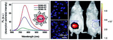
Near-infrared emitting semiconductor quantum dots (NIRQDs) are popular fluorescent probes due to better penetration depth and elimination of tissue autofluorescence. Here, we demonstrate one pot aqueous synthesis of cytocompatible, strongly luminescent, cationic Ag2S NIRQDs utilizing a mixed coating composed of branched polyethyleneimine (PEI)-25 kDa and L-cysteine (Cys) as in vitro luminescent tags and in vivo optical imaging agents. Ultrasmall sizes, a clear first excitonic peak in the absorption spectra, relatively narrow emission peaks with maxima between 730 and 775 nm and a Stokes shift less than 100 nm were obtained. Lifetime measurements indicate excitonic and defect-related emissions. Interestingly, not the emission maxima but the intensity was influenced by the Cys amount more dramatically. PEI/Cys 60/40 mol ratio provided the highest quantum yield reported until now for Ag2S NIRQD (157%) emitting at such a short wavelength. Low molecular weight PEI failed to produce luminescent QDs. Cytotoxicity evaluation of the most strongly luminescing NIRQDs, revealed the PEI/Cys (mol mol−1) 50/50 composition as the non-toxic composition below 2.4 μg Ag per mL concentration. Others had low-toxicity. In vitro microscopy experiments showed endosomal distribution of NIRQDs in Hela cells and strong NIR signal. In vivo imaging study demonstrated that Ag2S NIRQDs could effectively be used as strong optical imaging agents.


Introduction

Quantum dots emitting in the near-infrared (NIR) region (NIR QDs) have been of great interest in recent years due to their great potential in bioimaging and biolabeling.1 The NIR region, especially between 700 and 900 nm, promises higher penetration depth and higher signal-to-noise ratio in comparison with the optical probes emitting in the visible region.2,3 Oxyhemoglobin, deoxyhemoglobin and lipids absorb and scatter the incoming light in the visible region (400–700 nm). Additionally, tissue components like collagen have a significant autofluorescence in this region. However, absorption coefficients of body fluids are at their minimum in the 700–900 nm. Therefore, living tissues have low autofluorescence and the photons propagate deeper into the tissue in this region.1 Hence, the 700–900 nm window is regarded as ideal for in vivo imaging.4

The most studied NIRQDs are PbS,5 CdHgTe,6 CdSeTe7 and PbSe8 but high toxicity of Pb, Cd and Hg pose a serious toxicity problem limiting their medical use.9 Thus, it is an urgent necessity to develop biocompatible and water-soluble NIRQDs for bioapplications.

Ag2S quantum dots are the most popular NIRQDs within the last 5 years due to their emission in near-infrared (NIR) window and high cytocompatibility.10–12 Unlike highly toxic Ag+ ions, Ag2S is quite non-toxic due to ultralow solubility (Ksp = 6.3 × 10−50).13 However, majority of Ag2S quantum dots in the literature have low quantum efficiency.11–16 For example, Jiang et al. demonstrated emission tunable (from 510 nm to 1221 nm) synthesis of Ag2S QDs by the decomposition of 3-mercaptopropionic acid but the quantum yield (QY) was only 2.1% (using indocyanine green (ICG) as a reference standard (QY = 13% in DMSO)).10 Zhao et al. reported a facile phase transfer based synthesis of Ag2S QDs with 2.5% of quantum yield.11 Hocaoglu et al. reported the first Ag2S QDs with relatively high QY which was about 5.5% and increased up to 39% in time.12 The synthetic approach was a very simple, single step aqueous method, providing 2-mercaptopropionic acid (2MPA) coated Ag2S QDs. They also reported emission tunable synthesis of Ag2S QDs via the decomposition of meso-2,3-dimercaptosuccinic acid with QY around 6.5%.14 All of these Ag2S QDs are anionic in nature.

In an effort to combine imaging and therapy, cationic QDs that can effectively condense and carry genes, and allow tracking the destiny of the transfection vehicle and the outcome of the therapy, are desired. Polyethyleneimine (PEI) (25 kDa, branched) is the most efficient and widely used cationic polymer in gene transfection, but has significant toxicity.17,18 We have recently reported the first cationic Ag2S NIRQDs with PEI (25 kDa, branched) and 2MPA mixed coating with the highest reported QY in the literature for Ag2S QDs, strong intracellular optical signal and efficient transfection of green fluorescence protein (GFP).19 PEI in the coating provides gene binding and delivery potential and 2MPA provides the strong luminescence in such a composition. As the 2MPA amount increased, the luminescence intensity increased. Without 2MPA, PEI coated Ag2S did not show any luminescence, most probably due to crystal surface defects. But, the Ag2S NIRQDs with the mixed coating had stronger emission than Ag2S coated only with 2MPA, and improved cytocompatibility compared to PEI, indicating a synergy. Yet, a broad range of coating compositions could not be studied due to limitations in stability above 40 mol% 2MPA.

Ag2S QDs are promising agents for in vivo imaging and therapy. Wang et al. provided a detailed in vivo biodistribution and pharmacokinetic study with PEGylated dihydrolipoic acid coated Ag2S emitting around 1200 nm. No significant toxicity (15 and 30 mg kg−1) in two months, accumulation mostly in the reticuloendothelial system (RES) and fecal excretion was reported as well as a strong in vivo optical signal.20,21 These particles were later shown as effective optical agents in screening lymphatic drainage, imaging vascular networks, tracking tumor angiogenesis, and implanted stem cells when tagged with Tat-peptide, in vivo.22,23 Targeted in vivo cancer imaging were also demonstrated with antiVEGF conjugated BSA-stabilized Ag2S QDs.24 Folic acid tagged PEGylated Ag2S was reported for targeted delivery of doxorubicin in vitro with strong endosomal NIR optical signal.25

Here, we developed cationic Ag2S NIRQDs with a mixed coating comprised of branched PEI of different molecular weights and cysteine (Cys) with the aim of understanding the effect of Cys as a part of the coating composition and investigated the influence of PEI/Cys ratio on emission tunability, stability, quantum yield and cytotoxicity. Cys is an aminoacid without any cytotoxin and is used for the synthesis of water-soluble nanocrystals such as CdS,26 CdSe,27 CdSe/CdS,28 CdTe,29 and CdTe/Cd(OH)2.30 The mercapto group of Cys provides a strong binding to the crystal surface. The polar side group (amine) and carboxylic acid provides solubility in water. Influence of this side chain amine on particle properties in comparison to 2MPA is one of the motivations behind this study. Since the toxicity of PEI decreases with the decreasing molecular weight, we have investigated the possibility of forming a highly biocompatible, strongly luminescent cationic transfection agents with lower molecular weight branched PEI, specifically 10 and 1.8 kDa, as well. Initially, the effect of the PEI/Cys, then coating/Ag, Ag/S ratios and reaction temperature on particle properties including stability, luminescence intensity and emission wavelength were studied. Cytotoxicity of three different particles with similar emission wavelengths but with different PEI/Cys ratio in the coating was determined via MTT assay to evaluate the impact of coating composition on the cytotoxicity of the QDs. In vitro and in vivo optical imaging of these QDs was performed to demonstrate the potential of these cationic Ag2S QDs as optical probes in biomedical applications.

Materials and methods

Materials

Silver nitrate (AgNO3) and Cys were obtained from Sigma-Aldrich (USA). Thioacetamide (TAA), sodium hydroxide (NaOH), ethanol and acetic acid (CH3COOH) were purchased from Merck. Branched polyethylenimine (PEI) (Mw 25 kDa) was provided by Aldrich (Germany). 1.8 kDa and 10 kDa PEI were obtained from Polysciences, Inc. (Germany). LDS 798 Near-IR laser dye was provided from Exciton Inc. Ultra-pure water (MilliQ, Millipore, 18.2 MΩ cm) was used for the preparation of all aqueous solutions.

Roswell Park Memorial Institute (RPMI) 1640 medium (with L-glutamine and 25 mM HEPES) was obtained from Multicell, Wisent Inc. (Canada). Fetal bovine serum was purchased from Capricorn Scientific GmbH (Germany). Trypsin–EDTA and penicillin–streptomycin solutions were provided from Wisent Inc. (Canada). Thiazolyl blue tetrazolium bromide (MTT) and phosphate buffered saline (PBS) tablets were obtained from Biomatik Corp. (Canada). Paraformaldehyde solution 4% in PBS was purchased from Santa Cruz Biotechnology, Inc. (USA). 6-well and 96-well plates were purchased from Nest Biotechnology Co. Ltd. (China). Dimethyl sulfoxide Hybri-Max™ and 4′,6-diamidino-2-phenylindole (DAPI) were provided by Sigma (USA). HeLa (human epithelial carcinoma cell line) cells were given as a gift from the Gozuacik Lab (Sabanci University, Istanbul, Turkey) for this study. Mice were purchased from Koç University Animal Research Facility (Istanbul, Turkey). All chemicals were analytical grade and used without any purification.

Preparation of PEI and Cys coated Ag2S QDs

For a typical synthetic procedure, desired amounts of PEI and Cys were dissolved in the 75 mL of deoxygenated water, under argon flow, then 42.5 mg of AgNO3 (0.25 mmol) was added to this solution (Fig. 1). The pH of the solution was adjusted to 10.5 using NaOH (2 M) to have a clear solution. Thioacetamide (TAA) in an amount appropriate for the desired Ag/S ratio (e.g. 4.7 mg, 0.0625 mmol for Ag/S ratio = 4/1) was dissolved in 25 mL of deoxygenated water, separately and added to the initial solution under vigorous mechanical stirring (5000 rpm). The particle growth was followed by absorption and emission spectrum of the aliquots taken from the reaction at different time points.
image file: c6ra13804g-f1.tif
Fig. 1 Aqueous synthesis of PEI/Cys Ag2S NIRQDs.

The impact of reaction variables such as PEI/Cys, coating (PEI and Cys)/Ag, Ag/S, reaction temperature and reaction duration on the optical properties and stability of the Ag2S QDs was studied. Table 1 lists all PEI/Cys ratios used for the synthesis of Ag2S QDs at fixed Ag/S (=4), coating (PEI/Cys)/Ag (=5), reaction pH (=10.5) at 70 °C in 1 h reaction. Here, moles of coating is equal to the total number of moles of 1° and 2° amines coming from PEI and thiol coming from Cys. This is based on the stronger affinity of the thiol to Ag2S than the amine in Cys and the assumption that primary and secondary amines of PEI would be more effective in binding to Ag2S surface. Hence, the PEI/Cys mol ratio is equal to the total number of moles of primary and secondary amines of the branched PEI (25 kDa)/moles of thiol coming from Cys. So, when 100% PEI was used as a coating at the coating/Ag ratio of 5, the amount of PEI used brought in total of 1.25 mmol primary and secondary amines in total. In a mixed coating, such as 60% PEI and 40% Cys, amounts of each material was adjusted to provide 0.75 mmol amine (primary and secondary) coming only from PEI and 0.50 mmol thiol coming from Cys. To study the effect of coating/Ag ratio independently, other parameters were fixed at Ag/S = 4, pH = 10.5 at 70 °C for 1 h reactions. Studied ratios were listed in Table S1. The effect of the Ag/S ratio on the properties of Ag2S QDs were studied at fixed Ag+ concentration (0.25 mmol) by changing the number of moles of thioacetamide between 0.025 mmol (for Ag/S, 10/1) and 0.50 mmol (for Ag/S, 1/2) keeping all other parameters identical as summarized in Table S2. In order to study the impact of reaction temperature, QD60 synthesis was repeated at different reaction temperatures as listed in Table 2, keeping all other parameters unchanged. QD50 and QD80 was also synthesized at room temperature. The obtained brown quantum dot solutions were washed using Amicon-Ultra centrifugal filters (30[thin space (1/6-em)]000 Da cut off) with deionized water and kept in the dark at 4 °C.

Table 1 Effect of the PEI/Cys ratio on the properties of Ag2S QDsd
Rxn code PEI/Cys (mol%) λem(max) (nm) Sizea (nm) Band gap (eV) λabs(cutoff)b (nm) FWHM (nm) Dhc (nm) Zeta pot. (mV)
a Calculated by Brus equation.b Absorbance onset determined from the absorbance spectrum (Fig. S1a).c Hydrodynamic diameter measured by DLS and reported as the number average.d Reaction conditions: Ag[thin space (1/6-em)]:[thin space (1/6-em)]S = 4, coating (PEI/Cys)[thin space (1/6-em)]:[thin space (1/6-em)]Ag = 5, reaction pH = 10.5, T = 70 °C, 1 h reaction.
QD100-70 100/0 3.12 1.30 955 26.5 ± 4.1 41.2 ± 1.2
QD90-70 90/10 2.70 1.48 837 9.2 ± 0.8 41.2 ± 2.0
QD80-70 80/20 731 2.52 1.60 779 106 10.6 ± 1.2 45.8 ± 1.7
QD70-70 70/30 725 2.50 1.62 768 101 11.1 ± 1.3 41.9 ± 0.7
QD60-70 60/40 775 2.64 1.52 816 105 49.3 ± 14.1 63.6 ± 4.9
QD50-70 50/50 730 2.74 1.47 846 91 10.4 ± 2.7 28.4 ± 1.1
QD40-70 40/60 734 2.73 1.47 844 98 72.6 ± 11.1 44.7 ± 2.2
QD20-70 20/80 740 2.74 1.47 846 93 38.8 ± 6.7 47.3 ± 1.2
QD0-70 0/100 3.18 1.28 968 69.8 ± 16.7 −31.5 ± 0.8


Table 2 Effect of reaction temperature on the properties of PEI/Cys coated Ag2S QDse
Rxn code T (°C) Reaction time λem(max) (nm) Sizea (nm) Band gap (eV) λabs(cutoff)b (nm) FWHM (nm) Dhc (nm) Zeta pot. (mV) QY (%)
a Calculated by Brus equation.b Absorbance onset determined from the absorbance spectrum (Fig. S1d).c Hydrodynamic diameter measured by DLS and reported as the number average.d Not calculated.e Reaction conditions: Ag[thin space (1/6-em)]:[thin space (1/6-em)]S = 4, coating[thin space (1/6-em)]:[thin space (1/6-em)]Ag = 5, PEI[thin space (1/6-em)]:[thin space (1/6-em)]Cys = 60[thin space (1/6-em)]:[thin space (1/6-em)]40, reaction pH = 10.5.
QD60-RT 25 3 h 763 2.56 1.57 790 104 41.7 ± 3.0 68.6 ± 2.0 157
QD60-50 50 2 h 769 2.58 1.56 795 102 21.8 ± 8.6 38.6 ± 2.4 136
QD60-70 70 1 h 775 2.64 1.52 816 105 49.3 ± 14.1 63.6 ± 4.9 NCd
QD60-90 90 30 min 773 2.73 1.47 845 116 40.6 ± 3.8 40.6 ± 1.2 NCd


Characterization methods

Absorbance spectra of the quantum dots were measured by a Shimadzu 3101 PC UV-Vis-NIR spectrometer in the 300–1100 nm range. Brus equation was used for the calculation of crystal sizes of the QDs from the absorption edge determined from absorption spectra of the particles (eqn (1)).
 
image file: c6ra13804g-t1.tif(1)
R symbolizes the radius of the nanocrystal, me (0.286m0) and mh (1.096) show the respective effective electron and hole masses, εAg2S (5.95) is the dielectric constant for bulk Ag2S and ΔE is the band gap energy difference between the nanocrystal and the bulk semiconductor.

Photoluminescence measurements in near-infrared region were performed with a homemade set up composed of a 1/8 Newport Cornerstone 130 monochromator equipped with a 600 L mm−1 grating that functions in the 400–1100 nm range, a femtowatt sensitive Si detector (Thorlabs PDF10A, 1.4 × 10⁻¹⁵ W Hz−1/2) and a frequency doubled output of a DPSS laser (532 nm). The slit width was adjusted to 0.2 mm and a 590 nm long pass filter was used in data acquisition.

Quantum yield (QY) of the QDs was calculated by comparing the integrated emission of QD solution (in water) with that of a NIR dye (LDS 798 in methanol solution, quantum yield reported as 14% by the producer). Four different concentrations of QD and the dye solution were prepared with similar absorption (below 0.1) at the excitation wavelength and integrated areas of the emission curves were plotted against the absorbance (Fig. S2). The ratio of the slope of these plots with the refractive index of water and MeOH based on eqn (2) gave the QY of the prepared QDs.31,32

 
image file: c6ra13804g-t2.tif(2)

The microstructure of QDs (QD60-RT) was examined with a transmission electron microscope (TEM, Philips-FEI-Tecnai G2 F30) operating at an accelerating voltage of 200 kV. The hydrodynamic size and zeta potential of the QDs were conducted in water using a Malvern Zetasizer Nano-ZS.

Lifetime of Ag2S QDs (QD60-RT) in chloroform was measured using Horiba Fluorolog equipped with TCSPC Triple Illuminator. 456 nm Horiba Nanoled pulsed diode light source (pulse duration 1.2 ns) was used.

Ag amount in QD solutions was quantified by Agilent 7700x Inductively Coupled Plasma Mass Spectrometry (ICP-MS) after digestion of QDs with suprapur nitric acid 65% and suprapure sulphuric acid 96%. Amount of the organic content of QDs was measured on a DTA/TG (SII EXSTAR 6300) under argon gas flow from room temperature to 600 °C at the heating rate of 10 °C min−1. Organic elemental analyses of QDs were conducted with a CHNS/O Analyzer (Thermo Scientific Flash 2000 Organic Elemental Analyzer) to determine the S and N content of the coating. In each test 2–3 mg dried sample was loaded in to a tin capsule with the addition of vanadium pentoxide which is a typical “oxygen donor” that allows the total conversion of sulphur. Then, the capsules were placed on the combustion reactor via the Thermo Scientific MAS 200R autosampler. The standard 2,5-bis-(5-tert-butyl-2-benzoxazol-2-yl)thiophene (BBOT) was used to draw calibration curve.

FTIR spectrum was recorded on a Thermoscientific Nicolet iS10 instrument (ATR-FTIR) in the wavenumber region from 650 to 4000 cm−1 with resolution 1 cm−1. For XPS measurements Thermo Scientific K-Alpha XPS with Al K-alpha monochromatic radiation (1486.3 eV) was used. Dried samples (QD60-RT) were placed on an adhesive carbon tape and exposed to 400 μm X-ray spot size with 50.0 eV pass energy corresponding to a resolution of roughly 0.5 eV. The experimental pressure and the base pressure were kept below about 1 × 10⁻⁷ mbar and 9 × 10⁻⁹ mbar, respectively. All spectra were corrected with respect to C 1s peak at 284.5 eV.

Cell culture

All culture media were supplemented with 10% fetal bovine serum and 1% penicillin–streptomycin. HeLa cells were cultured in RPMI 1640 complete medium, supplemented with 10% FBS and 1% penicillin–streptomycin antibiotic solution, in a 5% CO2-humidified incubator at 37 °C.

Cytotoxicity assay

Cytotoxicity of the bPEI (25 kDa)/Cys was evaluated using the colorimetric MTT metabolic activity assay. HeLa cells growing in log phase were seeded into a 96-well plate at a density of 7500 cells per well and cultured at 37 °C in an atmosphere of 5% CO2. After overnight incubation, the medium was replaced with a fresh medium containing Ag2S quantum dots in concentrations of 0.8–10 μg mL−1 Ag as determined by the ICP analysis of the particles. Cells with no treatment were used as controls. After 24 h incubation, the medium was discarded and cells were washed with 1 × PBS, pH 7.4. 50 μL of MTT reagent (5 mg mL−1) and 150 μL medium were added to each well. Purple formazan crystals formed after 4 hours in each well were dissolved with ethanol[thin space (1/6-em)]:[thin space (1/6-em)]DMSO (1[thin space (1/6-em)]:[thin space (1/6-em)]1 v/v) solution shaking gently for 15 min. A microplate reader (BioTek ELx800 Absorbance Microplate Reader) was used to record the absorbance intensity at 600 nm with a reference wavelength of 630 nm. Untreated cells were taken as positive controls under the assumption of 100% viability and treated cells without MTT reagent were used for the correction of absorbance intensities (n = 8). The relative cell viability was calculated as [Asample/Acontrol] × 100.

In vitro cell imaging

The in vitro cell imaging of Ag2S QDs was performed on an Inverted Life Science Microscope (Olympus-Xcellence RT Life Science Microscopy). HeLa cells were grown on a 6-well plate at a density of 1.5 × 10⁵ cell per well in complete RPMI 1640 medium, incubated at 37 °C under 5% CO2 for 12 hours. Ag2S QDs at 10 μg mL−1 Ag dose were introduced to the cells and after 1 hour incubation, the cells were washed with 1 × PBS three times to remove QDs in the medium. Then, the cells were fixed using paraformaldehyde solution (4% in PBS) for 20 min, and after being washed three times, the nuclei were stained with a fluorescent dye, DAPI. The washing step was repeated for three times and 2 mL PBS was left in each well to protect the cells against drying. The fixed cells were visualized by inverted Life Science Microscope using specific filters for nuclei and NIR emitting QDs:DAPI region (λexc: 352–402 nm and λem: 417–477 nm) and NIR region (λexc: 550 nm and λem: 650 nm long pass), respectively. Same experimental process was performed with control cells. Images were processed and merged using ImageJ analysis program.33

In vivo imaging

The in vivo imaging of Ag2S QDs in NIR region were performed using BALB/c mouse purchased from Animal Research Laboratory of Koc University. All experiments were performed according to the protocols approved by Koc University Animal Research Local Ethics Committee. 2.25 mg of PEI/Cys coated Ag2S QDs (QD60-RT) (15 mg mL−1 in PBS) were administered to the mouse (27 g) by intraperitoneal injection. 1 h after injection, mouse was anesthetized with 2.5% isofluorane in oxygen and transferred to a Perkin Elmer IVIS® Lumina III In Vivo Imaging System (excitation: 740 nm, emission filter: 840 nm long-pass). An untreated mouse weighting 25 g was also imaged to determine the background signal.

Statistical analysis

For statistical analysis, one-way ANOVA with Tukey's multiple comparison test of GraphPad Prism 6 software package from GraphPad Software, Inc., USA was used. All data expressed as mean ± standard deviation (SD), and p < 0.05 was considered statistically significant.

Results and discussion

Synthesis and optical characterization of PEI/Cys Ag2S QDs

Near IR emitting Ag2S QDs were prepared directly in water from AgNO3 and TAA using branched 25 kDa PEI and Cys as a mixed coating. The reaction variables were studied to evaluate the emission tunability and the effects on particle properties with a motivation to find the best composition and size for high cytocompatibility and strong emission. All reaction mixtures were set to pH 10.5 before the addition of TAA, in order to deprotonate the amine groups of PEI and Cys and obtain a clear solution. However, after the purification steps, all aqueous QD solutions were adjusted to pH 7.4 to prevent any pH effect on the photoluminescence of QDs, for the purpose of comparison.

The organic coating passivates the surface of the growing crystal and often is effective in controlling the crystal size. Yet, it is also responsible from colloidal stability and surface quality. The later impacts the luminescence intensity and quantum yield dramatically. PEI coated Ag2S QDs were colloidally stable but did not luminesce in near IR region (Fig. S1e). Polymeric coatings are usually inferior to small thiolated molecules since they lack dense packing on the crystal surface leaving uncoordinated sites, hence do not provide strong luminescence. On the other hand, polymers provide enhanced colloidal stability due to multidentate binding.19 However, addition of Cys along with PEI provided both strong luminescence and colloidal stability as in the case of PEI/2MPA (Fig. 2a).19 Cys binds to crystal surface strongly with the thiol and since it is a small molecule, it can diffuse to the crystal surface and passivate the uncoordinated sites left behind by PEI. To investigate the effect of coating composition, PEI/Cys ratio was varied keeping the total Ag/coating ratio constant at 1/5. Cys was effective in generating different compositions and incorporated between 10 and 80 mol%. Overall, all particles with the mixed coating had absorption onset at shorter wavelengths than PEI coated Ag2S QD, indicating smaller crystal size possibly due to the presence of strongly binding small Cys which prevented crystal growth and strong luminescence due to better surface passivation (Table 1). 10 mol% Cys in the coating formulation shifted the absorption onset to shorter wavelength indicating smaller crystals but was not sufficient enough to provide detectable luminescence. All other mixed coatings provided luminescent QDs. By changing the coating composition it was possible to tune the emission between 725 and 775 nm with a full-width at half-maximum (FWHM) of around 100 nm which is relatively narrow compared to other aqueous Ag2S QDs. With increasing Cys amount up to 30 mol%, crystal size decreased from 3.12 to 2.50 nm shifting the emission maxima to shorter wavelengths. Beyond 40 mol%, emission peaks shifted slightly to longer wavelengths again. For the comparison, Ag2S coated only with Cys was prepared under identical conditions. Interestingly, Cys–Ag2S QDs synthesized under these conditions are the largest ones with no significant luminescence (Fig. S1e). Thus, there is a clear synergy in the combination of these two coating materials which results from different growth kinetics and different binding types. From a broader perspective, all Ag2S QDs had emission maxima between 725 and 740 nm. QD60-70 with emission max at 775 nm showed more than four times stronger intensity than the others. Interestingly, excluding the 60/40 ratio, in general the luminescence intensity decreased with the increasing amount of Cys which is opposite to what was seen with PEI/2MPA coating.19 Cys coated Ag2S has a featureless absorption profile, whereas all QDs with mixed coatings have a clear first excitonic peak (Fig. S1) and significant luminescence with Stoke's shift which is about 100 nm or less.


image file: c6ra13804g-f2.tif
Fig. 2 Photoluminescence spectra of PEI/Cys coated Ag2S QDs synthesized at (a) different PEI/Cys ratios, (b) Ag/coating (PEI + Cys), (c) Ag/S mole ratios and (d) at different temperatures.

All Ag2S QDs with PEI in the coating had positive zeta potential, between +28 and 63 meV (Table 1), which is good for binding negatively charged species such as drugs and oligonucleotides and for colloidal stability due to charge repulsion.34,35

Hydrodynamic sizes of these Ag2S QDs are all below 100 nm and mostly below 50 nm which is very valuable for long blood circulation time and molecular targeting in the in vivo use of such particles. Several studies have reported that optimal size range should be in between 10 and 100 nm for in vivo applications of a particle.36 Larger particles (>200 nm) accumulate in the spleen and are processed by the phagocytic cells or by the macrophages in blood,37 while smaller particles (<10 nm) undergo a rapid renal clearance.38

Ag2S QDs prepared with 60/40 PEI/Cys (QD60-70) coating formulation stood out as the most luminescent particle with a strongly cationic nature (+63.6 meV) and small hydrodynamic size (about 50 nm). Hence, keeping PEI/Cys ratio constant at 60/40, the effect of coating amount on particle properties was studied. Total coating/Ag ratio which was initially 5 was changed to 2.5 and 10 (Table S1). Absorption spectra of these three particles indicate (Fig. S1b) decreasing band gap with the increasing coating amount and hence, the inorganic core size increases. However, photoluminescence spectra of the QDs with coating/Ag ratio of 2.5 and 10 showed two peak maxima: one in 700–800 nm region and another one around 900 nm which is possibly defect-related emission (Fig. 2b).39 But, the QD prepared with coating/Ag ratio of 5, has relatively sharper peak centred at 775 nm with dramatically better intensity.

In terms of the hydrodynamic size and zeta potential, all of these three Ag2S NIRQDs are colloidally stable and in appropriate size range for in vivo applications (Table S1).

To tune the crystal size, and therefore emission wavelength of Ag2S QDs further, different Ag/S ratios between 10/1 and 1/4 were studied using PEI/Cys ratio of 60/40 (QD60-70), at coating/Ag ratio of 5. As shown in Fig. 1c and Table S2, decreasing Ag/S ratio causes a red shift in emission maxima from 716 (10/1 Ag/S) to 775 nm (4/1 Ag/S) with enhanced luminescence intensity. At the lower ratios, no detectable luminescence was observed.

Reaction temperature is usually a significant factor affecting the growth kinetics and hence the particle size. Particles were tried to be grown to a similar size at different temperatures (keeping the formulation constant). It is clearly seen in Table 2 that as the temperature increased from 25 to 90 °C, crystals grew to relatively similar sizes at shorter times but in the expense of emission intensity (Fig. 1d). The quantum yield of QD60-RT, for instance, is 157% (with respect to LDS-798 NIR dye) (Fig. S2) which is the highest value reported for Ag2S QD until now. Thioacetamide (TAA) is a slow sulphur releasing agent. It decomposes faster at high temperatures into H2S and acetamide, hence causes faster particle growth. This may produce larger number of defects responsible from non-radiative relaxation processes of the excited photons causing the observed reduction in the emission intensity. At low temperatures, nucleation and growth of the crystals may be slow but are more defect free.40 Emission peak of the QD prepared at 90 °C is also much broader, may be indicating a broader size distribution and defect related emissions, as well.41

Based on the dramatic increase in the luminescence intensity of QD60 at RT, QD80-70 and QD50-70 were also prepared at RT. Low reaction temperature improved the emission intensity of QD50 as well, but the opposite was seen with QD80. This may be due to slower diffusion of polymeric component at lower temperature, which is critical for growth and adsorption to crystal surface. This may cause more defects on crystal surface hence decrease the amount of radiative coupling. In the final case, three particles (QD80-70, QD60-RT, QD50-RT) with different composition but with strong luminescence at similar wavelengths (Fig. 3) were subjected to in vitro studies to evaluate the effect of composition on particle toxicity. Their properties are summarized in Tables 1, 2 and S3.


image file: c6ra13804g-f3.tif
Fig. 3 Photoluminescence spectra of the best luminescing PEI/Cys coated Ag2S QDs.

In addition, the reactions were repeated with 1.8 kDa and 10 kDa branched PEI, again in combination with Cys, to investigate the molecular weight effect of the polymer on the QD properties. However, no colloidally stable and luminescent particles were obtained.

Characterization of PEI/Cys coated Ag2S QDs

Photoluminescent lifetime measurements were performed on QD60-RT, which showed the strongest luminescence. The decay curve found to be multiexponential with an average lifetime calculated as 75.78 ns with 1.096 value of chi-square (Fig. 4 and Table S4). This is somewhat similar to literature values.2,3 Actually, the fit of the decay curve indicates four different events with 1.55 (1.60%), 11.66 (17.54%), 42.17 (45.69%) and 154.79 (35.17%) ns lifetimes. Usually, 1 ns and faster decays may be attributed to non-radiative decay.42 For a 4.5 nm Ag2S (3–6.5 nm) with emission at 850 nm, biexponential decay with 1 and 11 ns was reported and the later was attributed to the excitonic recombination.42 In another study, 1.5 nm Ag2S with emission maxima at 813 nm was reported to have an average lifetime of 57 ns, which was attributed to the radiative decay related to surface inhomogeneities.3 Zhang et al. reported increasing lifetimes for Ag2S–dodecanethiol with increasing crystal size.2 Actually, they have mostly focused on the long lifetime component increasing from 57 to 181 ns as the crystal size increased from 2.4 to 7.0 nm, may be because that was the major component in each case, but they have seen a slow component which was between 11 and 14 ns for 2.4–3.7 nm and 26–31 ns for 3.6–7.0 nm size Ag2S. Looking at all of these data, we may suggest that the relatively fast component (around 11 ns) may be due to radiative excitonic coupling in each of these cases. Slower components, which is around 42 ns, may be due to the lattice defects which creates mid-gap absorption,42 and the slowest component may be related to surface defects. Of course, considering the values reported for different sized particles2 and the size distribution of these particles (Fig. 5), such multicomponent decay may be at least partially due to the size distribution. Detailed studies are underway.
image file: c6ra13804g-f4.tif
Fig. 4 Time resolved photoluminescence decay curve of QD60-RT. Black dots are scattered and red line is fitted data.

image file: c6ra13804g-f5.tif
Fig. 5 XPS spectra of Ag2S (QD60-RT) QDs. (a) Ag 3d, (b) S 2p and (c) N 1s.

XPS analysis confirmed the suggested Ag2S structure as well (Fig. 5). Ag 3d peaks at the binding energies (BE) of 366.45 (3d5/2) and 372.44 (3d3/2) eV fit to Ag–S of Ag2S (Fig. 5a). Typically S 2p comes out as a doublet, and there are two different S in these QDs (Fig. 5b). The doublet at BE of 159.98 eV and 161.18 eV fits to the 2p3/2 and 2p1/2 of S–Ag, respectively. The other doublet at 166.84 eV and 168.04 eV belongs to the S of Cys. Two peaks of N 1s at BE of 397.73 and 399.29 eV originates from the secondary and primary nitrogens of PEI (Fig. 5c). The tertiary nitrogen peak is hindered by the peak of secondary N.

TEM images of Ag2S QDs revealed well dispersed spherical structures with broad size distribution (1–7 nm) (Fig. 6). Focused images (Fig. 6c) clearly showed crystalline structure with 0.220 nm interplanar distance, which fits well to the reported values for the [031] plane of monoclinic Ag2S.43,44


image file: c6ra13804g-f6.tif
Fig. 6 TEM analyses of PEI/Cys coated Ag2S QDs (QD60-RT). (a) and (b) TEM images of nanoparticles at different magnifications, (c) interplanar spacing and corresponding plane and (d) size distribution of nanoparticles.

The IR spectrum of PEI/Cys coated Ag2S QDs (QD60-RT) confirms the coexistence of Cys and PEI in the particle coating (Fig. 7). Strong bands at 1551 and 1386 cm−1 are typical for asymmetric and symmetric carboxylate stretching modes of Cys and the bands at 3276 cm−1 and 2940–2827 cm−1 point out the presence of N–H and C–H stretching modes of PEI, respectively. In addition, the disappearance of S–H stretching band which is around 2560 cm−1 confirms the binding of Cys to Ag2S core from the thiol.


image file: c6ra13804g-f7.tif
Fig. 7 IR spectrum of PEI/L-cysteine coated Ag2S QDs (QD60-RT).

The organic content of the best emitting QDs which is QD60-RT was determined as 55.6% by TGA (Fig. S3).

Evaluation of in vitro cytotoxicity of Ag2S QDs

The cytotoxicity of Ag2S QDs on HeLa cervical carcinoma cell line was determined with MTT assay. Three strongly luminescing QDs with different ratio of PEI/Cys (QD80-70, QD60-RT, QD50-RT: the first number indicates the PEI mol% in feed and the second indicates the synthesis temperature) with emission maxima around 750 nm and the one coated only with PEI (QD100-70) were studied. The emission wavelength and the size were tried to be kept similar in order to study mainly the effect of coating composition. Fig. 8 shows the viability of HeLa cells after 24 h incubation with these Ag2S QDs. Incubation dose was based on the Ag concentration determined with ICP-MS (Table S5) and was studied in 0.8–10 μg mL−1 range. According to ISO 10993-5, cell viability above 80% is accepted as non-cytotoxic, between 80 and 60% as weakly, 60–40% as moderately and below 40% as strongly cytotoxic.45,46 As seen in Fig. 8 and S4, the most toxic QD (viability at and below 50%) at all concentrations is the one coated only with PEI. However, addition of Cys to the coating improved cytocompatibility, dramatically. All Ag2S with the mixed coating compositions are weakly toxic below 2.4 μg mL−1. Ag2S QDs with 50/50 PEI/Cys (QD50-RT) emerged as the most cytocompatible formulation with no significant cytotoxicity upto 3.2 μg mL−1 and become strongly cytotoxic above 5.6 μg mL−1 Ag dose (Fig. S4). Ag2S QD with 80/20 PEI/Cys (QD80-70) is weakly toxic and the major difference between QD80-70 and QD50-RT starts at 3.2 mg Ag per mL dose. The high cytocompatibility of QD50 may be related to low zeta potential, low PEI content (Tables S3 and S5) and small hydrodynamic size. QD80-70 and QD50-RT have the highest and the lowest PEI content in the final particle as determined by the elemental analysis (Table S5) of the four particles studied for toxicity evaluation. Surface charge increased with the PEI content, rendering particles slightly more toxic even at similar particle sizes. Surprisingly, Ag2S QDs with 60/40 PEI/Cys (QD60-RT) which is the best luminescing particle, is the most toxic among the three mixed coating composition studied here. It may be due to high surface positive charge compared to the other two. It was weakly toxic at and below 2.4 μg Ag per mL dose and significantly less cytocompatible than the other two QDs with mixed coatings at all concentrations. All were toxic at and above 5.6 μg Ag per mL dose.
image file: c6ra13804g-f8.tif
Fig. 8 Viability of HeLa cells treated with PEI/Cys coated Ag2S QDs after 24 hours incubation measured by MTT assay compared to untreated HeLa cells. The data are expressed as mean ± S.D. (n = 8).

In vitro and in vivo optical imaging

HeLa cells were incubated with the Ag2S QDs (QD60-RT) to evaluate their potential as NIR imaging agents. Microscopy images of the HeLa cells treated with PEI/Cys Ag2S QDs revealed high uptake and endosomal localization of QDs in the cells (Fig. 9). Such a strong signal from the QD with almost no autofluorescence is very critical for biomedical applications. This was further shown with the strong optical signal detected from a BALB/c mouse which was subjected to an intraperitoneal injection of QD60-RT (Fig. 10). With QDs emitting in the visible region autofluorescence of the tissue is a very serious handicap and most of the time animals should be subjected to a special diet to reduce the autofluorescence. Provided in vitro and in vivo images indicate a strong potential for these QDs as an in vivo imaging agents. Considering that the peak maximum of this QD at 775 nm and the longpass filter used which allows signals above 840 nm reach to detector, indicates that such a strong luminescence signal is detected with only a small portion of the possible emission that can be obtained by these QDs.
image file: c6ra13804g-f9.tif
Fig. 9 Cellular localization of PEI/Cys coated Ag2S NIRQDs (QD60-RT) by HeLa cells. DIC (a), DAPI and NIR (b), and merged (c) images of control cells and DIC (d), DAPI and NIR (e), and merged (f) images of QD treated cells. The scale bar shows 100 μm.

image file: c6ra13804g-f10.tif
Fig. 10 In vivo fluorescence image of a BALB/c mice (left image) 1 h after intraperitoneal injection of the QD60-RT and (right image) the control mice with no QD injection (excitation at 740 nm. Emission filter: 840 nm long-pass).

Conclusions

Cationic NIR emitting Ag2S quantum dots were prepared successfully with PEI/Cys mixed coating approach, using 25 kDa branched PEI. It was shown here that lower molecular weight PEI even with the mixed coating approach does not provide luminescent QDs, at least under the studied conditions. On the contrary to the Ag2S NIRQDs coated with PEI/2MPA19 where the highest 2MPA amount was 40 mol% to ensure stability, a broad range of PEI/Cys compositions could be studied here. This provided an opportunity to produce smaller particles with emission maxima below 800 nm and to tune the emission in a slightly broader range with a narrower particle size distribution and FWHM. This is desirable with the current imaging instruments which usually lose sensitivity dramatically above 800 nm. Additional amine units contributed by Cys provided a stronger positive charge on the particle surface, as well. Increasing amount of Cys, reduced the luminescence intensity without affecting the emission maxima dramatically. Interestingly, PEI/Cys ratio of 60/40 (QD60-RT) produced the particle with the dramatically increased emission intensity (QY 157%) and at a significantly longer wavelength (775 nm) than others (725–740 nm). To the best of our knowledge, this unique combination of the coating molecules provided the first examples of Ag2S NIR QDs with the emission at the low wavelength range of NIR (below 800 nm) with an exceptionally high QY.

The reaction conditions like PEI/Cys, Ag/S, Ag/coating ratios and temperature play key roles in obtaining high PL emission and stability of Ag2S quantum dots. All compositions were colloidally stable. Other than the QD60 case, Ag/S ratio was the only effective parameter to tune the emission wavelength further. Temperature of the synthesis showed its influence mostly on the emission intensity. Particle growth and stabilization was influenced by the composition and hence for each composition the strong emission was obtained at different temperatures. Higher polymer content required higher reaction temperature for better passifization and stronger luminescence.

Source of the luminescence was explored with time resolved luminescence. Multiexponential decay curves indicated four different life times between 1.55 and 154.79 ns which gives an average lifetime of 75.78 ns. These values indicate non-radiative, excitonic, lattice and surface defect related decays. Effect of size distribution may be also considered for such variety in luminescence lifetimes.

The cytotoxicity analysis indicated dramatically better cytocompatibility of QDs with the mixed coating over pure PEI coating, in agreement with our previous findings.19 At and below 2.4 μg mL−1 Ag dose these QDs did not show much dose dependent toxicity. Yet, there is clear difference between the toxicity of QDs with about similar emission wavelengths but different coating compositions, favouring 50, 20, 60 mol% Cys in the coating composition in the decreasing order. It seems like size and surface charge has an important impact on this outcome.

Strong NIR signals obtained from the endosomes of HeLa cells and from the mice injected with QDs intraperitoneally, signify a great and effective contrast enhancement and hence potential as optical imaging agents. Strong cationic nature of these particles can be easily exploited for delivery of drugs and genes, as well.

Overall, these new cationic Ag2S NIR QDs with such a high quantum yield and cytocompatibility are new promising theranostic nanoparticles designed specifically for gene delivery but also suitable for drug delivery.

Acknowledgements

This work was supported by Scientific and Technological Research council of Turkey (TUBITAK, Project number 113Z164).

Notes and references

  1. R. G. Aswathy, Y. Yoshida, T. Maekawa and D. S. Kumar, Anal. Bioanal. Chem., 2010, 397, 1417–1435 CrossRef CAS PubMed.
  2. Y. Zhang, Y. Liu, C. Li, X. Chen and Q. Wang, J. Phys. Chem. C, 2014, 118, 4918–4923 CAS.
  3. P. Jiang, Z.-Q. Tian, C.-N. Zhu, Z.-L. Zhang and D.-W. Pang, Chem. Mater., 2012, 24, 3–5 CrossRef CAS.
  4. R. Weissleder, Nat. Biotechnol., 2001, 19, 316–317 CrossRef CAS PubMed.
  5. L. Bakueva, I. Gorelikov, S. Musikhin, X. S. Zhao, E. H. Sargent and E. Kumacheva, Adv. Mater., 2004, 16, 926–929 CrossRef CAS.
  6. M. T. Harrison, S. V. Kershaw, M. G. Burt, A. Eychmüller, H. Weller and A. L. Rogach, Mater. Sci. Eng., B, 2000, 69–70, 355–360 CrossRef.
  7. G. X. Liang, M. M. Gu, J. R. Zhang and J. J. Zhu, Nanotechnology, 2009, 20, 415103 CrossRef PubMed.
  8. B. L. Wehrenberg, C. Wang and P. Guyot-Sionnest, J. Phys. Chem. B, 2002, 106, 10634–10640 CrossRef CAS.
  9. P. Zrazhevskiy, M. Sena and X. Gao, Chem. Soc. Rev., 2010, 39, 4326–4354 RSC.
  10. P. Jiang, C.-N. Zhu, Z.-L. Zhang, Z.-Q. Tian and D.-W. Pang, Biomaterials, 2012, 33, 5130–5135 CrossRef CAS PubMed.
  11. Y. Zhao and Z. Song, Mater. Lett., 2014, 126, 78–80 CrossRef CAS.
  12. I. Hocaoglu, M. N. Cizmeciyan, R. Erdem, C. Ozen, A. Kurt, A. Sennaroglu and H. Y. Acar, J. Mater. Chem., 2012, 22, 14674–14681 RSC.
  13. Y. Zhang, G. Hong, Y. Zhang, G. Chen, F. Li, H. Dai and Q. Wang, ACS Nano, 2012, 6, 3695–3702 CrossRef CAS PubMed.
  14. I. Hocaoglu, F. Demir, O. Birer, A. Kiraz, C. Sevrin, C. Grandfils and H. Y. Acar, Nanoscale, 2014, 6, 11921–11931 RSC.
  15. L. Tan, A. Wan and H. Li, ACS Appl. Mater. Interfaces, 2014, 6, 18–23 CAS.
  16. R. Gui, J. Sun, D. Liu, Y. Wang and H. Jin, Dalton Trans., 2014, 43, 16690–16697 RSC.
  17. J. T. K. M. Laird Forrest and D. W. Pack, Bioconjugate Chem., 2003, 14, 934–940 CrossRef PubMed.
  18. H. Lv, S. Zhang, B. Wang, S. Cui and J. Yan, J. Controlled Release, 2006, 114, 100–109 CrossRef CAS PubMed.
  19. F. D. Duman, I. Hocaoglu, D. G. Ozturk, D. Gozuacik, A. Kiraz and H. Yagci Acar, Nanoscale, 2015, 7, 11352–11362 RSC.
  20. G. Hong, J. T. Robinson, Y. Zhang, S. Diao, A. L. Antaris, Q. Wang and H. Dai, Angew. Chem., 2012, 124, 9956–9959 CrossRef.
  21. Y. Zhang, Y. Zhang, G. Hong, W. He, K. Zhou, K. Yang, F. Li, G. Chen, Z. Liu, H. Dai and Q. Wang, Biomaterials, 2013, 34, 3639–3646 CrossRef CAS PubMed.
  22. G. Chen, F. Tian, Y. Zhang, Y. Zhang, C. Li and Q. Wang, Adv. Funct. Mater., 2014, 24, 2481–2488 CrossRef CAS.
  23. C. Li, Y. Zhang, M. Wang, Y. Zhang, G. Chen, L. Li, D. Wu and Q. Wang, Biomaterials, 2014, 35, 393–400 CrossRef CAS PubMed.
  24. Y. Wang and X.-P. Yan, Chem. Commun., 2013, 49, 3324 RSC.
  25. D. Asik, M. B. Yagci, F. Demir Duman and H. Yagci Acar, J. Mater. Chem. B, 2016, 4, 1941–1950 RSC.
  26. Y. Chen and Z. Rosenzweig, Anal. Chem., 2002, 74, 5132–5138 CrossRef CAS.
  27. J. Chen, Y. Gao, Z. Xu, G. Wu, Y. Chen and C. Zhu, Anal. Chim. Acta, 2006, 577, 77–84 CrossRef CAS PubMed.
  28. F. Huang and G. Chen, Spectrochim. Acta, Part A, 2008, 70, 318–323 CrossRef PubMed.
  29. J. Su, J. Zhang, L. Liu, Y. Huang and R. P. Mason, J. Nanosci. Nanotechnol., 2008, 8, 1174–1177 CAS.
  30. Z.-X. Cai, Y.-J. Chen and X.-P. Yan, Chin. J. Chem., 2008, 26, 1848–1852 CrossRef CAS.
  31. C. Mansilla, P. Berraondo, M. Durantez, M. Martinez, N. Casares, L. Arribillaga, F. Rudilla, J. Fioravanti, T. Lozano, L. Villanueva, P. Sarobe, F. Borras, C. Leclerc, J. Prieto and J. J. Lasarte, Int. J. Cancer, 2012, 131, 641–651 CrossRef CAS PubMed.
  32. J. Laverdant, W. D. d. Marcillac, C. Barthou, V. D. Chinh, C. Schwob, L. Coolen, P. Benalloul, P. T. Nga and A. Maître, Materials, 2011, 4, 1182 CrossRef CAS.
  33. P. J. M. Michael, D. Abramoff and S. J. Ram, Biophotonics International, 2004, 11, 36–42 Search PubMed.
  34. Zetasizer Nano Series User Manual.
  35. N. Gibson, O. Shenderova, T. J. M. Luo, S. Moseenkov, V. Bondar, A. Puzyr, K. Purtov, Z. Fitzgerald and D. W. Brenner, Diamond Relat. Mater., 2009, 18, 620–626 CrossRef CAS.
  36. C. Boyer, M. R. Whittaker, V. Bulmus, J. Liu and T. P. Davis, NPG Asia Mater., 2010, 2, 23–30 CrossRef.
  37. Y. Tabata and Y. Ikada, in New Polymer Materials, Springer, Berlin Heidelberg, 1990, vol. 94, ch. 4, pp. 107–141 Search PubMed.
  38. H. Soo Choi, W. Liu, P. Misra, E. Tanaka, J. P. Zimmer, B. Itty Ipe, M. G. Bawendi and J. V. Frangioni, Nat. Biotechnol., 2007, 25, 1165–1170 CrossRef PubMed.
  39. Y. Wang, H. Yang, Z. Xia, Z. Tong and L. Zhou, Bull. Korean Chem. Soc., 2011, 32, 2316–2318 CrossRef CAS.
  40. K. Sivasankar and N. Padmavathy, Micro Nano Lett., 2011, 6, 144–146 CAS.
  41. Q. Sun, S. Fu, T. Dong, S. Liu and C. Huang, Molecules, 2012, 17, 8430–8438 CrossRef CAS PubMed.
  42. W. J. Mir, A. Swarnkar, R. Sharma, A. Katti, K. V. Adarsh and A. Nag, J. Phys. Chem. Lett., 2015, 6, 3915–3922 CrossRef CAS PubMed.
  43. O. Madelung, Semiconductors: Data Handbook, 3rd edn, 2004 Search PubMed.
  44. H. F. M. Marlene, C. Morris, E. H. Evans, B. Paretzkin, H. S. Parker and N. C. Panagiotopoulos, Standard X-ray Diffraction Powder Patterns, UNT Digital Library, Washington D.C., 1960 Search PubMed.
  45. J. Lopez-Garcia, M. Lehocky, P. Humpolicek and P. Saha, J. Funct. Biomater., 2014, 5, 43–57 CrossRef PubMed.
  46. I. O. f. Standardization, Geneva, Switzerland, 2009.

Footnote

Electronic supplementary information (ESI) available. See DOI: 10.1039/c6ra13804g

This journal is © The Royal Society of Chemistry 2016
Click here to see how this site uses Cookies. View our privacy policy here.