
Preparation of amino-functionalized magnetic nanoparticles for enhancement of bacterial capture efficiency†
Weijun Fang*ab,
Chen Hanc,
Huabing Zhanga,
Wenmei Weia,
Rui Liua and
Yuxian Shen*ab
aSchool of Basic Medical Sciences, Anhui Medical University, Hefei 230032, P. R. China. E-mail: wjfang81@163.com; shenyx@ahmu.edu.cn
bBiopharmaceutical Research Institute, Anhui Medical University, Hefei 230032, P. R. China
cInstitute of Quality Inspection of Light Industry & Chemical Products, Shanghai Institute of Quality Inspection and Technical Research, Shanghai 201114, P. R. China
First published on 4th July 2016
Abstract
Pathogenic bacteria can lead to food poisoning and serious infectious diseases, and place a large burden on human health. A method for rapid capture and removal of the harmful bacteria from the contaminated source is essential and important. In this study, two types of amine group functionalized magnetic nanoparticles were successfully fabricated for effective capture of bacterial pathogens. These magnetic nanoparticles with a positive-charge surface strongly interacted with the negative-charge bacterial cell wall via electrostatic attractions to exhibit efficient adsorptive ability of a broad spectrum of bacterial pathogens. Owing to more cationic amine groups, the bacterial capture capacity of Fe3O4@sSiO2-PEI nanoparticles (PEI-MNPs) for E. coli BL21 was three times that of Fe3O4@sSiO2-NH2 nanoparticles (NH-MNPs), making them promising for practical applications. More importantly, the PEI-MNPs also showed excellent bacterial capture efficiency at a low concentration due to the multivalency effect that appeared in our system.
1. Introduction
Bacterial pathogens can distribute in a wide range of substrates, such as water, food and medical equipment, which greatly affect human health and cause significant morbidity, mortality and healthcare expenditures.1–3 Therefore, rapid capture and decontamination of bacterial pathogens are strongly required to avoid or minimize contamination of the environment.Currently, many methods are developed for rapid capture and removal of bacteria from the contaminated source.4–8 For instance, magnetic nanoparticles functionalized with biomolecules9–25 (such as antibody, carbohydrates, aptamers, vancomycin and gentamicin) for selectively capturing bacteria have been widely reported and demonstrated promising applications for removal of bacterial pathogens. However, under actual conditions two main questions should be taken into account: (1) microorganisms are likely present at low concentrations in environmental samples, which make the bacteria hard to capture and detect by traditional methods. (2) Many contaminated samples are polymicrobial, so affinity biomolecule functionalized nanoparticles are ineffective to interact with all bacterial pathogens. Therefore, it is very urgent to develop a bacterial capture system with a broad range of bacterial capture ability under low concentration.
The aim of this study is to develop magnetic nanoparticles that can capture a broad spectrum of bacteria at low concentration. We have successfully fabricated magnetic Fe3O4@sSiO2-PEI nanoparticles (PEI-MNPs) enriched with cationic amine groups on their surface, which can strongly interact with a broad spectrum of bacteria by electrostatic interactions since many kinds of bacterial cell walls are negatively charged.26–28 As compared to Fe3O4@sSiO2-NH2 nanoparticles (NH-MNPs) with triaminopropylalkoxysilanes directly modified on the silica, the as-prepared PEI-MNPs significantly improve bacterial capture efficiency under low concentration.
2. Experimental section
2.1 Materials
Tetraethoxysilane (TEOS), N-[3-(trimethoxysilyl)propyl]diethylenetriamine (TMPDT), N-hydroxysuccinimide (NHS), 1-[3-(dimethylamino)propyl]-3-ethylcarbodiimide hydrochloride (EDC·HCl) and fluorescein isothiocyanate (FITC) were purchased from Alfa Aesar. Polyethyleneimine (PEI, branched, MW = 70![[thin space (1/6-em)]](https://www.rsc.org/images/entities/char_2009.gif) 000, 50% w/w aqueous solution) was purchased from Aladdin (Shanghai, China). Ammonium aqueous solution (∼28 wt%), ethanol, N,N′-dimethylformamide (DMF) and hydrochloric acid (∼36 wt%) were obtained from Sinopharm Chemical Reagent Co. Ltd. (Shanghai, China). HeLa cells were purchased from the cell storeroom of Chinese Academy of Science. Cell culture medium, bovine serum albumin (BSA) and penicillin–streptomycin compound were purchased from Hyclone Laboratories Inc. 3-(4,5)-Dimethylthiahiazo-2-yl-2,5-diphenyltetrazolium bromide (MTT) was purchased from Sigma. All reagents were used as received without further purification. All bacteria (E. coli BL21, E. coli JM109, B. subtilis and S. aureus) were obtained from the department of microbiology, Anhui Medical University (China).
000, 50% w/w aqueous solution) was purchased from Aladdin (Shanghai, China). Ammonium aqueous solution (∼28 wt%), ethanol, N,N′-dimethylformamide (DMF) and hydrochloric acid (∼36 wt%) were obtained from Sinopharm Chemical Reagent Co. Ltd. (Shanghai, China). HeLa cells were purchased from the cell storeroom of Chinese Academy of Science. Cell culture medium, bovine serum albumin (BSA) and penicillin–streptomycin compound were purchased from Hyclone Laboratories Inc. 3-(4,5)-Dimethylthiahiazo-2-yl-2,5-diphenyltetrazolium bromide (MTT) was purchased from Sigma. All reagents were used as received without further purification. All bacteria (E. coli BL21, E. coli JM109, B. subtilis and S. aureus) were obtained from the department of microbiology, Anhui Medical University (China).
2.2 Synthesis of Fe3O4@sSiO2 nanoparticles
The monodispersed Fe3O4 nanoparticles were firstly synthesized following the reported method. And then, 4.0 mL of ethanol solution containing 40 mg of Fe3O4 was added to a solution of 80 mL of ethanol, 40 mL of deionized water and 500 μL of TEOS. Under ultrasonication, 12 mL was added drop-wise and reacted for 1 hour. The product was then collected by a magnet, washed with water and ethanol, and dissolved in ethanol for further use.2.3 Preparation of NH-MNPs
30 mg of Fe3O4@sSiO2 nanoparticles was dispersed in 10 mL of water, followed by adding 150 μL of TMPDT and 100 μL of CH3COOH. The mixture was stirred at room temperature for 5 h. The obtained NH-MNPs were separated by a magnet and washed with ethanol and DMF, and dissolved in DMF for further use.2.4 Preparation of PEI-MNPs
20 mg of NH-MNPs was dispersed in 20 mL of dry DMF and incubated with 80 mg of succinic anhydride at 50 °C for 12 h. The carboxyl group modified magnetic nanoparticles were collected by magnetic separation and washed with dry DMF several times, and mixed with 80 mg of NHS and 120 mg of EDC·HCl. After 12 h of stirring at room temperature, the magnetic nanoparticles were collected and redispersed in 2.0 mL of dry DMF. Then, the 2.0 mL of DMF solution was dropwise added to 20 mL of PEI solution (10 mg mL−1, MW = 70000, pH = 6.0). The mixture was stirred at room temperature for 10 h. The resulting PEI-MNPs were separated and then washed with distilled water and redispersed in distilled water by ultrasonication.
2.5 Bacteria labeled with FITC
The bacterial strain E. coli BL21 and B. subtilis were used as the model bacteria. First, the bacteria were suspended in phosphate-buffered saline (pH = 7.4), and adjusted to an OD600 of 1.0 about 5 × 10⁸ CFU mL−1 (5 × 10⁸ colony forming units per mL). Then, 100 μL of dry DMF containing 50 μg of FITC was added to 10 mL of the above bacterial suspension, and stirred for 4 h on ice. The FITC-labeled bacteria were collected by centrifugation, washed four times with PBS buffer, and redispersed in PBS buffer for further use.2.6 Capability of magnetic nanoparticles for capture of bacteria
1.0 mL of bacterial suspension (approximately 2.5 × 10⁸ CFU mL−1, 0.5 OD) was incubated with different amounts of Fe3O4@sSiO2, NH-MNPs or PEI-MNPs (0.1 mg mL−1, 0.2 mg mL−1, 0.4 mg mL−1, 0.6 mg mL−1, 0.8 mg mL−1, 1.0 mg mL−1, 1.5 mg mL−1, and 2.0 mg mL−1) for 5 min. After magnetic separation by the nanoparticles, the bacterial-capture efficiency of the magnetic nanoparticles was determined by measuring the optical density (OD600) at 600 nm.2.7 Bacterial capture efficiency of magnetic nanoparticles at different pH values
Briefly, 2.0 mg of NH-MNPs or 0.6 mg of PEI-MNPs were mixed with 1.0 mL of bacterial suspension (∼2.5 × 10⁸ CFU mL−1, 0.5 OD) in PBS buffer at pH values of 3, 5, 7, 9 and 11, and incubated for 5 min. After magnetic separation by the nanoparticles, the bacterial-capture efficiency of the magnetic nanoparticles was tested by measuring the OD at 600 nm.2.8 Binding affinity of magnetic nanoparticles to bacteria at low concentrations
0.1 mg of NH-MNPs or PEI-MNPs were incubated with approximately 1.0 × 10⁶ CFU bacteria in different concentrations. In brief, 0.1 mg of nanoparticles were incubated with ∼1.0 × 10⁶ CFU bacteria (FITC-labeled E. coli BL21 or B. subtilis) in 10 μL, 100 μL, 1000 μL and 10.0 mL distilled water at room temperature. After magnetic separation by the nanoparticles, the total volume of the reactive solution was adjusted to 10 mL for testing the fluorescent intensity.2.9 Recyclability of magnetic nanoparticles for the capture of bacteria
In a typical experiment, 1.0 mg of PEI-MNPs (Fe3O4@sSiO2-PEI) or 1.0 mg of NH-MNPs (Fe3O4@sSiO2-NH2) were washed with 250 μL of aqueous ammonia solution (1.5 wt%) two times to release the captured bacteria and incubated with 250 μL of hydrochloric acid solution (0.1 M). After washing with distilled water, the magnetic nanoparticles were incubated with the bacterial suspension to investigate their bacterial-capture performance.2.10 In vitro assessment of magnetic nanoparticle cytotoxicity
HeLa cells (1 × 10⁴) were seeded in a 96-well plate for 12 h at 37 °C in 5% CO2. Then, the cells were treated with nanoparticles at a determined concentration. After incubation for another 24 h, cell viabilities were tested by the standard MTT (3-(4,5)-dimethylthiahiazo-2-yl)-2,5-diphenyltetrazolium bromide assay.3. Results and discussion
The magnetic Fe3O4 nanoparticles prepared by the hydrothermal method were directly coated with a layer of amorphous silica through the Stöber method.29 As shown in scanning electron microscopy (SEM) images (Fig. 1), the Fe3O4 nanoparticles had an average size of 230 ± 15 nm with a rough surface. After being coated with the silica shell, the overall size of the Fe3O4@sSiO2 nanoparticles increased ∼330 nm and the nanoparticles’ surface become smooth. The silica shell with a thickness of ∼50 nm (Fig. 1b, inset) was favourable for the dispersion of the magnetic core in aqueous solution and also served as a versatile group for further organic functionalization. To achieve effective capture of a wide range of bacteria, the surface of silica was functionalized with cationic amino groups which have been widely applied in bacterial separation via electrostatic interaction. In this work, two approaches were applied to achieve such surface functionalization. In one approach, triaminopropylalkoxysilanes were directly introduced on the surface of Fe3O4@sSiO2 nanoparticles by hydrolysis of TMPDT (designated as NH-MNPs). In the other approach, branched polyethyleneimine (PEI) were coated onto the silica surface by covalent bonds (designated as PEI-MNPs, see the Experimental section for details). The zeta potential of the magnetic nanoparticles after surface functionalization through these two approaches was tested. As revealed by Fig. 2, the zeta potential of Fe3O4@sSiO2, NH-MNPs and PEI-MNPs in ultra-pure water was −24.8 mV, +21.5 mV and +37.2 mV, respectively. The PEI-MNPs had a higher zeta potential than NH-MNPs, which was attributed to the greater number of amines on the nanoparticles’ surface by PEI modification. To directly compare the amount of amine groups on the two types of nanomaterials, elemental analysis was carried out and the results are presented in Table 1. As can be seen in the table, the content of nitrogen in PEI-MNPs is about 1.79% (w/w), which is higher than that of NH-MNPs (1.12%). | ||
| Fig. 1 SEM images of (a) Fe3O4 and (b) Fe3O4@sSiO2 nanoparticles and TEM image of Fe3O4@sSiO2 nanoparticles (inset). | ||
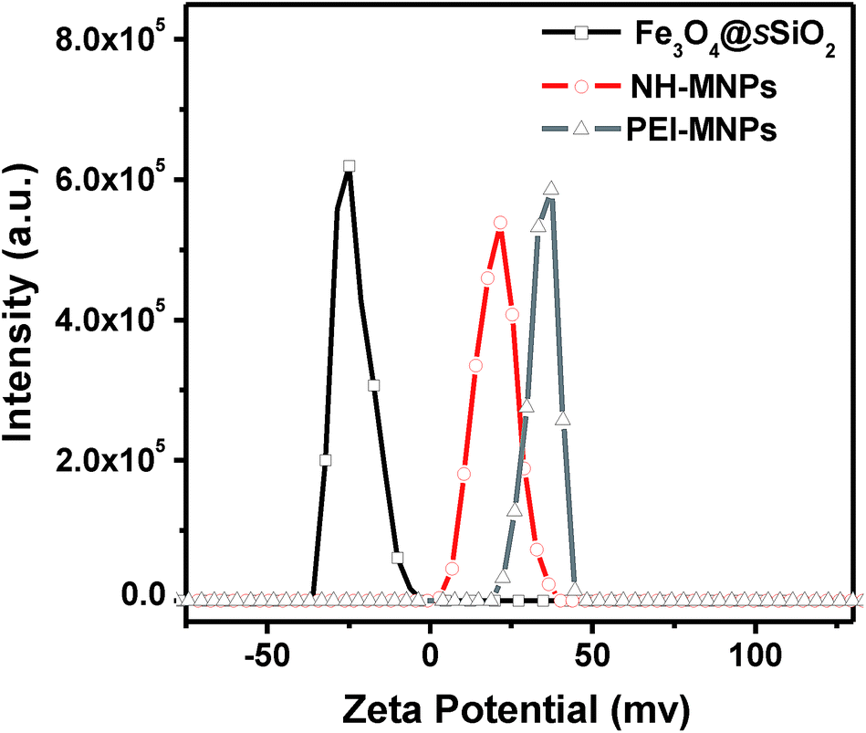 | ||
| Fig. 2 Zeta potential of Fe3O4@sSiO2 nanoparticles, NH-MNPs and PEI-MNPs in ultra-pure water. | ||
| Samples | Nitrogen content (w/w) |
|---|---|
| NH-MNPs | 1.12% |
| PEI-MNPs | 1.79% |
Fourier transform infrared spectroscopy (FTIR) was also used to characterize the Fe3O4@sSiO2 nanoparticles after surface functionalization. Fig. 3a is the IR spectrum of Fe3O4@sSiO2 nanoparticles. From the spectrum, it can be seen that there are two characteristic peaks around 590 cm−1 and 1091 cm−1, assigned to the Fe–O and Si–O–Si stretching vibrations, respectively.28 The IR spectrum of PEI-MNPs (Fig. 3c) shows a broad band at 3420 cm−1 corresponding to the vibration of the amine N–H bonds, while the bands at 2852 cm−1, and 2935 cm−1 are attributed to C–H stretching vibrations of the PEI.30 A similar IR spectrum was also observed on pure PEI, suggesting that PEI was successfully linked onto the silica surface. Subsequently, superconducting quantum interference measurements (SQUD) were used to determine the magnetic behavior of these magnetic nanoparticles. As illustrated in Fig. S1† the three samples were superparamagnetic at room temperature. The saturation magnetization of the Fe3O4 nanoparticles was 76.5 emu g−1 and decreased to 42.6 emu g−1 for NH-MNPs and 40.5 emu g−1 for PEI-MNPs after the coating process. This decrease could be attributed to the additional weight of the amorphous silica shell and the conjugated organic molecules. The above results suggested that these fabricated nanocomposites still possessed strong superparamagnetic properties and could be easily separated by a magnet.
 | ||
| Fig. 3 FTIR spectra of Fe3O4@sSiO2 nanoparticles, NH-MNPs, PEI-MNPs and PEI. | ||
The zeta potential of NH-MNPs and PEI-MNPs were measured in PBS buffer (170 mM NaCl, 3.4 mM KCl, 15 mM phosphate, the pH values of solutions were adjusted by 1.0 M sodium hydroxide or hydrochloric acid) at different pH values and the results are provided in Fig. 4. For the two samples, the zeta potential changed from positive to negative when the pH value was increased from 3 to 11. The PEI-MNPs (IP = 10.3) exhibited a higher isoelectric point (IP) than the NH-MNPs (IP = 8.4). The higher isoelectric point of PEI-MNPs could enhance the electrostatic interactions between nanoparticles and bacteria (the zeta potential of examined bacterial pathogens are presented in Fig. S2†), which helped to improve the bacterial capture efficiencies.
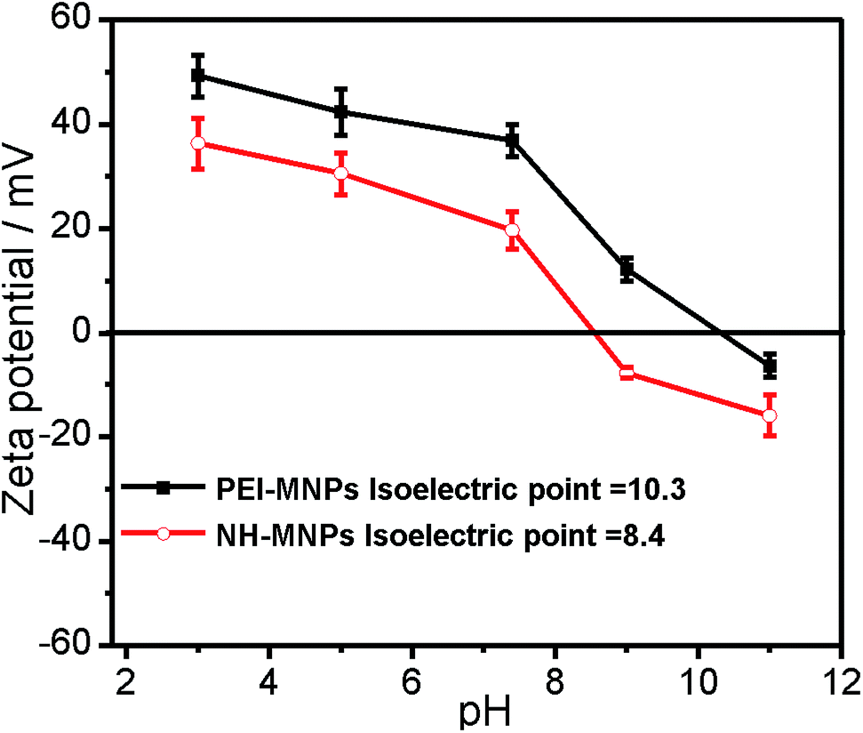 | ||
| Fig. 4 The pH-dependent zeta potential of NH-MNPs and PEI-MNPs. | ||
Unlike a bacterial selective capture system, our cationic amine group functionalized magnetic nanoparticles are able to capture a broad range of bacteria via electrostatic attractions which has been widely reported.31–37 One other advantage in this system is that the PEI-functionalized nanoparticles have more amine groups, which were approved to capture more bacteria than triaminopropylalkoxysilane directly modified nanoparticles. To compare their bacterial capture efficiencies and investigate the broad-spectrum bacterial capture properties, we employed two Gram-negative bacteria (E. coli BL21 and E. coli JM109, both are gene-engineering bacteria and widely used as protein expression systems) and two Gram-positive bacteria (B. subtilis and S. aureus, B. subtilis are not pathogenic rod-shape bacteria, and S. aureus are highly pathogenic spherical bacteria) as models. The concentrations of the four bacteria were adjusted to an OD600 of 0.5. As a control experiment, the Fe3O4@sSiO2 nanoparticles without amine group functionalization were also examined. As illustrated in Fig. 5, the bacterial capture efficiencies increased with the dosage of materials. In the case of Gram-negative bacteria, 2.0 mg mL−1 NH-MNPs were required to achieve 96.1% capture efficiency for E. coli BL21 and 95.7% capture efficiency for E. coli JM109, respectively. However, only 0.6 mg mL−1 PEI-MNPs were able to capture the same amount of the both bacteria, which meant that the bacterial capture capacity of PEI-functionalized nanoparticles was 3.3 times higher than that of triaminopropylalkoxysilane directly modified nanoparticles. For Gram-positive bacteria, with 2.0 mg mL−1 nanoparticles, the capture efficiency of NH-MNPs was 85% for B. subtilis and 83.9% for S. aureus, respectively. In contrast with NH-MNPs, the PEI-MNPs still showed higher bacterial removal capacities and could capture more than 98% of B. subtilis and 97.7% of S. aureus even at a dosage decreased to 0.8 mg mL−1, respectively. As can be seen from Fig. 5, the Fe3O4@sSiO2 nanoparticles also displayed low bacterial capture abilities for the four bacterial species, which could be explained by the nonspecific adsorption between the bacterial cell wall and the silica surface such as hydrogen bonds. Overall, all these observations demonstrated that the PEI-MNPs have a higher adsorption capacity for both Gram-negative and Gram-positive bacteria than the NH-MNPs.
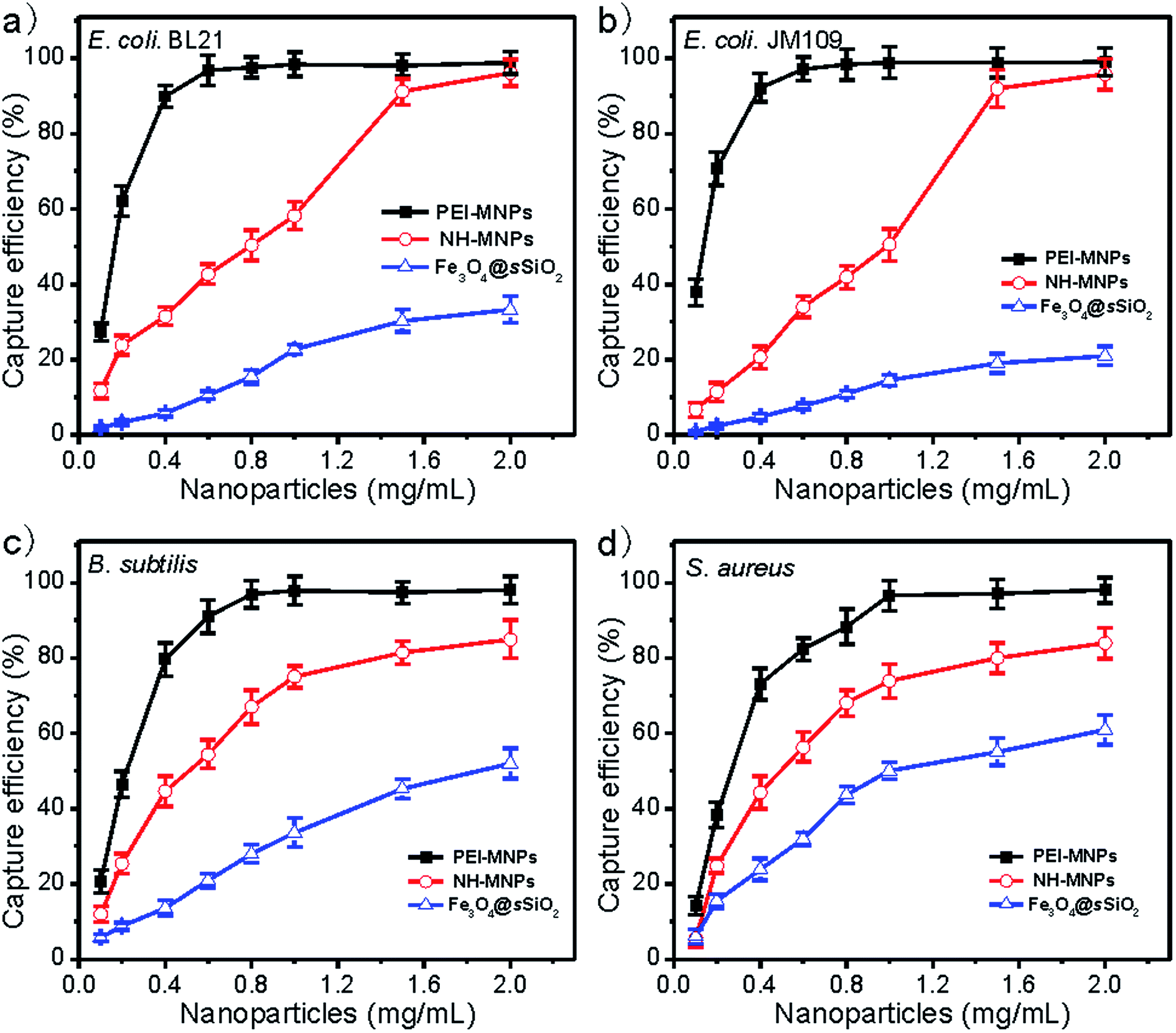 | ||
| Fig. 5 Effects of Fe3O4@sSiO2 nanoparticles, NH-MNPs and PEI-MNPs concentrations on capture efficiency of four bacterial species in water. (a) E. coli BL21; (b) E. coli JM109; (c) B. subtilis; and (d) S. aureus. The concentration of the bacteria is ∼2.5 × 10⁸ CFU mL−1 (0.5 OD600). | ||
More importantly, we found out that the more amine groups in PEI-MNPs resulted in higher bacterial capture efficiency from their low-concentration solutions. At a bacterial concentration of 1 × 10⁸ CFU mL−1 E. coli BL21 suspensions (OD600 = 0.2, Fig. 6a and b), both NH-MNPs and PEI-MNPs captured a similar amount of bacteria, 97.5% and 98.7%, respectively. Meanwhile, when the bacterial concentration was decreased to 1 × 10⁷, 1 × 10⁶ and 1 × 10⁵ CFU mL−1, the capture efficiency with NH-MNPs was significantly dropped to 92%, 44.4% and 18.8%, respectively. However, the capture efficiency with the PEI-MNPs was only decreased to 94%, 80.8% and 73.7%, respectively, for the solutions under the same experimental conditions. For B. subtilis (Fig. 6c and d), similar results were also observed. These results suggested that PEI-MNPs have a strong affinity for bacteria, which could be explained by the widely reported multivalency effect in bacterial capture.38–41 In order to clearly compare their affinity for bacteria, transmission electron microscopy (TEM) technology was used. In Fig. 6, there are a few NH-MNPs found on the surface of E. coli BL21 (Fig. 6d). Meanwhile for PEI-MNPs, lots of nanoparticles were observed to aggregate on the bacterial cell wall (Fig. 6e).
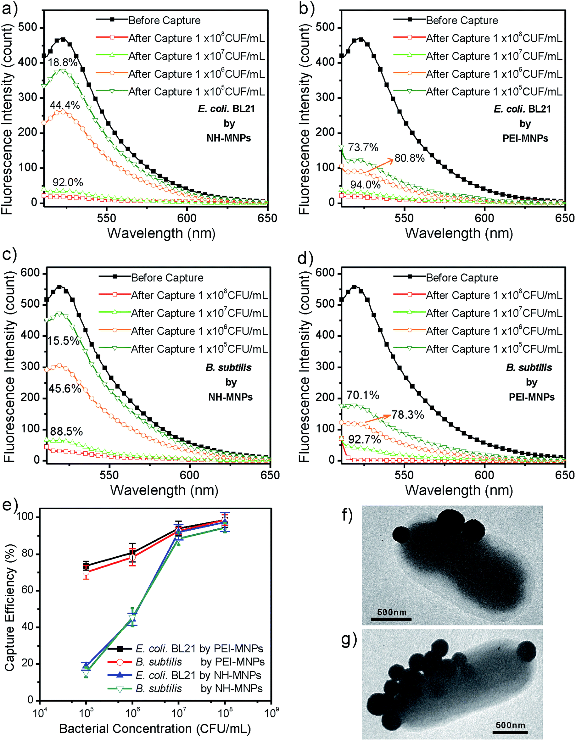 | ||
| Fig. 6 Fluorescence spectra showing the change of emission intensity of the solution of FITC-labeled E. coli BL21 (a, b) and B. subtilis (c, d) before and after treatment with NH-MNPs (a, c) and PEI-MNPs (b, d) with different concentrations of bacteria. The curves with filled square symbols represent the fluorescence spectrum of the FITC-labeled bacteria solution before treatment with nanoparticles. The curves with open square symbols, open up triangle symbols, open circle symbols and open down triangle symbols represent the spectra of the FITC-labeled bacteria solution after treatment with nanoparticles in a 1 × 10⁸, 1 × 10⁷, 1 × 10⁶ and 1 × 10⁵ CFU mL−1 bacterial solution, respectively. All solutions examined were in the same volume. (e) Bacterial capture efficiency of the magnetic nanoparticles with different concentrations of bacteria based on the fluorescence spectra of (a), (b), (c) and (d). (f) TEM image of E. coli BL21 interacting with NH-MNPs for 10 min. (g) TEM image of E. coli BL21 interacting with and PEI-MNPs for 10 min. | ||
To further examine the practical applications of the magnetic nanoparticles in aqueous solutions at different pH values. Here, the pH effects on the bacterial capture efficiencies were studied using E. coli BL21 as a model bacterium. It is clear from the data in Fig. S3† that high bacterial capture efficiencies of these two types of nanoparticles were obtained at low pH values. With a higher isoelectric point, the PEI-MNPs show high capture efficiencies under basic conditions. For instance, at pH 9 the capture efficiency was maintained at 93.6% with PEI-MNPs, whereas the capture efficiency dropped to 78.1% with the NH-MNPs. Nevertheless, these two types of magnetic nanoparticles still have more than 60% capture efficiency at a pH value equal to 11, indicating that the as-prepared magnetic nanoparticles could be applied to separate bacteria from water at a wide range of pH values.
The recyclability of the magnetic nanoparticles throughout five consecutive cycles was investigated with E. coli BL21 (Fig. 7). After each bacterial capture, the magnetic nanoparticles were rinsed with aqueous ammonia solution to release the captured bacteria, then incubated with hydrochloric acid solution and washed with distilled water for the next bacterial separation. After five consecutive cycles, the bacterial capture efficiency was maintained at 68.5% for NH-MNPs and 70.5% for PEI-MNPs, respectively. These observations suggested that the magnetic nanoparticles exhibited good recyclability for capturing bacteria from water.
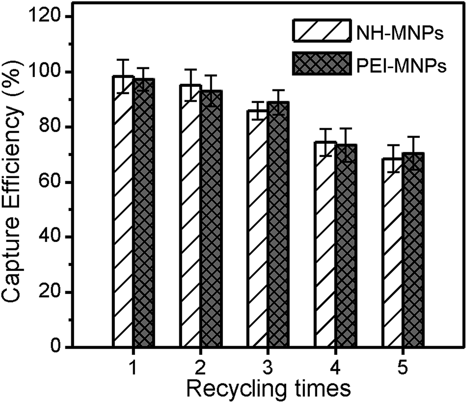 | ||
| Fig. 7 Recycling of NH-MNPs and PEI-MNPs in the capture of E. coli BL21. | ||
Finally, we also tested the biocompatibility of the magnetic nanoparticles by incubating HeLa cells with the nanoparticles at different concentrations for 24 h. As revealed by MTT assay (Fig. 8), the cell viability was only reduced to 92.5% for NH-MNPs and 82.7% for PEI-MNPs, respectively, even with the concentration of the nanoparticles as high as 200 ppm, demonstrating that these magnetic nanoparticles had a good biocompatibility.
 | ||
| Fig. 8 Viability of HeLa cells after a 24 h incubation with different concentrations of NH-MNPs and PEI-MNPs. | ||
4. Conclusions
In conclusion, we have successfully fabricated two types of amine group functionalized magnetic nanoparticles with excellent capture performance of bacteria. More amine groups were efficiently incorporated onto the surface of PEI-MNPs as compared to NH-MNPs. Such PEI-MNPs possessed a high bacterial adsorption capacity and strong affinity for bacteria from their low-concentration solutions making the as-prepared magnetic nanoparticles promising for practical applications.Acknowledgements
We thank the Natural Science Foundation of Anhui Province (No. 1508085QB39, 1408085MH153), the China Postdoctoral Science Foundation Funded Project (No. 2016M590559) and the Open Project of State Key Laboratory of Physical Chemistry of Solid Surfaces (Xiamen University, No. 201416) for the financial support.Notes and references
- D. I. Andersson and D. Hughes, Nat. Rev. Microbiol., 2010, 8, 260–271 CAS.
- N. Woodford and D. M. Livermore, J. Infect., 2009, 59, S4–S16 CrossRef PubMed.
- D. C. Angus, W. T. Linde-Zwirble, J. Lidicker, G. Clermont, J. Carcillo and M. R. Pinsky, Crit. Care Med., 2001, 29, 1303–1310 CrossRef CAS PubMed.
- L. R. Bakken, Appl. Environ. Microbiol., 1985, 49, 1482–1487 CAS.
- Y.-Q. Li, B. Zhu, Y. Li, W. R. Leow, R. Goh, B. Ma, E. Fong, M. Tang and X. Chen, Angew. Chem., Int. Ed., 2014, 53, 5837–5841 CrossRef CAS PubMed.
- X. Liu, Z. Lei, F. Liu, D. Liu and Z. Wang, Biosens. Bioelectron., 2014, 58, 92–100 CrossRef CAS PubMed.
- W. Jing, W. Zhao, S. Liu, L. Li, C.-T. Tsai, X. Fan, W. Wu, J. Li, X. Yang and G. Sui, Anal. Chem., 2013, 85, 5255–5262 CrossRef CAS PubMed.
- T. Ishikawa, T. Shioiri, K. Numayama-Tsuruta, H. Ueno, Y. Imai and T. Yamaguchi, Lab Chip, 2014, 14, 1023–1032 RSC.
- X. Zhang, J. Zhou, C. Zhang, D. Zhang and X. Su, RSC Adv., 2016, 6, 1279–1287 RSC.
- V. Kumar, G. Nath, R. K. Kotnala, P. S. Saxena and A. Srivastava, RSC Adv., 2013, 3, 14634–14641 RSC.
- M. Colombo, S. Carregal-Romero, M. F. Casula, L. Gutierrez, M. P. Morales, I. B. Bohm, J. T. Heverhagen, D. Prosperi and W. J. Parak, Chem. Soc. Rev., 2012, 41, 4306–4334 RSC.
- M. Varshney, L. Yang, X.-L. Su and Y. Li, J. Food Prot., 2005, 68, 1804–1811 CAS.
- Z. Fan, D. Senapati, S. A. Khan, A. K. Singh, A. Hamme, B. Yust, D. Sardar and P. C. Ray, Chem.–Eur. J., 2013, 19, 2839–2847 CrossRef CAS PubMed.
- P. Bakthavathsalam, V. K. Rajendran, U. Saran, S. Chatterjee and B. M. Jaffar Ali, Microchim. Acta, 2013, 180, 1241–1248 CrossRef CAS.
- Z. H. Wei, Z. J. Zhou, M. Yang, C. H. Lin, Z. H. Zhao, D. T. Huang, Z. Chen and J. H. Gao, J. Mater. Chem., 2011, 21, 16344–16348 RSC.
- V. C. Ozalp, G. Bayramoglu, M. Kavruk, B. B. Keskin, H. A. Oktem and M. Y. Arica, Anal. Biochem., 2014, 447, 119–125 CrossRef CAS PubMed.
- D. Cheng, M. Yu, F. Fu, W. Han, G. Li, J. Xie, Y. Song, M. T. Swihart and E. Song, Anal. Chem., 2016, 88, 820–825 CrossRef PubMed.
- J.-J. Lee, K. J. Jeong, M. Hashimoto, A. H. Kwon, A. Rwei, S. A. Shankarappa, J. H. Tsui and D. S. Kohane, Nano Lett., 2014, 14, 1–5 CrossRef CAS PubMed.
- A. J. Kell, G. Stewart, S. Ryan, R. Peytavi, M. Boissinot, A. Huletsky, M. G. Bergeron and B. Simard, ACS Nano, 2008, 2, 1777–1788 CrossRef CAS PubMed.
- J. J. Lee, K. J. Jeong, M. Hashimoto, A. H. Kwon, A. Rwei, S. A. Shankarappa, J. H. Tsui and D. S. Kohane, Nano Lett., 2014, 14, 1–5 CrossRef CAS PubMed.
- T. Y. Liu, K. T. Tsai, H. H. Wang, Y. Chen, Y. H. Chen, Y. C. Chao, H. H. Chang, C. H. Lin, J. K. Wang and Y. L. Wang, Nat. Commun., 2011, 2, 538 CrossRef PubMed.
- H. Gu, P.-L. Ho, K. W. T. Tsang, L. Wang and B. Xu, J. Am. Chem. Soc., 2003, 125, 15702–15703 CrossRef CAS PubMed.
- L. Y. Chen, F. S. Razavi, A. Mumin, X. X. Guo, T. K. Sham and J. Zhang, RSC Adv., 2013, 3, 2390–2397 RSC.
- J. Chen, B. Duncan, Z. Wang, L.-S. Wang, V. M. Rotello and S. R. Nugen, Nanoscale, 2015, 7, 16230–16236 RSC.
- J. Sun, J. Li, H. Fan and S. Ai, J. Mater Chem. B, 2013, 1, 5436–5442 RSC.
- S. C. Hayden, G. X. Zhao, K. Saha, R. L. Phillips, X. N. Li, O. R. Miranda, V. M. Rotello, M. A. El-Sayed, I. Schmidt-Krey and U. H. F. Bunz, J. Am. Chem. Soc., 2012, 134, 6920–6923 CrossRef CAS PubMed.
- A. F. Radovic-Moreno, T. K. Lu, V. A. Puscasu, C. J. Yoon, R. Langer and O. C. Farokhzad, ACS Nano, 2012, 6, 4279–4287 CrossRef CAS PubMed.
- G. Chen, Z. Li, X. Wang, L. Xie, Q. Qi and W. Fang, Mater. Lett., 2014, 134, 290–294 CrossRef CAS.
- W. Fang, X. Chen and N. Zheng, J. Mater. Chem., 2010, 20, 8624–8630 RSC.
- P. M. Reddy, K.-C. Chang, Z.-J. Liu, C.-T. Chen and Y.-P. Ho, J. Biomed. Nanotechnol., 2014, 10, 1429–1439 CrossRef CAS PubMed.
- M. L. Bhaisare, H. N. Abdelhamid, B.-S. Wu and H.-F. Wu, J. Mater. Chem. B, 2014, 2, 4671–4683 RSC.
- H. N. Abdelhamid and H.-F. Wu, J. Mater. Chem. B, 2013, 1, 3950–3961 RSC.
- Y.-F. Huang, Y.-F. Wang and X.-P. Yan, Environ. Sci. Technol., 2010, 44, 7908–7913 CrossRef CAS PubMed.
- Y. Jin, J. Deng, J. Liang, C. Shan and M. Tong, Colloids Surf., B, 2015, 136, 659–665 CrossRef CAS PubMed.
- Y. Jin, F. Liu, C. Shan, M. Tong and Y. Hou, Water Res., 2014, 50, 124–134 CrossRef CAS PubMed.
- L. Bromberg, S. Raduyk and T. A. Hatton, Anal. Chem., 2009, 81, 5637–5645 CrossRef CAS PubMed.
- D.-T. Vo, C. G. Whiteley and C.-K. Lee, Ind. Eng. Chem. Res., 2015, 54, 9270–9277 CrossRef CAS.
- S. Lata, M. Gavutis, R. Tampé and J. Piehler, J. Am. Chem. Soc., 2006, 128, 2365–2372 CrossRef CAS PubMed.
- R. J. Pieters, Med. Res. Rev., 2007, 27, 796–816 CrossRef CAS PubMed.
- D. Cui, M. Li, X. Xu, J. Lu, J. Qian and S. Liu, Langmuir, 2014, 30, 11833–11840 CrossRef CAS PubMed.
- R. J. Pieters, Org. Biomol. Chem., 2009, 7, 2013–2025 CAS.
Footnote |
| † Electronic supplementary information (ESI) available. See DOI: 10.1039/c6ra13070d |
| This journal is © The Royal Society of Chemistry 2016 |