Monoclonal antibody-tagged polyethylenimine (PEI)/poly(lactide) (PLA) nanoparticles for the enhanced delivery of doxorubicin in HER-positive breast cancers

Ning Cuia and Su-Hua Zhu*b
aDepartment of General Surgery, The First People's Hospital of Henan, Shangqiu, 476000, China
bDepartment of Traditional Chinese Medicine Surgery, The First People's Hospital of Henan Shangqiu, No. 292, Kaixuan Road, Shangqiu, Henan 476000, P. R. China. E-mail: zhusuhuash@hotmail.com; Fax: +86-370-3255757; Tel: +86-370-3255757

Received 15th May 2016 , Accepted 17th July 2016

First published on 15th August 2016


Abstract

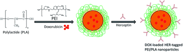
Poor therapeutic response and adverse side effects of chemotherapeutic agents are the major obstacles for effective chemotherapy against breast cancers. In this study, we reported a successful application of an Herceptin-tagged polyethylenimine (PEI) and poly (lactide) PEI/PLA nanoparticle system (hPPD) to deliver the chemotherapeutic drug doxorubicin (DOX) to HER2-positive breast cancer cells for improved efficacy of chemotherapy. The prepared nanoparticles were nanosized and exhibited a typical controlled drug release pattern suggesting its suitability for biomedical applications. The presence of Herceptin on the nanoparticle surface enhanced the accumulation of carrier on the cellular cytoplasm as is evident from the CLSM image. Our data showed that hPPD could significantly inhibit the proliferation of breast cancer cells compared to that with free DOX. Furthermore, Herceptin-based nanocarrier-mediated delivery of DOX could not only enhance the therapeutic outcome of using the anticancer drug, but also reduced the side effects of DOX in an effective manner in a xenograft tumor model. Overall, the Herceptin-tagged nanosystem demonstrates great potential as a novel therapeutic strategy in the anticancer treatment of HER2-positive breast cancers.


Introduction

Breast cancer is one of the most commonly diagnosed malignancies in women and among the leading cause of cancer-related death in women worldwide.1 Despite the significant advances in the treatment and diagnosis, mortality rate due to breast cancers kept increasing with every year.2 Specifically, resistance to conventional chemotherapeutics poses a big challenge towards the effect breast cancer treatment. Among all the treatment options, chemotherapy is an effective option to treat the malignancy; however it often suffers from severe drug-related side effects.3–5 Therefore, to overcome the adverse effects and to increase the therapeutic efficacy, alternative strategies have to be designed.

Doxorubicin (DOX) is one of the widely used anticancer agents for the treatment of multiple cancers including breast cancers.6 However, clinical application of DOX is severely limited by its potentially lethal adverse side effects in normal tissues, especially in the heart and kidneys.7 Therefore effective ways need to be developed to target the drug to the cancer cells and to reduce its distribution in the cardiac tissues.

In this regard, nanoparticles based drug delivery has gained increasing attention in the treatment of various diseases including cancers. The use of nanocarriers enhances the therapeutic efficacy of anticancer drugs and at the same time reduces its side effects. These nanoparticles provide the long term stability in the blood circulation and control the release of drugs.8–10 Although these nanomedicines enhanced the antitumor effect and reduced the drug originated side effect, the outcome is still modest. Commonly, nanomedicines could passively accumulate in tumor because of the enhanced permeability and retention (EPR) effect.11,12 Therefore, several efforts were made to increase the specificity of nanoparticles towards the cancers. For example, active targeted delivery has been achieved by the conjugation of antibody, folic acid, galactose and peptides.13,14 Among all these active targeting strategies, antibodies are most promising and have been applied in clinical settings.

Herceptin, a recombinant monoclonal antibody targets the human epidermal growth factor receptor 2 (HER2) which is overexpressed in most of the breast cancers. The Herceptin has been indicated in the treatment of HER2-positive breast cancers along with the chemotherapy.15 Herceptin acts by blocking the HER2 domain cleavage; block signaling pathways and inhibit the DNA repair process.16 Many anticancer drugs have been combined with Herceptin and recently it was conjugated on the nanoparticles and delivered along with paclitaxel.17 The Herceptin could be conjugated on the nanoparticle surface through chemical crosslinking.18 Herceptin has also been conjugated onto the surface of nanocarriers for use as a targeting ligand to direct the delivery of Paclitaxel to HER2-positive breast cancer cells. The combination of Herceptin and Paclitaxel not only endows the nanocarriers with targeting behavior, but also sensitizes cancer cells to Paclitaxel and thus achieves enhanced tumor suppression.19–21 In this study, polymeric nanoparticles have been selected owing to its unique properties such as high drug loading, high stability in the systemic circulation and controlled release pattern. Polymer NPs for biological applications are typically based on biodegradable polyesters such as poly(lactic acid) (PLA), poly(caprolactone) (PCL), and poly(lactic-co-glycolic acid) (PLGA), whose biocompatibility is widely assessed.22,23 Therefore, we have selected PLA to load the anticancer drug. In order to further increase the nanoparticle uptake in the cancer cells, we have modified the surface of nanoparticle with a positively charged polyethylenimine (PEI). The high positive charge of PEI can cause this polymer to exhibit electrostatic interactions with negatively charged biological molecules (nucleic acids or proteins) and thus facilitate cellular uptake. It has been reported that PEI coated nanoparticles were used as drug vectors and exhibited enhanced cellular uptake efficiency. The high positive charge of PEI is expected to enhance the accumulation of nanoparticles in the negatively charged cancer cells.24,25

In this study, PEI-supported PLA nanoparticles (PP) was prepared and then conjugated with Herceptin on the surface using the chemical cross-linking. The main aim of present study was to increase the therapeutic efficacy of DOX by targeting it specifically to the breast cancers. The targeting was achieved by the conjugation of monoclonal antibody (Herceptin) on the nanoparticle surface which will interact with the HER2 overexpressing breast cancer cells. The nanoparticles were characterized for particle size, shape and release kinetics. The cellular uptake efficiency of targeted and non-targeted nanoparticle was studied in SKBR3 cells and anticancer effect was evaluated by MTT assay. Importantly, anticancer efficacy of different formulations was studied in SKBR3 cancer cell bearing xenograft nude mice.

Materials and methods

Materials

Doxorubicin hydrochloride salt (DOX) was purchased from Beijing HuaFeng Co. LTD (Beijing, China). Polyethylenimine (PEI, Mw 25 kDa, branched), polylactide, and poloxamer 407 were purchased from Sigma-Aldrich, China. All other chemicals are of reagent grade and used without further purifications.

Preparation of Herceptin (HER)-tagged PLA/PEI nanoparticles

HER-tagged PP nanoparticles were prepared by emulsion-solvent evaporation method. Briefly, 100 mg of PLA and 15 mg of DOX was dissolved in dichloromethane (DCM) and stirred for 15 min. Aqueous phase was prepared by dissolving 0.5% (w/v) of PEI in poloxamer 407 (2%) solution and stirred for 15 min. The organic phase was dissolved in aqueous phase in a drop-wise manner and immediately homogenized (T25, IKA, Germany) at 14[thin space (1/6-em)]000 rpm for 3 min. 10 mL of distilled water was added to the above solution and again homogenized for 2 min at 14[thin space (1/6-em)]000 rpm. The emulsion was allowed to stir overnight to remove the organic solvent. The so-formed nanoparticle was separated and washed and lyophilized. The HER was chemically conjugated to DOX-loaded PP nanoparticles (PPD) after activating the HER.26 For this EDC and NHS was dissolved in HER solution and allowed to activate for 1 h. Briefly, 40 mg EDC and 9.7 mg NHS were dissolved in 4 mL of PBS (0.1 M, pH 5.8) followed by the addition of 250 μL of antiHER2 (1 mg mL−1 anti-HER2 in 0.1 M PBS, pH 7.4) in the suspension. Followed by, lyophilized PPD nanoparticle (10 mg) was dissolved in specified manner in antibody solution and left at room temperature for 12 h. The amine groups present at the surface of PPD (originates from PEI moiety) were made to covalently couple with the activated HER2 in the presence of EDC/NHS by forming an amide linkage. The excess EDC and NHS were removed by dialysis method and final product was stored at 4 °C. The amount of DOX loaded in the nanoparticle was determined by fluorescence spectrometer (Shimadzu, Japan). The nanoparticle dispersion was centrifuged at a high speed of 15[thin space (1/6-em)]000 rpm min−1 and supernatant was used to determine the amount of drug unentrapped.

Characterization of nanoparticles

The particle size and size distribution of nanoparticle were determined by dynamic light scattering (DLS) method using ZetaSizer (ZS Nano, Malvern Instruments, UK). The morphology of nanoparticles was determined by transmission electron microscope (TEM; JEM-100CX JEOL, Japan) at 25 °C. The nanoparticles were appropriately dilute for the both the experiments. For TEM, nanoparticles were stained with 2% phosphotungstic acid (PTA).

In vitro drug release

To determine the amount of drug release from respective nanoparticles, 15 mg of lyophilized nanoparticles (equivalent to 2 mg DOX) suspended in 1 mL of water and sealed in a dialysis membrane (molecular weight cut off: 3500 Da) and in turn placed in a phosphate buffered saline (pH 7.4). The samples were collected at predetermined time intervals and fluorescence intensity of released DOX was detected by fluorescence spectrophotometer (Shimadzu, Japan). All the above procedures were performed at 37 °C in the dark.

Cellular uptake study

The cellular uptake of nanoparticles was studied by means of confocal laser scanning microscopy (CLSM). The SKBR3 cells at seeding density of 3 × 10⁵ cells per well were plated in 6-well plate containing a cover slip. The cells were allowed to attach for 24 h. Next day, old media was replaced with new media containing the PPD and hPPD nanoparticles and incubated for 2 h at 37 °C in dark conditions. The cells were washed with PBS and fixed with 4% paraformaldehyde (PFA). The cells were then stained with DAPI as a nuclear staining agent for 10 min. The cells were again washed and mounted on a glass slide and observed under CLSM (LSM710, Carl Zeiss, Germany).

Cytotoxicity assay

SKBR3 breast cancer cell was cultured in RPMI-1640 medium that was supplemented with 10% FBS, 100 units per mL of penicillin, and 100 mg mL−1 of streptomycin at 37 °C in the presence of 5% CO2. SKBR3 cells were seeded in a 96-well plate at a seeding density of 2 × 10³ cells per well and allowed to incubate overnight. The cells were treated with different concentration of blank NP, free DOX, PPD, and hPPD and incubated for 24 h. After incubation for 24 h, 20 μL of MTT (5 mg mL−1) was added to each cell and incubated for another 4 h. Then the medium was removed, the cells were washed by PBS once and replaced by 150 μL of dimethyl sulfoxide (DMSO). Finally, the absorbance was measured by a microplate reader (Thermo Scientific, USA) at 490 nm. Cell viability was calculated from the following equations:
Cell viability (%) = Atreated/Acontrol × 100

Breast tumor induction and antitumor efficacy study

The animal study was approved by the ‘Institutional Animal Ethics Committee’, The First People's Hospital of Henan Shangqiu, China. Female Balb/C mice (6–7 weeks old) were cared as in accordance with the guidelines framed by Provincial Animal Ethics Advisory Committee (PAEAC) at our University. The animals were given free access to food and water and exposed to 12 light/dark cycle.

SKBR3 breast cancer cells were grown in RPMI-1640 culture media with 10% fetal bovine serum (FBS), 100 μg mL−1 penicillin and 100 μg mL−1 streptomycin in a humidified 5% CO2 incubator at 37 °C. 1 × 10⁷ cells in 100 μL was mixed with 50 μL of matrigel and subcutaneously injected into the flank of anesthetized mouse. Anesthesia was induced by intraperitoneal injection of xylazine and ketamine (10 and 100 mg kg−1/BW, respectively). The tumors were allowed to grow for 50–80 mm3 and the mice were randomly divided into 4 experimental groups: (control); free DOX; PPD, and hPPD, respectively. The formulations were injected 4 times with injection every third day. Ellipsoidal tumor volume was calculated using the following formula: V = π × l × d × w, where l, w, and d represent the length, width, and depth, respectively. Measurements were performed using a digital caliper (Kanon, Nakamura Mfg. Co., Tokyo, Japan).

Histopathology study

At the end of antitumor study, mice were sacrificed and tumor tissues were removed and fixed in 10% neutral buffered formalin. The tissues were embedded in paraffin and serially sectioned into 5 mm-thick sections. The sections were then stained with hematoxylin and eosin (H&E). Histological slides were evaluated using bright field microscopy (Nikon, Japan). Tumor malignancy was graded between 3 and 9 on the basis of three features: nuclear pleomorphism, number of mitotic figures/10 high-power fields, and tubular formation, which were scaled as 1, 2, or 3. A tumor with a sum of 3–5 was considered as grade 1 (well differentiated). A tumor with a sum of 6 or 7 was considered as grade 2 (moderately differentiated), and a tumor with a sum of 8 or 9 was considered as grade 3 (poorly differentiated).

Statistical analysis

Statistical analyses were performed by one-way ANOVA, and Tukey's HSD was used as post hoc test. Values of P < 0.05 were considered significant.

Results

Characterization of hPPD nanosystem

The particle size and size distribution was determined by dynamic light scattering (DLS) technique. The particle size of PPD nanoparticle was ∼140 nm (PDI ∼ 0.185) with a strong positive charge of +34.5 mV (Fig. 2a). The conjugation of Herceptin was confirmed by the decrease in the surface charge of PP nanoparticles. The final surface charge of hPPD was +10.5 mV indicating the successful conjugation of Herceptin on the nanoparticle surface. The final particle size of hPPD was ∼220 nm with a PDI of 0.254. The morphology of hPPD was confirmed by transmission electron microscope (TEM) (Fig. 2b). The particles were spherical and uniformly distributed on the copper grid. As the particles were observed in the dried state, the presence of thin layer of Herceptin could not be visualized clearly. Nevertheless, spherical shaped particles are expected to increase the overall antitumor efficacy of the encapsulated drug. The particles were highly stable upon storage up to 30 days indicating its excellent colloidal as well as storage stability (Fig. 3a).
image file: c6ra12616b-f1.tif
Fig. 1 Schematic illustration of construction of Herceptin-tagged PEI/PLA-based nanoparticles for the delivery of DOX in breast cancer cells.

image file: c6ra12616b-f2.tif
Fig. 2 (a) Dynamic light scattering (DLS) histograms of hPPD nanoparticles as determined from ZetaSizer; (b) transmission electron microscope (TEM) of hPPD nanoparticles.

image file: c6ra12616b-f3.tif
Fig. 3 (a) Stability analysis of PPD and hPPD in PBS medium; (b) in vitro drug release profile of PPD and hPPD nanosystem in phosphate buffered saline (pH 7.4) at 37 °C.

Drug loading and in vitro drug release

The hPPD nanoparticles showed a high entrapment efficiency of more than 95% with an active drug loading of 14.5% w/w. High drug loading capacity of nanocarrier is beneficial for cancer targeting applications. The in vitro drug release study was performed in phosphate buffered saline (PBS, pH 7.4) at 37 °C. As seen (Fig. 3b), hPPD and PPD nanosystem exhibited a controlled release of drug throughout the study period. Approximately ∼35% of DOX was released within 24 h of study period while ∼80% of drug released at the end of 72 h.

Cytocompatibility of blank nanoparticles

It can be seen that the cell viability of SKBR3 cells remained very high even when exposed with the maximum concentration (200 μg mL−1) of blank nanoparticles (Fig. 4). The cell viability was more than 90% when treated with 100 μg mL−1 of nanoparticle dispersion, however, cell viability slightly decreased when the concentration of blank nanoparticles was increased to 200 μg mL−1. Nevertheless, ∼75% of cells were viable indicating its excellent biocompatibility profile.
image file: c6ra12616b-f4.tif
Fig. 4 The cellular uptake of PPD and hPPD nanoparticles in SKBR3 breast cancer cells. The cellular uptake of nanoparticles was monitored in CLSM upon incubation for 2 h.

Cellular uptake efficiency of nanoparticles

In this study, we have employed HER as a targeting moiety and tagged at the surface of nanoparticles. Therefore, we have compared the cellular uptake efficiency of nanoparticles using confocal microscopy. For this purpose, cells were exposed with both the nanoparticles and incubated for 2 h. The nucleus was stained with DAPI and red fluorescence originated from DOX itself (Fig. 5). It can be seen that red fluorescence concentrated largely on the cytoplasmic region indicating a typical endocytosis-mediated cellular uptake. Especially, hPPD showed a bright red fluorescence on the cytoplasm compared to that of PPD nanosystem indicating a higher internalization of nanoparticle via HER-based receptor targeting.
image file: c6ra12616b-f5.tif
Fig. 5 Effect of blank PPD and blank hPPD nanosystem on the cell proliferation of SKBR3 cancer cells. The cells were treated with a concentration up to 200 μg mL−1. Untreated cells were used as control and represent 100% of the proliferation.

Cytotoxicity potential of drug-loaded nanoparticles

Cytotoxicity of drug-loaded nanoparticle is a primary concern in the development of drug delivery system. As shown in Fig. 6, free drug and drug-loaded nanoparticles exhibited a dose-dependent and time-dependent cytotoxic effect in SKBR3 cancer cells. It should be noted that throughout the entire dose, PPD (drug-loaded nanoparticles) showed a higher cytotoxic effect compared to that of free DOX. Importantly, HER-tagged nanoparticles exhibited a superior cytotoxic effect in this cancer cells than any other formulations indicating a typical receptor-mediated endocytosis uptake and effective targeting ability of the tagged moiety.
image file: c6ra12616b-f6.tif
Fig. 6 In vitro cell viability of SKBR3 breast cancer cells treated with different formulations and incubated for 24 h and 48 h, respectively. Cell viability was quantitatively measured using MTT assay upon incubation with different concentrations of free DOX, PPD, and hPPD. Data were shown as the mean of triplicate measurements with standard deviations (n = 5).

In vivo antitumor efficacy

As presented in Fig. 7a, DOX treatment could only slightly decrease the tumor growth speed compared with saline group because of the low targeting efficiency of free DOX. Since PPD could passively accumulate in tumor through EPR effect, this group displayed better antitumor effect than free DOX group. Importantly, hPPD treated mice group showed a significant decrease in the tumor volume indicating its excellent antitumor efficacy. The final tumor volumes were ∼2100 mm3, ∼1300 mm3, ∼1000 mm3, and ∼400 mm3 for control, free DOX, PPD, and hPPD treated mice groups, respectively.
image file: c6ra12616b-f7.tif
Fig. 7 In vivo antitumor efficacy of different formulations in breast cancer tumor model. (a) Tumor volumes were measured at indicated time-points and shown as average of duplicate measurements with standard error bars (n = 8 for each group). (b) The body weights of mice were measured at indicated time-points and shown as average of duplicate measurements with standard error bars (n = 8 for each group).

The body weight was calculated to observe the toxicity profile of all formulations (Fig. 7b and c). No noticeable change in body weight was observed after administration of both PPD and hPPD formulations suggesting that they did not have obvious systemic toxicity to mice. However, the weight loss of DOX group was obviously observed in the early administration mainly owing to the severe side effects of DOX.

Histopathological study

H&E staining was used to examine the histological feature of tumor induced by different components. As shown in Fig. 8, tumors extracted from the untreated mice group were densely cellular with no sign of cellular death. In contrast, free drug and drug-loaded formulations treated group were less cellular compared to that of control group. Especially, hPPD treated mice group showed a significant apoptosis of cancer cells indicating the great therapeutic potential of Herceptin conjugated nanocarrier system.
image file: c6ra12616b-f8.tif
Fig. 8 Representative data of H&E staining of tumor tissues.

Discussion

Breast cancer is one of the most commonly diagnosed malignancies in women and despite the significant advancement in the diagnosis and treatment, mortality rate due to breast cancers kept increasing with every year. Chemotherapy is an effective option to treat the malignancy, however, severe adverse effects limits its clinical potential. Therefore, we have designed a novel monoclonal antibody (Herceptin) conjugated PEI/PLA nanoparticles to increase the therapeutic efficacy of DOX by targeting it specifically to the breast cancers. For this purpose, DOX-loaded PEI-supported PLA nanoparticle was prepared where PLA acts as a polymer core and PEI was used to provide the positive charge to the nanocarrier (Fig. 1). Besides, presence of PEI on the surface was used to conjugate the Herceptin as a targeting ligand to the cancer cells. PEI is most widely investigated because of its ability to effectively transfect/internalizes a broad range of cell lines with high efficiency. However, PEI has been reported to cause systemic concerns due to toxicity and stability in biological fluids. Therefore, in the present research, we have formed an emulsion by combining with PLA with more favorable properties for systemic delivery. Once a complex NP is formed, it is less likely that a surface-bound PEI will leach toward the external contacting biological medium and, thus, cause toxic effects; so even though a lower quantity of PEI is found on the surface, the toxic effect will be reduced. Furthermore, PEI provides a means of escaping the late endosome through the proton sponge effect; however, as noted above, the positive charge that makes PEI so efficient is also likely toxic to cells through disruption of the cellular membrane. The formation of PLA/PEI complex NP is expected to lessen PEI's toxicity by providing a physical barrier between the cell and PEI.

The particle size of PPD nanoparticle was ∼140 nm (PDI ∼ 0.185) with a strong positive charge of +34.5 mV. The final particle size of hPPD was ∼220 nm with a PDI of 0.254. A small particle size of around ∼200 nm with a targeting moiety would increase the internalization of nanoparticles in the cancer cells.27 The final surface charge of NP was slightly positive. The decrease in the surface charge of PLA–PEI NP was due to the conjugation of antibody to the PEI surface. The slight positive will facilitate the internalization of NP in the cancer cells while it will reduce the associated systemic toxic effects of PEI. The in vitro drug release study was performed in phosphate buffered saline (PBS, pH 7.4) at 37 °C. The data clearly indicated that despite the difference in the particle size of PPD and hPPD, release profile was similar with almost same amount of drug released at the end of 72 h. Both the nanoparticle system exhibited zero-order kinetics. Due to spatial confinement within a core, crystalline drug molecules are normally unable to form highly ordered crystals, instead remaining in amorphous or noncrystalline forms, which is known to improve the drug dissolution rate. In this case, drug solubility is an important factor regarding the amount of the drug that was loaded inside and, therefore, released from the nanoparticles. The results also reveal that presence of antibody on the surface did not inhibit the diffusion of drug from the nanoparticle core to the release medium. As is generally known, the zero order kinetics models are used to describe the release of drugs from degradable matrices. The slow and sustained release of DOX could benefit for constant exposure of drug to the cancer cells and reaching the effective treatment concentration. Overall, drug release from the NPs is a complex process, which could be attributed to drug diffusion in the polymeric matrix followed by polymer degradation, which are affected by constituents and architectures of the copolymers, surface erosion properties of the NPs, and the physico-chemical properties of the drugs. The controlled release of drug in the physiological medium indicates its ability to minimize the drug-related adverse effects while it can be expected that much of the drug will be available to release in the tumor tissue. It is important to note that conjugation of Herceptin on the nanoparticle surface did not change the release pattern of DOX from the PP nanoparticles. One of the main aims for nanobased cancer therapy is to develop nanosystems for sustained drug release and, consequently, to improve the therapeutic efficiency and minimize the toxicity of the anticancer drug molecules.28,29

The biocompatibility of nanocarriers is important when the formulation is intended for systemic administration. The ability of the nanoparticles being non-toxic to cells will be of clinical significance. The cell viability was more than 90% when treated with 100 μg mL−1 of nanoparticle dispersion indicating its non-toxic nature. The slight toxicity at highest concentration was due to the presence of PEI on the nanoparticle surface. In contrast, free drug and drug-loaded nanoparticles exhibited a dose-dependent and time-dependent cytotoxic effect in SKBR3 cancer cells. Importantly, HER-tagged nanoparticles exhibited a superior cytotoxic effect in this cancer cells than any other formulations indicating a typical receptor-mediated endocytosis uptake and effective targeting ability of the tagged moiety. IC50 value was calculated to quantitate the difference between the effects of formulations. The IC50 value of free DOX, PPD, and hPPD were 1.25 μg mL−1, 1.12 μg mL−1, and 0.59 μg mL−1, respectively after 24 h incubation. Similar trend followed after 48 h incubation with 0.96 μg mL−1, 0.84 μg mL−1, and 0.089 μg mL−1, respectively for these formulations. The results therefore clearly demonstrate the effect of HER on the surface of nanoparticles. The presence of targeting moiety increased the accumulation of drug in the cancer cells and sustained release property of nanoparticles exposed the drug in a constant manner which increased the cytotoxic effect of the loaded anticancer drugs.

The cellular uptake studies showed a typical endocytosis-mediated cellular uptake indicated by the high red fluorescence. The stronger red fluorescence clearly indicates that Herceptin could function as a targeting agent to enhance the uptake and accumulation of PPD into the cancer cells. Based on the result, it could be expected that DOX could be accumulated in the cancer cells in the preferential manner when conjugated with the Herceptin and thereby promotes the DNA damage and induces the apoptosis of cancer cells.

Subcutaneous tumor model of breast cancer was used to evaluate the antitumor efficacy of different formulations. The hPPD treated mice group showed a significant decrease in the tumor volume indicating its excellent antitumor efficacy. The final tumor volumes were ∼2100 mm3, ∼1300 mm3, ∼1000 mm3, and ∼400 mm3 for control, free DOX, PPD, and hPPD treated mice groups, respectively. The excellent antitumor efficacy could be due to the better targeting ability of Herceptin to the corresponding receptors overexpressed in breast cancer cells that might contributed to the higher internalization of nanoparticles and therapeutic effect of DOX.25 The major deficiency of conventional chemotherapy in cancer treatment is non-specificity. Administrated chemotherapeutic agents demonstrate general systemic distribution, which can lead to well-known side effects of chemotherapy. In this regard, excellent antitumor efficacy of hPPD achieved our aim of enhancing the tumor accumulation and improving the efficacy of cancer therapy.

Conclusion

In this study, we reported a successful application of Herceptin-tagged PEI/PLA nanoparticle system to deliver chemotherapeutic drug DOX into the breast cancer cells for improved efficacy of chemotherapy. The presence of Herceptin on the nanoparticle surface enhanced the accumulation of carrier on the cellular cytoplasm as evident from CLSM image. Our data showed that hPPD could significantly inhibit the proliferation of breast cancer cells compared to that of free DOX. Furthermore, Herceptin-based nanocarrier-mediated delivery of DOX could not only enhance the therapeutic outcome of anticancer drug, but also reduced the side effects of DOX in an effective manner in xenograft tumor model. Overall, Herceptin-tagged nanosystem could be an promising strategy for the treatment of breast cancers.

Acknowledgements

This study was supported by the Research grant of ‘The First People's Hospital of Henan’, China.

Notes and references

  1. R. Siegel, E. Ward, O. Brawley and A. Jemal, Ca-Cancer J. Clin., 2011, 61, 212–236 CrossRef PubMed.
  2. F. Kamangar, G. M. Dores and W. F. Anderson, J. Clin. Oncol., 2006, 24, 2137–2150 CrossRef PubMed.
  3. H. M. Coley, Cancer Treat. Rev., 2008, 34, 378–390 CrossRef CAS PubMed.
  4. L. Carey, E. Winer, G. Viale, D. Cameron and L. Gianni, Nat. Rev. Clin. Oncol., 2010, 7, 683–692 CrossRef PubMed.
  5. L. A. Carey, Breast Cancer Res., 2007, 9(suppl. 1), S1 Search PubMed.
  6. K. C. Weng and C. O. Noble, Nano Lett., 2008, 8, 2851–2857 CrossRef CAS PubMed.
  7. L. L. Bao, A. Haque, K. Jackson, S. Hazari, K. Moroz and R. Jetly, et al., Am. J. Pathol., 2011, 178, 838–845 CrossRef CAS PubMed.
  8. S. Vrignaud, J. P. Benoit and P. Saulnier, Biomaterials, 2011, 32, 8593–8604 CrossRef CAS PubMed.
  9. S. L. Luo, E. L. Zhang, Y. P. Su, T. M. Cheng and C. M. Shi, Biomaterials, 2011, 32, 7127–7138 CrossRef CAS PubMed.
  10. Y. G. Seo, D. H. Kim and T. Ramasamy, et al., Int. J. Pharm., 2013, 452, 412–420 CrossRef CAS PubMed.
  11. H. B. Ruttala and Y. T. Ko, Colloids Surf., B, 2015, 128, 419–426 CrossRef CAS PubMed.
  12. H. B. Ruttala and Y. T. Ko, Pharm. Res., 2015, 32, 1002–1016 CrossRef CAS PubMed.
  13. S. I. Kang, K. Na and Y. H. Bae, Colloids Surf., A, 2003, 231, 103–112 CrossRef CAS.
  14. T. H. Tran, T. Ramasamy and J. Y. Choi, et al., Int. J. Nanomed., 2015, 10, 5249 CAS.
  15. Y. Mi, X. Liu, J. Zhao, J. Ding and S. S. Feng, Biomaterials, 2012, 33, 7519–7529 CrossRef CAS PubMed.
  16. J. Zhao and S. S. Feng, Biomaterials, 2014, 35, 3340–3347 CrossRef CAS PubMed.
  17. S. Parveen, M. Mitra, S. Krishnakumar and S. K. Sahoo, Acta Biomater., 2010, 6, 3120–3131 CrossRef CAS PubMed.
  18. Y. Jung, H. J. Park, P. H. Kim and J. Lee, et al., J. Controlled Release, 2007, 123, 164–171 CrossRef CAS PubMed.
  19. G. S. Hamilton, Biologicals, 2015, 43, 318–332 CrossRef CAS PubMed.
  20. J. K. H. Liu, Ann. Med. Surg., 2014, 3, 113–116 CrossRef PubMed.
  21. K. Yu, J. Zhao, Z. Zhang, Y. Gao, Y. Zhou, L. Teng and Y. Li, Int. J. Pharm., 2016, 497, 78–87 CrossRef CAS PubMed.
  22. S. Acharya and S. K. Sahoo, Adv. Drug Delivery Rev., 2011, 63, 170–183 CrossRef CAS PubMed.
  23. R. Ferrari, C. Colombo, M. Dossi and D. Moscatelli, Macromol. Mater. Eng., 2013, 298, 730–739 CrossRef CAS.
  24. K. C. T. Nguyen, M. Muthiah and M. A. Islam, et al., Colloids Surf., B, 2014, 119, 126–136 CrossRef CAS PubMed.
  25. S. Nimesh, Gene Therapy, Woodhead Publishing, 2013, pp. 197–223 Search PubMed.
  26. G. Arya, M. Vandana, S. Acharya and S. K. Sahoo, J. Nanomed. Nanotechnol., 2011, 7, 859–870 CrossRef CAS PubMed.
  27. F. Kratz and A. Warnecke, J. Controlled Release, 2012, 164, 221–235 CrossRef CAS PubMed.
  28. T. Ramasamy and Z. S. Haidar, et al., Acta Biomater., 2014, 10, 5116–5127 CrossRef CAS PubMed.
  29. S. Diermeier, G. Horváth, R. Knuechel-Clarke, F. Hofstaedter, J. SzöllÅsi and G. Brockhoff, Exp. Cell Res., 2005, 304, 604–619 CrossRef CAS PubMed.

This journal is © The Royal Society of Chemistry 2016
Click here to see how this site uses Cookies. View our privacy policy here.