The influence of protonation on the structure and spectral properties of porphine: UV-vis, 1H NMR and ab initio studies

Saeed Zakavi*a, Reza Omidyan*b and Sadegh Talebzadeha
aDepartment of Chemistry, Institute for Advanced Studies in Basic Sciences (IASBS), Zanjan 45137-66731, Iran. E-mail: zakavi@iasbs.ac.ir
bDepartment of Chemistry, University of Isfahan, 81746-73441 Isfahan, Iran

Received 13th May 2016 , Accepted 23rd August 2016

First published on 23rd August 2016


Abstract

In this study, protonation of porphine (H2P) with a range of weak and strong acids was investigated. The dication of H2P was water soluble and therefore UV-vis and 1H NMR studies were performed in water and D2O as well as in dichloromethane and dimethylformamide. In contrast to the dication of other porphyrins, protonated species of H2P were completely decomposed upon evaporation of solvent at room temperature and therefore were studied in solution. Also, high level ab initio calculations were used to predict the structure, frontier molecular orbitals and transition energies of H2P dication. The Soret band of H2P was only slightly shifted to longer or shorter wavelengths in reaction with weak and strong acids, respectively. The results show that the presence of aryl or at least alkyl substituents at the meso positions of porphyrin macrocycle is necessary for the observation of significant red shifts of the Soret band. In the case of the Q(0,0) band, large blue shifts of the band were observed for the dications that correlate with the absence of any π electron-donating group at the meso position. In 1H NMR spectra, signals for both the β and meso protons were shifted downfield, which shows the negligible decrease in the porphine ring current caused by the out-of-plane distortion of the macrocycle. While ab initio calculations show a saddle shaped conformation for [H4P]2+, H4P(CF3COO)2 was found to adopt an unusual wave conformation that clearly differs from that of previously characterized dications of meso-tetra(aryl)- and meso-tetra(alkyl)porphyrins. Also, the calculations on monoprotonated species show that [H3P]+ and H3P(CF3COO) adopt a nearly saddle type and dome shaped conformation, respectively. On the other hand, the energies of absorption bands of H4P(CF3COO)2 calculated at the TD-B3LYP/cc-pVDZ level of theory predicted the red shift of the Soret band and the blue shift of the Q(0,0) bands that are in accord with the results of UV-vis studies. The results revealed the role played by the acid molecules on the blue shift of the Q(0,0) band of the diprotonated species. Furthermore, the calculated changes in the bond lengths and bond angles show that the involvement of in-plane nuclear reorganization (IPNR) in the observed red shifts of the Soret and Q(0,0) bands cannot be excluded.

Introduction

Nonplanar porphyrins and the nonplanarity induced by protonation, alkylation and metallation of the aromatic macrocycle have been the subject of many theoretical and experimental studies.1–4 From a biological point of view, the nonplanarity of the porphyrin cofactor of hemoproteins and the porphyrinoid one of the pigments of photosynthetic proteins and F430 of methylreductase is believed to affect the biological activity of the proteins.5 The redox properties of the aromatic macrocycle and the metal complexes, basicity of the inner nitrogen atoms and axial ligand binding affinity of the metal center which can influence the biological function of porphyrin cofactors in proteins are known to be modified by nonplanar distortion of porphyrins.1 While protonation of planar meso-tetraaryl- and meso-tetraalkylporphyrins with strong acids involves the distortion of the porphyrins to give a saddled structure, in the case of nonplanar porphyrins such as meso-tetra(tert-butyl)porphyrin, H2T(tert-Bu)P, switching between different conformational states has been observed.6 The acid molecules are attached to the two pyrrolenine nitrogen donors from above and below the mean plane of the porphyrins and therefore, the type of counteranion has a determining effect on the structure of the dication.3 In this regard, the formation of hydrogen bonds between the porphyrin dication and the counteranions has been revealed to play a crucial role in stabilizing the adducts.3,7–9 The basicity of central nitrogen atoms of porphyrin macrocycle depends on the electronic effects of the substituents at the meso positions; for the a2u orbitals, the electron densities are largest on the methene carbons and pyrrole nitrogens and accordingly the resonance effects are predominantly transmitted from the meso aryl substituents to the porphyrin system in the meso-tetra(aryl)porphyrins and their corresponding dications.10,11 The dications of porphyrins with strong acids have been extensively studied for more than five decades.3,4,6–10,12–14 Also, we have reported the interaction of porphyrins bearing alkyl and aryl substituents at the meso position with weak carboxylic acids.8,15–17 In the case of porphine (H2P, Fig. 1), the lack of meso groups at the meso positions of the aromatic macrocycle probably decreases the basicity of the macrocycle relative to meso-tetra(aryl)porphyrins and leads to the low stability of the H2P–CF3COOH adducts. Accordingly, there have been no reports to date concerning the crystal structure of porphyrin diacid. The 1H NMR spectrum of H2P in neat CF3COOH (D) was reported by Abraham et al.18 in 1961. However, proton NMR studies on the protonation of porphyrins with CF3COOH in chloroform showed that the chemical shifts of different protons of the H2P dication are remarkably dependent on the concentration of the acid.19 In 1985, Kobayashi et al. reported the luminescence of some porphyrins and metalloporphyrins including the CF3COOH dication of H2P.20 In this study, the dications of H2P with CF3COOH and HCOOH were studied under nearly stoichiometric conditions (1[thin space (1/6-em)]:[thin space (1/6-em)]2 ratio of H2P to acid) in different solvents by 1H NMR and UV-vis spectroscopy. Interestingly, the UV-vis and 1H NMR spectral changes upon diprotonation of H2P with HCOOH and CF3COOH are different from those observed in the case of the corresponding dications of meso-tetra(alkyl)- and meso-tetra(aryl)porphyrins. Also, ab initio calculations were used to determine the optimized structure of porphine dication with CF3COOH. Interestingly, H4P(CF3COO)2 adopts a waved conformation rather than a saddle one.
image file: c6ra12520d-f1.tif
Fig. 1 Porphine and its different positions.

Experimental

H2T(tert-Bu)P was prepared and purified according to the modified Lindsey method21 using DDQ as oxidant. The porphyrin was purified by two successive alumina column using CH2Cl2 and CH2Cl2/n-hexane (2[thin space (1/6-em)]:[thin space (1/6-em)]1, v/v) as eluent for the first and second columns, respectively. In the UV-vis spectrum, the absorption bands appeared at (λmax.) 446, 552, 596, 628 and 691 nm (see ESI, Fig. S6 for 1H NMR spectrum). H2P has been prepared and purified by treatment of H2T(tert-Bu)P with sulfuric acid/1-butanol at 90 °C for 15 min following the method published by Neya et al.22 UV-vis spectrum is shown in Fig. 2. Also, the 1H NMR spectrum has been shown in the ESI, Fig. S1. The dication of H2P with CF3COOH was prepared by adding ca. 4 equivalents of the respective acid to the dichloromethane solution of H2P; apparently, due to the lower basicity of H2P relative to other porphyrins, more than 2 equivalents of CF3COOH should be added for completeness of the protonation reaction. While the dichloromethane or chloroform solutions of the dications of para-substituted and meso-tetra(alkyl)porphyrins meso-tetra(aryl)porphyrins with strong acids such as CF3COOH may be dried,6,8,15 the dication of H2P with CF3COOH was unstable and readily decomposed upon evaporation of solvent at room temperature. Accordingly, the solutions of dication in CH2Cl2 and CDCl3 were used for spectrophotometric and 1H NMR studies, respectively. In the case of the dications with HCOOH and H3CH2COOH, excess amounts of acid were needed to complete the reaction.17 Also, treatment of H2P with concentrated H2SO4 and HCl in DMF gave the corresponding dications.13
image file: c6ra12520d-f2.tif
Fig. 2 UV-vis spectrum of H2P (dashed curve) and H2P(CF3COOH)2 (solid curve) in CH2Cl2.

Instrumental

UV-vis absorption spectra were measured on an Ultraspec 3100 Pro spectrophotometer. 1H NMR spectra were obtained on a Bruker Avance DPX-400 MHz spectrometer.
The Soret band. The protonation of meso-tetra(alkyl)- and meso-tetra(aryl)porphyrins with strong and weak acids generally leads to the shift of the Soret band to higher wavelengths.6,8,10,15,17 We have previously reported that the protonation of porphyrins with different carboxylic acids is accompanied with small and large red shifts of the Soret band of meso-tetra(alkyl)- and meso-tetra(aryl)porphyrins with non-bulky alkyl or aryl substituents, respectively.15 In other words, the presence of π-donating aryl groups at the meso positions results in the large red shifts of the Soret bands. It should be noted that for meso-tetra(alkyl)porphyrins, weak hyperconjugative interaction is considered between the alkyl groups and the porphyrin π-system.10 According to Table 1, upon the protonation of H2P with CF3COOH (Fig. 2) and HCOOH the Soret band may either shifts to higher or lower wavelengths. On the basis of the reduced shifts of the Soret band on going from the dications of meso-tetra(alkyl)porphyrins to those of meso-tetra(aryl)porphyrins, small red shifts of the H2P Soret band was expected. However, due to the absence of any substituents at the meso position of H2P, the porphyrin core of H2P is probably more flexible13 than the other porphyrins. The red shift of the Soret band of porphyrins depends mainly on the extent of out-of-plane deformation of porphyrin core and electron-donating or withdrawing ability of the meso and β substituents.1,13,15,23 Cheng et al. showed that the increased steric hindrance by the introduction of bulky substituents such as mesityl or 2,6-dichlorophenyl at the meso positions leads to decreased flexibility and core distortion of porphyrins.13 On the other hand, the hydrogen bond formation between the porphyrin dication and the counteranion has a pronounced effect on magnitude of the out-of-plane deformation of porphyrin core.3 It is observed (Table 1) that there are large differences between the Soret band of H2P dication of the used acids in organic solvents. The Soret band of H2P(HCl)2 is significantly red shifted compared to that of the other dications. This observation is probably due to the increased size of chloride anion3 compared to the hydrogen bond forming atom (the oxygen atom) of the other counteranions (formate, trifluoroacetate and hydrogen sulfate). The Soret band of H2P(H2SO4)2 and H2P(H2SO4)2 was the same in water. As we have shown elsewhere,9 due to the large dielectric constant and hydrogen bond forming ability of water, H2O molecules form hydrogen bond with the [H4P]2+ moiety instead of the corresponding anion of the acids. Interestingly, in contrast to the dications of H2P with the other acids (Table 1) and the dications of meso-tetra(alkyl)- and meso-tetra(aryl)porphyrins with HCOOH,15–17 the Soret band of H2P(HCOOH)2 appears at a shorter wavelength than that of H2P. Apparently, the lower basicity of H2P relative to the meso-tetra(alkyl)- and meso-tetra(aryl)porphyrins10 prevents the formation of strong adducts with weak acids such as HCOOH (pKa = 3.75 (ref. 24)). In other words, HCOOH forms a weak adduct with H2P and consequently cannot impose large distortion in the porphyrin core. To confirm the point, the interaction of H2P with excess amounts of propionic acid was also examined and nearly the same wavelength was observed for the Soret band. It is noteworthy that due to the weaker acidity of propionic acid (pKa = 4.87 (ref. 24)), an excess amount of the acid should be added to complete the protonation reaction.
Table 1 UV-vis spectral data of H2P and the dications with CF3COOH and HCOOH in different solvents
Porphine and the dications Soret band (λ/nm) Q(0,0) band (λ/nm) Solvent
a Δν (cm−1) = (1/λ2 − 1/λ1)107.b Due to insolubility of H2P in water, the shift of the band measured relative to that in CH2Cl2 may be misleading.
H2P 393 615 CH2Cl2
H2P(CF3COOH)2 396 574
Δν (cm−1)a −192 1108
H2P(HCOOH)2 391 590 CH2Cl2
Δν (cm−1) 130 636
H2P(CH3CH2COOH)2 392 589 CH2Cl2
Δν (cm−1) 130 636
H2P(HCl)2 412 580 DMF
Δν (cm−1) −1173 928
H2P(H2SO4)2 400 574 DMF
Δν (cm−1) −445 1108
H2P(H2SO4)2 394 571 H2O
Δν (cm−1) −64b 1200
H2P(HCl)2 394 571 H2O
Δν (cm−1) −64b 1200


The Q(0,0) band. Protonation of H2P with different acids in organic solvents and water shifts the Q(0,0) band (Table 1) to lower wavelengths. High level ab initio calculations showed that the out-of-plane deformation of H2P causes the red shift of the Q(0,0) band.25 Accordingly, the blue shift of the Q(0,0) band of H2P diprotonated species seems to be due to shift of electron density15 from the pyrrolenine nitrogens as electron donor to the acid molecules as electron acceptor. It should be noted that the protonation of meso-tetra(alkyl)porphyrins also results in the blue shift of their Q(0,0) band.6,15 In the case of meso-tetra(aryl)porphyrins, the enhanced π electron donation10,13 from the meso aryl groups to the porphyrin core resulted in by the increased coplanarity of the meso aryl substituents and porphyrin mean plane leads to the red shift of the Q(0,0) band. It should be noted that the red shift of the Q(0,0) band was much remarkable for the dications of meso-tetra(aryl)porphyrins with strong π-electron donors such as 4-OCH3 phenyl group at the meso position.15
1H NMR spectra. Diprotonation of porphyrins with weak16 and strong6,19,23 acids is generally accompanied by an upfield shift of the β protons and downfield shifts of the protons of meso aryl substituents and the central NH groups. 1H NMR resonances of H2P (ESI, Fig. S1) and its dication with CF3COOH and HCOOH are summarized in Table 2. In contrast to the dications of other porphyrins, the β and meso protons of H2P(CF3COOH)2 (Fig. 3) and H2P(HCOOH)2 (ESI, Fig. S2 and S3) show a downfield shift compared to the corresponding protons of the free base porphyrin. The upfield shift of the β protons has been attributed to the shielding effects caused by the decreases in the porphyrin ring current effect19 which is evident by strongly upfield shifted β protons of nonplanar porphyrins such as H2T(tert-Bu)P.21 The observed downfield shifts of the β and meso protons of H2P(CF3COOH)2 and H2P(HCOOH)2 shows the dominance of the σ inductive effects of the coordinated acid molecules over the ring current effects that deshield the β-protons and cause a downfield shift of their signals. According to Table 2, the downfield shifts of the β and meso protons are significantly larger in the case of H2P(CF3COOH)2. This observation is in agreement with the greater acidity of CF3COOH.
Table 2 1H NMR spectral data of porphine and its dications with CF3COOH and HCOOH at room temperaturea
Porphine and dications Solvent δN–H δH-β δH-meso
a Chemical shifts in ppm relative to CHCl3 (δ 7.26 ppm) impurity in CDCl3 and H2O (δ 4.79) impurity of D2O.b The NH signal may be seen at low temperature at δ 0.5 ppm.16c The Δδ values are relative to the corresponding signals in CDCl3.d A signal with a large integration value was also observed at δ −0.71 ppm (see the text).
H2P CDCl3 −3.94 9.54 10.38
H2P(CF3COOH)2 CDCl3 0.45 9.89 11.16
Δδ   4.48 0.35 0.78
H2P(HCOOH)2 CDCl3 b 9.60 10.51
Δδ   0.06 0.13
H2P(CF3COOH)2 D2O 0.50d 9.13 10.53
Δδc   3.23 −0.41 0.15


image file: c6ra12520d-f3.tif
Fig. 3 1H NMR of (A) H2P and (B) H2P(CF3COOH)2 in CDCl3. The inset (C) shows the NH signal of H2P.

On the other hand, the 1H NMR spectrum of H2P(CF3COOH)2 in D2O (Fig. 4) prepared by addition of excess amounts of CF3COOH shows a remarkably upfield shifted signal for the β protons of H2P(CF3COOH)2. Apparently, the formation of strong and numerous hydrogen bonds between [H4P]2+ species and H2O (Fig. 5) molecules (formed by H/D exchange with CF3COOH) led to significant out-of-plane deformation of the porphyrin core and remarkable upfield shift of the β protons. Interestingly, a signal with a very large integration (ESI, Fig. S4) value was also observed at −0.71 ppm that may be attributed to the OH protons of water molecules hydrogen bonded to [H4P]2+ dication; several water molecules seems to be located at the centre of ring current and consequently their OH protons are significantly upfield shifted. This in turn led to a decreased downfield shift of the meso protons compared to that observed for H2P(CF3COOH)2 in CDCl3.


image file: c6ra12520d-f4.tif
Fig. 4 1H NMR spectrum of H2P(CF3COOH)2 in D2O; excess amounts of CF3COOH was added to complete the reaction.

image file: c6ra12520d-f5.tif
Fig. 5 Formation of hydrogen bonds between [H4P]2+ and H2O molecules (formed by H/D exchange with CF3COOH).

Ab initio calculations

The ab initio calculations have been performed with the TURBOMOLE program suit (V 6.2),26,27 making use of the resolution-of-identity, (RI) approximation28 for the evaluation of electron repulsion integrals. The equilibrium geometry of all systems at the ground state has been determined at the RI-MP2 (Møller–Plesset second order perturbation theory)29 level. The Dunning's correlation consistent split-valence double-ζ basis function (cc-pVDZ)30,31 was employed for determination equilibrium structures. The default convergence criteria for geometry optimization and energy evaluation were used for all quantum chemistry methods; (the thresholds for SCF and one-electron density convergence, have been defaulted to 10−7 and 10−6 au, respectively and the convergence thresholds for all structure optimizations were set to 10−6 au). The MP2 calculations for determination minimum structures were well converged and no difficulty has been arisen from possible multireference nature of the ground state of the investigated systems; (the D1 diagnostic32,33 values for MP2 method has been determined to be in the range of 0.03–0.05) at the TD-B3LYP/cc-pVDZ level of theory. Vertical excitation properties (energies and the oscillator strengths) of the S1 (the first singlet excitation energy) to S4 (the fourth excitation energy), have been determined at the TD-B3LYP/cc-pVDZ level of theory.

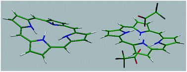
The porphine dications with different acids are readily decomposed when heated or even dried at room temperature. Accordingly, the optimum structure of H2P and H4P(CF3COO)2 was studied by ab initio calculations. Due to the crucial role played by the counter anion on the spectral features and the optimized structure3,14,15,17 of porphyrin dications, the calculations were performed on both the bare porphine dication, [H2P]2+ and H4P(CF3COO)2. Also, the calculations were performed on the dication of meso-tetra(n-butyl)porphyrin, H2T(n-Bu)P. X-ray crystallographic studies showed a symmetrically saddle distorted porphyrin core for H4T(n-Bu)P(CF3COO)2. Furthermore, the changes in different bond lengths and angles as well as the internal distances of porphyrin core were calculated; the changes in the macrocycle bond lengths and bond angles termed in-plane nuclear reorganization (IPNR) was proposed as a possible cause of the observed shifts in the electronic spectra of nonplanar porphyrins.1 The optimized structure of H2P, [H4P]2+ and H4P(CF3COO)2 are shown in Fig. 6; the results show a planar, saddle and waved conformation for H2P, [H4P]2+ and H4P(CF3COO)2, respectively. X-ray crystallographic studies show that protonation of porphyrins results in a saddled-shaped porphyrin core for most meso-tetra(aryl)- and meso-tetra(alkyl)porphyrins.7,13,21 As may be seen from Fig. 6, the saddle conformation of [H4P]2+ changes to a waved one in the presence of the two counter anions. In other words, H4P(CF3COO)2 adopts a conformation different from that of the other porphyrin dications; in this structure, instead of the two opposite pyrrole rings,3,7,13,21 the two adjacent rings are tilted above and below the porphyrin mean plane. The calculated bond lengths and angles of H2P, [H4P]2+ and H4P(CF3COO)2 are summarized in Table 3. The degree of saddle distortion of porphyrin core of [H4P]2+ is 9.3 (Fig. 6). Interestingly, the presence of two trifluoroacetate anions changes the saddle-shaped [H4P]2+ to a waved conformation.


image file: c6ra12520d-f6.tif
Fig. 6 The optimized structure of H2P, [H4P]2+ and H4P(CF3COO)2.
Table 3 Bond lengths of H2P, [H4P]2+ and H4P(CF3COO)2
Bond lengths H2P [H4P]2+ Bond length changea (%) H4P(CF3COO)2 Bond length changea (%)
a The values are relative to those of H2P.
N–H 1.022 1.022 0 1.056 3.32
N–Cα 1.366 1.397 2.26 1.374 0.5
Cα–Cβ 1.447 1.435 0.82 1.447 0
Cβ–Cβ 1.372 1.387 1.09 1.381 0.65
Cα–Cmeso 1.401 1.405 0.4 1.408 0.28


In this structure, a tilt angle of 10.1 degrees was found for the two oppositely directed pyrrole rings. The optimized structure of [H4T(n-Bu)P]2+ and H4T(n-Bu)P(CF3COO)2 are shown in Fig. 7. It is observed that the latter has an increased saddle distortion (20.4) relative to the former (13.5). In other words, the addition of two counter anions to the bare dication only increases the magnitude of saddle distortion of the aromatic macrocycle.


image file: c6ra12520d-f7.tif
Fig. 7 The optimized structure of [H4T(n-Bu)P]2+ and H4T(n-Bu)P(CF3COO)2.

Tables 3 and 4 summarize the changes in various bond angles and lengths of H2P upon protonation with CF3COOH. Also, the changes of internal distances are shown in Table 5. According to Tables 4–6, there is no changes larger than ca. 3% in the bond lengths and angles of [H4P]2+ and H4P(CF3COO)2 compared to those of H2P. However, diprotonation leads to remarkable changes in the internal distances of H4P(CF3COO)2.

Table 4 Bond angles of H2P, [H4P]2+ and H4P(CF3COO)2
Bond angles H2P [H4P]2+ Bond angle changea (%) H4P(CF3COO)2 Bond angle changea (%)
a The values are relative to those of H2P.
Cα–N–Cα 107.95 108.09 0.12 109.97 1.87%
N–Cα–Cβ 109.1 107.4 1.55 107.7 1.28%
N–Cα–Cmeso 125.55 125 0.43 126.1 0.43%
Cα–Cβ–Cβ 106.6 108 1.33 107.51 0.85%
Cα–Cmeso–Cα 126.9 127 0.07 128.52 1.27%
Cmeso–Cα–Cβ 125.41 127.5 1.66 126.14 0.58%


Table 5 The internal distances of H2P and H4P(CF3COO)2
Internal distances H2P H4P(CF3COO)2 Changea (%)
a The values are relative to those of H2P.b Hydrogen bonds between [H4P]2+ and the counteranions.
C10–C20 6.882 6.725, 7.144 2.28, 3.81
N1–N2 2.932 3.158 7.71
N2–N3 2.932 2.946 0.48
N3–N4 2.932 3.237 10.40
N4–N1 2.932 2.949 0.58
N3–N1 4.042 4.355, 4.343 7.74, 7.45
NH⋯Ob 1.485, 1.625


Table 6 Bond lengths of H2P, [H3P]+ and H3P(CF3COO)
Bond lengths H2P [H3P]+ Bond length changea (%) H3P(CF3COO) Bond length changea (%)
a The values are relative to those of H2P.b Non-protonated ring.c Hydrogen bonds between [H3P]+ and the counteranion.
N–H 1.022 0.993 2.84 1.003 1.86
N–Cα 1.366 1.405 2.85 1.399 2.41
N–Cα 1.366 1.385b 1.39 1.385b 1.39
Cα–Cβ 1.447 1.459 0.83 1.462 1.04
Cα–Cβ 1.447 1.495b 3.32 1.385b 4.28
Cβ–Cβ 1.372 1.392 1.46 1.391 1.38
Cβ–Cβ 1.372 1.363b 0.65 1.363b 0.65
Cα–Cmeso 1.401 1.399 0.14 1.400 0.07
Cα–Cmeso 1.401 1.403b 0.14 1.402b 0.07
NH⋯Oc 1.953, 2.050, 2.085  


Although due to the increased basicity of porphyrin ring caused by the loss of planarity, the monocation of planar and sterically uncrowded porphyrins is hard to observe,34–36 the ab initio calculations were performed on [H3P]+ and H3P(CF3COO) to investigate the influence of the second acid molecule on their structure. The results are summarized in Tables 6–8.

Table 7 Bond angles of H2P, [H3P]+ and H3P(CF3COO)
Bond angles H2P [H3P]+ Bond angle changea (%) H3P(CF3COO) Bond angle changea (%)
a The values are relative to those of H2P.b Non-protonated ring.
Cα–N–Cα 107.95 109.2 1.15 109.47 1.41
Cα–N–Cα 107.95 106.8b 1.06 107.0 0.88
N–Cα–Cβ 109.1 107.5 1.46 107.48 1.48
N–Cα–Cβ 109.1 109.9b 0.73 109.7b 0.55
N–Cα–Cmeso 125.55 126.1 0.44 126.7 0.91
N–Cα–Cmeso 125.55 126.5b 0.76 126.45b 0.72
Cα–Cβ–Cβ 106.6 107.9 1.22 107.76 1.08
Cα–Cβ–Cβ 106.6 106.7b 0.09 106.8b 0.18
Cα–Cmeso–Cα 126.9 126.4 0.39 127.2 0.24
Cα–Cmeso–Cα 126.9 125.8b 0.87 125.75b 0.90
Cmeso–Cα–Cβ 125.41 123.3 1.68 125.5 0.07
Cmeso–Cα–Cβ 125.41 123.5b 1.52 123.8b 1.28


Table 8 The internal distances of H2P and H3P(CF3COO)
Internal distances H2P H3P(CF3COO) Changea (%)
a The values are relative to those of H2P.b Non-protonated ring.
C10–C20 6.882 6.997 1.70
N1–N2 2.932 3.022 3.07
N2–N3 2.932 2.970 1.29
N3–N4 2.932 2.924b 0.27
N4–N1 2.932 2.950b 0.61
N3–N1 4.042 4.196, 4.189b 3.81, 3.64b


The optimized structures of [H3P]+ and H3P(CF3COO) are demonstrated in Fig. 8. The two opposite pyrrole rings of the former are tilted above the porphyrin mean plane and the other one is tilted below this plane. Also, the unprotonated pyrrole ring is nearly in the plane formed by the meso carbon atoms. In the optimized structure of H3P(CF3COO), the average tilting angle of pyrrole rings is greater than that of [H3P]+. Furthermore, the three pyrrole rings are tilted above the porphine mean plane and form a nearly dome type conformation that probably occurs to optimize the hydrogen bonds3 with CF3COO anions. In comparison with the optimized structure of [H4P]2+ and H4P(CF3COO)2, a decrease in the average degree of saddle distortion as well as the symmetry of the structure is observed. On the other hand, the optimized structure of H3P(CF3COO) shows the crucial role played by hydrogen bonds in determining the structure of porphyrin protonated species.


image file: c6ra12520d-f8.tif
Fig. 8 The optimized structure of [H3P]+ and H3P(CF3COO). The numbers show the degree of saddle distortion of different pyrrole rings.

The calculated frontier molecular orbitals of H4P(CF3COO)2 are shown in Fig. 9. For the HOMO−1 the electron densities are largest on the α and β carbon atoms. Also, the electron densities of the H orbital are largest on the meso carbon atoms and pyrrole nitrogens. Therefore, the HOMO−1 and H orbital are indeed the a1u and a2u orbitals obtained by the four orbital model.11


image file: c6ra12520d-f9.tif
Fig. 9 Calculated frontier molecular orbitals of H4P(CF3COO)2.

Also, the estimated wavelengths of the π to π* transitions and the involved orbitals for the different dihedral angles are demonstrated in Table 9. The comparison of the results with those calculated for H2P25 clearly shows the red shift of the Soret band (S4, Table 9) and the blue shift of the Q(0,0) band (S2) with respect to the corresponding bands of free base H2P.25 It is noteworthy that in a previous work, we have shown that increasing the dihedral angle from 0 to 30° is accompanied with the red shift of the Soret and Q(0,0) bands of H2P.25 Herein, the vertical transition energies, determined at the TD-B3LYP/cc-pVDZ level of theory demonstrates that the presence of two acid molecules above and below the H2P mean plane led to the blue shift of the Q(0,0) band. Accordingly, the observed blue shift of the band is attributed to the shift of electron density from the pyrrolenine nitrogen atoms to the acid molecules.15

Table 9 Vertical transition energies, oscillator strengths and configurations determined at the TD-B3LYP/cc-pVDZ level of theory
Vertical transition eV λ (nm) Oscillator strengths Contributed orbitals Weight (%)
a H and L show HOMO and LUMO orbital, respectively.
S1 2.29 540.8 6 × 10⁻⁴ H–L+1a 53
H−1–L 45
S2 2.40 516.7 3 × 10⁻⁴ H–L 53
H−1–L+1 45
S3 3.35 369.5 0.43 H−1–L 33
H−3–L+1 33
S4 3.44 360.1 0.62 H−1–L+1 39
H–L 35


Conclusion

In summary, protonation of H2P with weak and strong acids in different solvents was studied by UV-vis and 1H NMR spectroscopy. Interestingly, in contrast to the dication of porphyrins with no cationic or anionic substituents at the β and meso positions, the dication of H2P is water soluble and therefore was studied in water as well as in dichloromethane and DMF. While in the UV-vis spectra of all known porphyrin diacids, the Soret band shifts to red, in the case of H2P, the band was found to shift to higher or lower wavelengths depending on the used acid. Also, large blue shifts were observed in the position of the Q(0,0) band of H2P. These observations are probably due to the absence of any substituents at the porphine periphery. Furthermore, ab initio calculations predicted an unusual wave conformation of H4P(CF3COO)2 that is different from the common saddle (or nearly saddle) distortion observed in the structure of the previously reported porphyrin diacids. The energies of absorption bands of H4P(CF3COO)2 calculated at the TD-B3LYP/cc-pVDZ level of theory are in accord with the red shift of the Soret band and the blue shift of the Q(0,0) bands. The results of this calculations also reveal the role played by the acid molecules on the blue shift of the Q(0,0) band of the diprotonated species. In the 1H NMR spectra, the β and meso protons of H2P shifted to downfield which shows the dominance of the σ-inductive effects of the acid molecules over the ring current effects. This observation is in accord with the relatively slight out-of-plane deformation of porphine core (ca. 10 degrees), calculated by the ab initio studies. The 1H NMR spectrum of H4P(CF3COO)2 in D2O gave evidence of the formation of strong hydrogen bonds between [H4P]2+ and water molecules instead of the trifluoroacetate anions. Ab initio calculations performed on the monoprotonated and diprotonated H2P in the presence and the absence of the counteranion (CF3COO) revealed the crucial role played by the counteranion on the optimized conformation of the protonated species, especially in the case of the diprotonated one. On the other hand, changes of approximately 0–4% in the bond lengths and angles and 0–10% in the internal distances of H2P were found upon protonation with CF3COOH. Accordingly, changes in the macrocycle bond lengths and bond angles, termed in-plane nuclear reorganization (IPNR) may be also involved in the observed changes in the optical absorption bands of H2P upon protonation with different acids.

Acknowledgements

Financial Support by the Institute for Advanced Studies in Basic Sciences (IASBS) and University of Isfahan is gratefully acknowledged. Also, the computational support for this work provided by the computer centre of the Paris sud-11 University is gratefully acknowledged.

References

  1. R. E. Haddad, S. Gazeau, J. Pe'caut, J. C. Marchon, C. J. Medforth and J. A. Shelnutt, J. Am. Chem. Soc., 2003, 125, 1253 CrossRef CAS PubMed.
  2. A. Y. Lebedev, M. A. Filatov, A. V. Cheprakov and S. A. Vinogradov, J. Phys. Chem. A, 2008, 112, 7723 CrossRef CAS PubMed.
  3. A. Rosa, G. Ricciardi, E. J. Baerends, A. Romeo and L. M. Scolaro, J. Phys. Chem. A, 2003, 107, 11468 CrossRef CAS.
  4. O. S. Finikova, A. V. Cheprakov, P. J. Carroll, S. Dalosto and S. A. Vinogradov, Inorg. Chem., 2002, 41, 6944 CrossRef CAS PubMed.
  5. J. A. Shelnutt, X. Z. Song, J. Ma, S. L. Jia, W. Jentzena and C. J. Medforth, Chem. Soc. Rev., 1998, 27, 31 RSC.
  6. M. O. Senge, Z. Naturforsch., B: J. Chem. Sci., 2000, 55, 336 CAS.
  7. A. Stone and E. B. Fleischer, J. Am. Chem. Soc., 1968, 90, 2735 CrossRef CAS.
  8. S. Zakavi, H. Rahiminezhad and R. Alizadeh, Spectrochim. Acta, Part A, 2010, 77, 994 CrossRef PubMed.
  9. S. Rayati, S. Zakavi, A. Ghaemi and P. J. Carroll, Tetrahedron Lett., 2008, 49, 664 CrossRef CAS.
  10. M. Meot-Ner and A. D. Adler, J. Am. Chem. Soc., 1975, 97, 5107 CrossRef CAS.
  11. M. Gouterman, J. Mol. Spectrosc., 1961, 6, 138 CrossRef CAS.
  12. S. Aronoff, J. Phys. Chem., 1958, 62, 428 CrossRef CAS.
  13. B. Cheng, O. Q. Munro, H. M. Marques and R. Scheidt, J. Am. Chem. Soc., 1997, 119, 10732 CrossRef CAS.
  14. A. Rosa, G. Ricciardi and E. J. Baerends, J. Phys. Chem. A, 2006, 110, 5180 CrossRef CAS PubMed.
  15. S. Zakavi and N. G. Gharab, Polyhedron, 2007, 26, 2425 CrossRef CAS.
  16. S. Zakavi and M. Najafi ragheb, Inorg. Chem. Commun., 2013, 36, 113 CrossRef CAS.
  17. S. Zakavi and S. Hoseini, RSC Adv., 2015, 5, 106774 RSC.
  18. R. J. Abraham, A. H. Jackson and G. W. Kenner, J. Chem. Soc., 1961, 3468 RSC.
  19. R. I. Walter, E. C. A. Ojadi and H. Linschitzll, J. Phys. Chem., 1993, 97, 13308 CrossRef CAS.
  20. O. Ohno, Y. Kaizu and H. Kobayashi, J. Chem. Phys., 1985, 82, 1779 CrossRef CAS.
  21. M. O. Senge, I. Bischoff, N. Y. Nelson and K. M. Smith, J. Porphyrins Phthalocyanines, 1999, 3, 99 CrossRef CAS.
  22. S. Neya and N. Funasaki, Tetrahedron Lett., 2002, 43, 1057 CrossRef CAS.
  23. S. Zakavi, R. Omydian, L. Ebrahimi and F. Heidarizadi, Inorg. Chem., 2011, 14, 1010 CAS.
  24. J. Lurie, Handbook of Analytical Chemistry, MIR Publishers, Moscow, 1975 Search PubMed.
  25. S. Zakavi, R. Omydian and S. Talebzadeh, Polyhedron, 2013, 49, 36 CrossRef CAS.
  26. R. Ahlrichs, M. Bär, H. Baron, R. Bauernschmitt, S. Böcker, M. Ehrig, K. Eichkorn, S. Elliott, F. Furche and F. Haase, TURBOMOLE V6. 2 2010, A Development of University of Karlsruhe and Forschungszentrum Karlsruhe GmbH, TURBOMOLE GmbH, 1989–2007, since 2007 Search PubMed.
  27. R. Ahlrichs, M. Bär, M. Häser, H. Horn and C. Kölmel, Chem. Phys. Lett., 1989, 162, 165 CrossRef CAS.
  28. R. Ahlrichs, Phys. Chem. Chem. Phys., 2004, 6, 5119 RSC.
  29. F. Haase and R. Ahlrichs, J. Comput. Chem., 1993, 14, 907 CrossRef CAS.
  30. T. H. Dunning Jr, J. Chem. Phys., 1989, 90, 1007 CrossRef.
  31. R. A. Kendall, T. H. Dunning Jr and R. J. Harrison, J. Chem. Phys., 1992, 96, 6796 CrossRef CAS.
  32. C. L. Janssen and I. M. B. Nielsen, Chem. Phys. Lett., 1998, 290, 423 CrossRef CAS.
  33. C. Hättig, J. Chem. Phys., 2003, 118, 7751 CrossRef.
  34. A. B. Rudine, B. D. DelFatti and C. C. Wamser, J. Org. Chem., 2013, 78, 6040 CrossRef CAS PubMed.
  35. T. Honda, T. Kojima and S. Fukuzumi, Chem. Commun., 2009, 4994 RSC.
  36. O. S. Finikova, A. V. Cheprakov, P. J. Carroll, S. Dalosto and S. A. Vinogradov, Inorg. Chem., 2002, 41, 6944 CrossRef CAS PubMed.

Footnote

Electronic supplementary information (ESI) available. See DOI: 10.1039/c6ra12520d

This journal is © The Royal Society of Chemistry 2016
Click here to see how this site uses Cookies. View our privacy policy here.