Investigating the metabolic fingerprint of term infants with normal and increased fetal growth

C. Fotakisa, M. Zogab, C. Baskakisa, Th. Tsiakaa, T. Boutsikoub, D. D. Brianab, K. Dendrinoub, A. Malamitsi-Puchner*b and P. Zoumpoulakis*a
aInstitute of Biology, Medicinal Chemistry and Biotechnology, National Hellenic Research Foundation, Athens, Greece. E-mail: pzoump@eie.gr; Fax: +30 210 7273 831; Tel: +30 210 7273 853
bDepartment of Neonatology, National and Kapodistrian University of Athens, Athens, Greece. E-mail: amalpu@med.uoa.gr; Fax: +30 210 3303110; Tel: +30 6944443815

Received 12th May 2016 , Accepted 11th August 2016

First published on 15th August 2016


Abstract

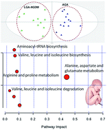
An NMR metabolomic approach was employed to highlight the metabolic changes underlying prenatal disorders and determine metabolites that could serve as potential markers in relation to large for gestational age (LGA) newborns. In this holistic study, multivariate data analysis elicited information from the NMR spectra and probed to metabolic signatures of macrosomic fetuses. Moreover, metabolic trends that characterize LGA from mothers diagnosed with gestational diabetes mellitus (LGA-GDM), as well as LGA from mothers not diagnosed with GDM (LGA-NGDM) were framed. Results obtained from maternal and umbilical cord (UC) samples indicated that LGA fetuses present alterations especially in the aminoacid metabolism as compared to Appropriate for Gestational Age (AGA) cases. Clear discrimination of LGA-NGDM from LGA-GDM was achieved both in maternal and in UC samples' blood. The role of glutamine and alanine together with four essential (valine, leucine, isoleucine, threonine) aminoacids, as well as the role of glycerol and glucose is emphasized for the case of maternal LGA samples' differentiation. Glycine and histidine only contributed to the differentiation of UC samples, the former characterized the AGA cases, while the latter was ascribed to both LGA-GDM and LGA-NGDM cases. Interestingly, both UC and maternal LGA-GDM samples were characterized by increased levels of N-acetylglutamic and acetoacetic acids. The OPLS-DA models were validated with permutation testing and ROC curves. In conclusion, this study indicates that NMR metabolomics may enable the detection of metabolic changes associated with LGA prenatal disorders.


1 Introduction

Fetal growth is influenced by a complex interplay of various genetic and environmental factors. There are several determinants of intrauterine growth that affect the intrinsic growth potential of a neonate, including maternal age, race, height and weight at the beginning of pregnancy, gestational age, parity as well as neonatal gender and weight.1 Under normal conditions infants develop appropriately for gestational age (AGA), while infants with a larger than expected growth (above the 90th percentile) are referred as large for gestational age (LGA).2 Fetal overgrowth is a risk factor for operative delivery, traumatic birth injury,3 and perinatal hypoxia, while it is implicated in long term morbidity, due to obesity, cardiovascular disease and metabolic syndrome in adult life.4 Moreover, it has been associated with increased risk of type 2 diabetes in the later life of the infant.5

Among causes for macrosomia are constitutional factors (parental BMI), gestational diabetes (GDM), maternal obesity or excessive weight gain during pregnancy, several syndromes (Beckwith–Wiedemann, Sotos) and erythroblastosis fetalis.

Lately, patterns of gestational weight gain (GWG) have been correlated with LGA and small for gestational age (SGA) births among overweight or obese women. Thus, the pattern of weight gain during pregnancy may be linked to abnormal fetal growth, with higher risk of LGA in women with consistently high GWG, (reflecting excessive nutrition and weight gain during pregnancy).6,7

GDM is a common metabolic abnormality which affects annually ∼2–5% of pregnancies. GDM is defined as any degree of glucose intolerance with onset, or first recognition during pregnancy.8 GDM patients are usually identified during the second or third trimester of pregnancy, while glucose tolerance usually returns to normal range within 6 weeks after delivery. GDM may lead to type 2 diabetes and if not identified and controlled in time, it may adversely affect renal and liver function (especially the creatinine, transaminases – ALT, ALP – and serum bilirubin levels).9 Risk factors for the development of GDM include advanced maternal age, race, history of previous macrosomic infants, a family history of non-insulin dependent diabetes mellitus (NIDDM) or GDM, poor glycemic control and a high pre-pregnancy body mass index (BMI).10,11

GDM is characterized by a cluster of symptoms resembling type 2 diabetes and metabolic syndrome, including insulin resistance, glucose intolerance, hyperlipidemia, impaired beta cell function and endothelial dysfunction.12 Maternal hyperglycemia increases fetal insulin levels according to the Pedersen hypothesis, leading to accelerated growth and macrosomia in the fetus.13 Many reports have signified the crucial role of fetal insulin as the most important growth hormone during intrauterine development, while elevated fetal insulin levels are related to accelerated growth.1,14

The role of the fetus and the prenatal metabolic environment known as “perinatal programming”, in relation to certain chronic diseases in the adult life has been previously reported. More specifically, pathological conditions including obesity, cardiovascular problems or metabolic syndrome may originate from intrauterine life.15,16 The significance of metabolic dysregulation in early pregnancy has been highlighted and correlated to inborn errors and fetal growth up to the increase of the risk of obesity at preschool age.17

In the advent of the –omics era, genomics, trancriptomics, proteomics and metabolomics, have been increasingly utilized in the field of early diagnosis of infant diseases. Particularly, the metabolomics approach implements state-of-the-art, high throughput and robust techniques, such as Nuclear Magnetic Resonance (NMR) spectroscopy and/or high resolution (HR) gas or liquid chromatography mass spectrometry (MS) in order to elicit valuable information regarding biological processes and systems.18,19

HR NMR spectroscopy elaborates minimally invasive procedures and yields high reproducibility, which may assess the metabolic profile of biofluids and other biological samples by relating the metabolic responses of living systems to disease, as well as to toxicological or nutritional stimuli. Thus, NMR metabolomics has hitherto acceded to a disease diagnostic tool by examining various tissues and elucidating biomarkers of prenatal health.20 For instance, an NMR metabolomic study focused on urine samples from premature neonates to investigate the role of possible confounders and signature specificity in order to frame the metabolic variations ascribing to prenatal disorders and pinpoint early biomarkers.21 Another NMR metabolomic research on umbilical cord plasma delineated the maternal–fetal nutrient exchange in preterm infants. Particularly, 1H NMR spectra of plasma acquired after birth from umbilical vein, umbilical artery and maternal blood successfully highlighted the metabolic adaptations associated with premature birth.22 Furthermore, NMR complemented with multivariate analysis discriminated between women with idiopathic recurrent spontaneous miscarriage and controls, thus providing useful information in understanding metabolic dysregulation during the implantation window.23 Moreover, another NMR-based metabolomic study assessed the time-related urinary metabolic profiles of appropriate for gestational age (AGA), with intrauterine growth restriction (IUGR) and large for gestational age (LGA) newborns.24

The importance of metabolomics towards the detection and prediction of metabolic alterations and diseases related to fetal growth as well as early diagnosis of metabolic syndrome has been highlighted in a recent review.25

Experimental studies have indicated that fetal malnutrition, either excessive or insufficient, may alter the metabolic processes of the fetus in a permanent manner and increase the risk of future chronic diseases.26,27

Research on biofluids for the identification of prenatal biomarkers highlights the strong correlation between biomarkers found in these tissues and fetal malformations, preterm delivery, premature rupture of membranes, gestational diabetes mellitus, preeclampsia, neonatal asphyxia, and hypoxic-ischemic encephalopathy.28

In such metabolomic studies the large amount of data produced, necessitates the use of multivariate data analysis. For this purpose, pattern recognition tools act as a filter to remove the noise and detect the “information” that resides in the collected data. Then, the resonance peaks of a metabolite can be related to the biological condition of the analyzed samples. The most commonly employed methods comprise principal component analysis (PCA), partial least squares (PLS), orthogonal projection to latent structures (O-PLS) and discriminant analysis (PCA-DA, PLS-DA, O-PLS-DA).29,30 Furthermore, special care should be taken to ensure that the utilized statistical methods do not predict randomly. Towards this aim internal and external validation steps have to be followed to avoid model over-fitting and ensure that the obtained results are unbiased.31–33

In the present holistic study, we hypothesized that alterations in the metabolome between LGA and AGA mothers/fetuses will provide insights for possible key metabolites related to LGA abnormalities. Furthermore, possible alterations in the metabolome of diabetic versus non diabetic LGA pregnancies might reflect the pathogenetic pathways linking diabetes to macrosomia.

2 Experimental

The study protocol was approved by the Ethics Committee of our teaching hospital and informed consent was signed by all recruited mothers.

2.1 Patients – methods

The characteristics of all the women participating in this study are provided in Table S1 (ESI). For each fetus/neonate the customized centile was calculated taking into consideration several parameters of fetal growth according to the computer generated GROW (Gestation Related Optimal Weight) program.

The main cause for deliveries by caesarean section in the AGA group was previous caesarean section or maternal request, while no pathological conditions were involved.

Blood was collected from full-term parturients being at the first stage of labor ​or before anesthesia in cases of elective cesarean section, as well as from the doubly clamped umbilical cord (UC-mixed arterio-venous blood) of their singleton infants at birth, representing fetal state.

Samples were left at room temperature for 45 min to allow clotting followed by centrifugation at 1500 × g for 10 min. Five hundred microliter aliquots of the supernatant (serum) were transferred into sterile cryo vials, frozen, and stored at −80 °C.

The study group was divided in training and test sets. The training set consists of LGA-NGDM (n = 20), LGA-GDM (n = 12), and AGA (n = 28) controls. The external validation test consists of 18 samples, specifically LGA-NGDM (n = 6), LGA-GDM (n = 4) and AGA (n = 8).

In summary, the AGA sample-set consists of 36 samples (18 from maternal and 18 from umbilical cord blood), divided in two sets (training set with 28 samples and test set with 8 samples). Similarly, the LGA sample-set (including LGA-GDM and LGA-NGDM) consists of 42 samples (21 from maternal and 21 from umbilical cord blood), divided in a training set (n = 32 including 20 LGA-NGDM and 12 LGA-GDM) and a test set (n = 10 including 6 LGA-NGDM and 4 LGA-GDM).

2.2 Nuclear magnetic resonance spectroscopy (NMR)

Nuclear Magnetic Resonance (NMR) spectroscopy and Carr–Purcell–Meiboom–Gill (CPMG) pulse sequence was employed on maternal and fetal (UC) serum. The samples were thawed at room temperature 30 min before performing the NMR experiments.

Samples were prepared adding 140 μL phosphate buffer in D2O to 400 μL of serum. After centrifugation at 4 °C for 10 minutes at 10[thin space (1/6-em)]000g, 50 μL sodium maleate was added as internal standard to 500 μL of the supernatant and transferred to 5 mm NMR tubes. Sodium maleate was chosen since it provides a distinct peak in the 1H NMR spectrum and does not interact with blood serum proteins which could cause spectra normalization problems when using CPMG pulse sequence, as in the case of trimethylsilyl propionate (TSP). NMR spectra were acquired on a Varian-600 MHz NMR spectrometer at 25 °C equipped with a triple resonance probe {HCN}. The CPMG pulse sequence was applied with 128 transients collected with 64k data points. Relaxation delay was set to 6 s. Proton spectra were referenced at the resonance peak of sodium maleate (5.95 ppm). Receiver gain was kept constant for all acquisitions.

A series of 2D experiments, gCOSY, zTOCSY, gHMBCad, gHSQCad were recorded at 25 °C and permitted the assignment of the existing metabolites. The acquisition parameters for gCOSY were: spectral width (SW) 7225.4 Hz, t1 increments 256, acquisition time 0.150 s, number of scans 128, 1084 data points, receiver gain 30 and relaxation delay 1 s. The acquisition parameters for zTOCSY was performed with spectral width (SW) 7225.4 Hz, t1 increments 256, number of scans 128, acquisition time 0.283 s, 2048 data points, receiver gain 30 and relaxation delay 1 s. The acquisition parameters for gHSQCad were: f2 spectral width (SW) 7225.4 Hz, f1 spectral width (SW) 30[thin space (1/6-em)]165.9 Hz, t1 increments 256, number of scans 128, acquisition time 0.150 s, 1084 data points, receiver gain 30 and relaxation delay 1 s. The acquisition parameters for gHMBCad were: f2 spectral width (SW) 7225.4 Hz, f1 spectral width (SW) 36[thin space (1/6-em)]199.1 Hz, t1 increments 256, number of scans 128, acquisition time 0.150 s, 1084 data points, receiver gain 40 and relaxation delay 1 s.

The interpretation of 2D spectra was performed with the use of MestReNova v.10.1 software. The identification procedure was also assisted by the combined use of a reference metabolic 1H NMR database (Chenomx Suite 7.6), spiking experiments and literature data.

2.3 Data reduction and spectral alignment

The NMR spectra were reduced into buckets of 0.001 ppm. The D2O (4.6–4.8 ppm) region was removed. The spectra were aligned, normalized to the standardized area of the sodium maleate peak and converted to ASCII format using the Mnova processing template.

2.4 Univariate data analysis

Students' t-test was used to determine whether there were any statistical differences in pairwise comparisons between the AGA, LGA-GDM and LGA-NGDM groups, either in maternal or in UC samples. Moreover, the Spearman correlation was applied to extract correlation coefficients among the patient characteristics. These calculations were performed with the SPSS (IBM SPSS Statistics, version 19.0, Chicago, IL, USA) statistical software for Windows.

2.5 Multivariate data analysis

The obtained spectra were submitted to the Simca 14 software for statistical analysis. First, the exploratory Principal Component Analysis (PCA) was employed in order to acquire a general insight and visualize any relation (trends, outliers) among the observations (samples). A PCA model estimates the systematic variation in a data matrix by a low dimensional model plane. The spectral data was mean-centered with no scaling (Ctr) and the PCA model was extracted at a confidence level of 95%.

Then classification analysis ensued by further subjecting the data set to SIMCA (Soft Independent Modelling of Class Analogy), PLS-DA, and OPLS-DA. In SIMCA the class homogeneity is examined by investigating each class with a unique PCA model.29,32

OPLS-DA, is an extension of the supervised PLS regression method that manage to increase the quality of the classification model by separating the systematic variation in X into two parts, one that is linearly related to Y (predictive information) and one that is unrelated to Y (orthogonal information). The mathematical background and applications of these methods has been extensively discussed.29,32 The extracted OPLS-DA models were Pareto scaled (Par) at a confidence level of 95%. Particularly, the application of Par scaling allows any metabolites of low-medium intensity to affect the analysis only if they represent systematic variation.

Loading and contribution plots were extracted to reveal the variables which bear class discriminating power. Moreover, in order to improve model visualization and interpretation, S-line plots were extracted to detect the metabolites that influence most the group membership. VIP plots were employed for variable selection as they display the overall importance of each variable (X) on all responses (Y) cumulatively over all components. Terms with VIP larger than 1, are the most relevant for explaining Y.

2.6 Model validation tools

The quality of models (PCA & PLS/OPLS-DA) was described by the goodness-of-fit R2 (0 ≤ R2 ≤ 1) and the predictive ability Q2 (0 ≤ Q2 ≤ 1) values. The R2 explains the variation, thus constituting a quantitative measure of how well the data of the training set was mathematically reproduced. The overall predictive ability of the model is assessed by the cumulative Q2 representing the fraction of the variation of Y that can be predicted by the model, which was extracted according to the internal cross validation default method of SIMCA-P software. The Q2 is considered as de facto the default diagnostic parameter to validate PLS-DA models in metabolomics. In particular, all OPLS-DA models demonstrated high statistical values (R2 > 0.7 and Q2 ≥ 0.54), the difference between the goodness-of-fit and the predictive ability remained always lower than 0.3 (R2X(cum) − Q2(cum) < 0.3) and the goodness-of-fit never equaled to one (R2X(cum) ≠ 1). Therefore, since the extracted models abide by these rules, their robustness and predictive response are enhanced and over-fitting is effaced.32

Regression models have been validated using cross validation-analysis of variance (CV-ANOVA), with a P-value < 0.05. CV scores plots were elicited in order to indicate the sensitivity of a model to the exclusion of an observation of the work set. Furthermore, permutation tests were employed (999 permutations) in order to evaluate whether the specific classification of two classes in a model are significantly better than any other models obtained by randomly permuting the original groups attribution. An additional measure of PLS-DA model validity included the extraction of receiver-operator characteristic (ROC) curves to assess the ability of the PLS latent variable Tpred to correctly classify the test set. The area under the ROC (AUROC) was calculated. A perfect discrimination corresponded to an AUROC equal to 1. Finally, classification lists, misclassification tables depicting the proportion of correctly classified observations in the prediction set, and most importantly external data set have been employed, further attesting to the generalizability of the models.31–33

In this context, 60 samples were selected and randomly divided into two equal subsets. Supervised models (PLS-DA/OPLS-DA) were extracted for each of the subsets and the markers highlighted were in agreement for the cases compared. All supervised models demonstrated high statistical values (R2 > 0.7 and Q2 ≥ 0.65), the difference between the goodness-of-fit and the predictive ability remained always lower than 0.2 (R2X(cum) − Q2(cum) < 0.2). Moreover, these regression models were validated using cross validation-analysis of variance (CV-ANOVA), with a P-value < 0.05. Permutation testing was applied (999 permutations) to check the validity and the degree of overfit for the supervised models and finally receiver-operator characteristic (ROC) curves were extracted. Then we merged the two subsets and rerun all the aforementioned tests. Finally, an external validation set was utilized and was successfully incorporated to the total sample set. Again we rerun all the aforementioned tests in order to verify the validity of our results.

2.7 Metabolic pathway analysis

Pathway analysis of the exported dataset was analyzed using the online software of Metaboanalyst 3.0 (available: http://www.metaboanalyst.ca/)34 which is a free, user-friendly, and easily accessible tool for biomarker discovery, classification and pathway mapping.

3 Results and discussion

3.1 Metabolite identification

Assignment of spectral lines was performed using 2D NMR TOCSY, COSY, HSCQC and HMBC experiments complemented with results from ChenomX software and the online database HMDB (http://www.hmdb.ca/). The assigned metabolites are presented in Fig. 1 and in the Table S2 (ESI).
image file: c6ra12403h-f1.tif
Fig. 1 Assignment of proton NMR spectral lines. (1) R–CH3, (2) valine, (3) isoleucine, (4) leucine, (5) alanine, (6) lactic acid, (7) (CH2)n, (8) 3-hydroxy butyrate, (9) citric acid, (10) acetic acid, (11) acetoacetate, (12) CH[double bond, length as m-dash]CHCH2, (13) CH2CH2CO, (14) glutamic acid, (15) glutamine, (16) lysine, (17) [double bond, length as m-dash]CH–CH2–CH[double bond, length as m-dash], (18) creatine, (19) creatinine, (20) glucose, (21) tyrosine, (22) L-phenyl alanine, (23) L-histidine, (24) formic acid, (25) –CH[double bond, length as m-dash]CH–, (26) glycogen, (27) urea, (28) unsaturated lipid, (29) 1-methyl histidine, (30) choline, (31) glycerol, (32) glycine, (33) threonine, (34) TMAO, (35) betaine.

3.2 Exploring the sample pool

Starting the sample pool investigation, correlation analysis was used in order to investigate and highlight any potential confounders among the patient characteristics, namely mode of delivery, stage of endometriosis, gestational age, gender, booking BMI and BMI after delivery in relation to growth percentile. Results did not probe to strong correlations among the patient characteristics, besides mode of delivery and growth percentile (significant at the 0.01 level), which is expected, since infants with higher growth percentiles are mostly delivered with caesarean section (CS).

The first step in a multivariate data analysis workflow is to implement exploratory analysis such as principal component analysis (PCA). Then, classification analysis ensues by further subjecting the data set to partial least-squares (PLS), orthogonal-PLS (OPLS), and dynamic extensions thereof which are efficient to unveil potent biomarkers. The concept of these methods has been extensively discussed.29

On these grounds, a PCA model provided a general overview on the 78 samples (Fig. 2). In particular, along the first component the maternal and UC samples tend to discriminate, while a trend of the diabetic samples to cluster at the second quadrant is observed. Evidently, the PCA model provides information mainly for the variables that discriminate maternal and umbilical cord blood samples, while the impact of the growth and development levels of the fetus is masked.


image file: c6ra12403h-f2.tif
Fig. 2 PCA model with 2 components and 78 samples, Par scaled with a 95% confidence interval. R2X(cum) = 0.52, Q2(cum) = 0.45. Squares = maternal samples, circles = UC samples, red = LGA-GDM, blue = AGA, green = LGA-NGDM.

The metabolic signature of the maternal and UC samples is presented in a volcano plot (ESI, Fig. S1). Specifically, increased content of glycerol, threonine and pyruvic acid is ascribed to the UC samples, while the maternal samples displayed higher concentration in lipids, histidine, glycine and citric acid. In agreement to our results, Tea et al.,22 also reported increased levels of lipoproteins in maternal samples.

Another important result from these PCA scores plot is the grouping of the GDM samples. A contribution plot (ESI Fig. S2) highlighted the differences of these samples to the rest of the sample pool. Specifically, the diabetic samples were characterized by increased concentration of glucose, glycerol, alanine, glutamine and glutamate. On the other hand, lipids, organic acids and the rest of amino acids exhibited low concentration in these samples compared to the rest of the samples.

3.2.1 Investigation of the GDM group. To a step further, two PCA-class models were extracted for the LGA-NGDM samples (ESI Fig. S3A) and LGA-GDM (ESI Fig. S3B) in order to examine homogeneity. In these models a clear separation of maternal and umbilical cord samples was obtained only in the model regarding the LGA-NGDM samples. This is an important finding which supports our initial hypothesis of different metabolic signatures for the LGA-NGDM sample set between mothers and newborns. This finding may indicate a relatively hindered transport from maternal to fetal blood through umbilical cord for the LGA-NGDM cases.

Then, the GDM set was further divided into maternal and UC samples. Interestingly, a trend was displayed in the maternal LGA-GDM sample pool (ESI Fig. S5C). Specifically, the samples from GDM mothers under diet were localized in the 2nd and 3rd quadrants, as compared to GDM mothers under insulin treatment, which were gathered in the 1st and 4th quadrants.

3.3 Classification analysis – metabolite elucidation

The next step of the study, involved an attempt to delineate distinct metabolic signatures for each group, either in maternal or UC samples. For this purpose, pairwise comparisons through OPLS-DA models were implemented.
3.3.1 Maternal samples (LGA-NGDM, LGA-GDM and AGA).
3.3.1.1 LGA-NGDM vs. AGA. Most importantly, discrimination between LGA-NGDM and AGA maternal blood samples was clearly achieved along the first principal component (Fig. 3A). An S-line plot (Fig. 3D) highlights the key metabolites that contribute to this discrimination. In fact, LGA-NGDM samples exhibited increased levels of alanine, isoleucine, leucine, valine, glutamine, lipid (CH2[double bond, length as m-dash]CH2CO), citric acid, threonine, glycerol and glucose compared to AGA ones.
image file: c6ra12403h-f3.tif
Fig. 3 OPLS-DA models of maternal samples with A = 1 + 1 components, Par scaled and a 95% confidence interval, (blue circles: LGA-NGDM, red circles: AGA, green circles: LGA-GDM) (A) LGA-NGDM vs. AGA, R2X(cum) = 0.54, R2Y(cum) = 0.85, Q2(cum) = 0.73; (B) LGA-GDM vs. AGA, R2X(cum) = 0.36, R2Y(cum) = 0.95, Q2(cum) = 0.87; (C) LGA-GDM vs. LGA-NGDM, R2X(cum) = 0.59, R2Y(cum) = 0.93, Q2(cum) = 0.80; (D) S-line plot, (1) valine, (2) leucine, (3): isoleucine, (4) alanine, (5) lipid (CH2CH2CO), (6) glutamine, (7) citric acid, (8) & (9) α-glucose & β-glucose & glycerol, (10) threonine; (E) S-line plot, (1) N-acetylglutamate, (2) acetoacetic acid, (3) glucose; (F) S-line plot, (1) LDL (R–CH3), (2) leucine, (3) alanine, (4) lipid (CH2[double bond, length as m-dash]CH2CO), (5) lipid (CCH2), (6) N-acetyl glycoprotein, (7) lipid (aCH2), (8) acetic acid, (9) glutamine/glutamic acid, (10) creatinine, (11) pyruvic acid, (12) lipid (–N(CH3)3), (13) glucose, (14) glycerol, (15) guanidoacetic acid.

Alanine has been correlated with higher blood pressure, high energy intake, high cholesterol levels, and increased body mass index.35 Alanine plays a key role in glucose–alanine cycle between tissues and liver, enabling pyruvate and glutamate to be removed from the muscle and be reformed in the liver. During this process, alanine is transported by the bloodstream from the muscle tissues to the liver.35

Leucine is another essential amino acid which regulates insulin secretion and synthesis36 as well as protein synthesis by acting on multiple tissues and at multiple levels of metabolism.37 Of the three branched chain amino acids, leucine seems to be the most potent with regard to the above mentioned effects and may therefore be the most physiologically relevant.38

Additionally, higher levels of citrate have been previously traced in urine samples of LGA newborns. This metabolite is an intermediate of the Krebs tricarboxylic acid (TCA) cycle and differences in LGA compared with AGA possibly probe to changes in energy metabolism. However, it has to be noted that citrate as a TCA cycle intermediate, is also a positive modulator of other metabolic pathways, such as in the lipid synthesis.


3.3.1.2 LGA-GDM vs. AGA. The discrimination between LGA-GDM and AGA samples is depicted in an OPLS-DA model (Fig. 3B). The S-line plot (Fig. 3E) reveals the contribution of N-acetylglutamate, glucose and acetoacetate which are increased in LGA-GDM.

In alignment to our results it has been reported that there were no differences in triglyceride, VLDL, LDL and HDL cholesterol concentrations between women with GDM and controls throughout the pregnancy.39

Interactions between maternal glucose and fetal growth are not completely understood but there may be a causal link between low maternal blood glucose and low birth weight.22


3.3.1.3 LGA-NGDM vs. LGA-GDM. Similar data were obtained when focusing on LGA-NGDM and LGA-GDM maternal blood samples. In the OPLS-DA model (Fig. 3C) a clear separation along the first component was accomplished. The corresponding S-line plot (Fig. 4F) attributed the presence of leucine, glucose, acetic acid, glutamine, creatinine and lipids (CH2[double bond, length as m-dash]CH2CO, LDL R–CH3, CCH2, aCH2, –N(CH3)3), N-acetyl glycoprotein and alanine in LGA-GDM maternal samples, while pyruvic acid, glycerol and guanidoacetic acid in LGA-NGDM maternal samples.
image file: c6ra12403h-f4.tif
Fig. 4 OPLS-DA models of UC samples with A = 1 + 1 components, Par scaled and a 95% confidence interval, (blue circles: LGA-NGDM, red circles: AGA, green circles: LGA-GDM). (A) LGA-NGDM vs. AGA, R2X(cum) = 0.44, R2Y(cum) = 0.81, Q2(cum) = 0.60; (B) LGA-GDM vs. AGA, R2X(cum) = 0.57, R2Y(cum) = 0.85, Q2(cum) = 0.63; (C) LGA-NGDM vs. LGA-GDM, R2X(cum) = 0.37, R2Y(cum) = 0.82, Q2(cum) = 0.56; (D) S-line plot, (1) lipid (–CCH2), (2) N-acetyl glycoprotein, (3) lipid (aCH2), (4) lipid (N(CH3)3), (5) betaine, (6) diethylamine, (7) choline of phospholipids, (8) glycine, (9) glycerol; (E) S-line plot, (1) valine, (2) leucine, (3) isoleucine, (4) lysine, (5) lipid (aCH2), (6) N-acetyl glutamine, (7) glutamine/glutamic acid, (8) acetoacetic acid, (9) creatinine, (10) threonine, (11) histidine; (F) S-line plot, (1) glucose, (2) valine, (3) alanine, (4) glutamine, (5) histidine.

N-acetyl signals from the glycoproteins in the maternal samples arise from acute phase glycoproteins known to reflect inflammatory status.22

High levels of creatinine in maternal LGA-GDM blood serum have been identified in accordance to previous findings, when compared to normal pregnant women.11,40 In general, creatinine is associated with muscle metabolism and is an indicator for kidney (dys)function as well as a warning sign of impending renal disease in GDM pregnancies.9 Glycerol is important for lipid metabolism and mitochondrial energy productions based on lipids. All these metabolic pathways are overexpressed in metabolic syndrome and its acute expression in late preeclampsia.28 Regarding the increased levels of glycerol in LGA-NGDM maternal samples, this may be related to fatty acids and glycerol metabolism disorders. Glycerol and free fatty acids (FFA) are produced from lipolysis of triglycerides stored in adipose tissue. The glycerol formed during the hydrolysis of glycerides is not reutilized in the adipose tissue, but is either oxidized in, or transformed to glucose, in glycerokinase containing tissues (kidneys, intestinal mucosa, liver).41 In an older study of UC venous blood samples from term infants, an increase of glycerol and FFA was evident within a short time after delivery.42 Increased levels of glycerol may be explained by its poor utilization by the adipose tissue mainly due to lack of glycerokinase from kidneys or liver.42

Second, the significant increase in acetate in the LGA-GDM group might be attributed to the lower levels of non-essential fatty acids, implying higher insulin sensitivity in adipose tissue, allowing for a stronger suppression of lipolysis.

Furthermore, increased levels of lipids were found for LGA-GDM cases. A previous study presented that in well-controlled GDM pregnancies, maternal lipids are strong predictors for fetal lipids and fetal growth. More specifically, LGA-NGDM newborns showed high FFA concentrations which may explain the increase observed in lipid NMR peaks. Interestingly, elevated concentration of leucine has been associated to GDM through an increased activity of the syncytiotrophoblast amino acid transporter system A in microvillous plasma membranes. Thus, leucine transport was found to be increased in LGA-GDM cases which may contribute to the accelerated fetal growth.43

The intrauterine signs of insulin resistance in LGA-NGDM infants could be responsible for the elevated FFA levels which might reflect a reduced effect of insulin inhibiting lipolysis or augmented lipolytic activity due to their increased adipose tissue mass.44

Apart from GDM which may lie behind LGA newborns, a question arises relatively to whether pregnant women with LGA newborns had been properly examined for GDM in regular time intervals. This is supported by the fact that biomarkers which characterise maternal LGA-NGDM samples, such as leucine and glycerol can be associated with diabetes.

3.3.2 UC samples (LGA-NGDM, LGA-GDM and AGA).
3.3.2.1 LGA-NGDM vs. AGA. The differentiation of LGA-GDM to AGA was accomplished with the OPLS-DA model as presented in Fig. 4A. In particular, the corresponding S-line plot (Fig. 4D) pinpoints lipids (CCH2, aCH2, N(CH3)3) N-acetyl glycoprotein, choline of phospholipids, betaine, diethylamine, glycine and glycerol, the metabolites responsible for this discrimination.

These results confirmed those found in previous studies using classical techniques, where an elevation of lipids was reported in the plasma of women during uncomplicated pregnancy, with an increase in triglyceride, total cholesterol, VLDL, HDL and LDL levels, along with lipid levels in fetal umbilical cord and neonatal plasma below those reported in adults, meeting maternal and developing fetal energy requirements in late gestation.

Glycine, an important precursor of proteins and inhibitory neurotransmitters in the central nervous system, can be used as conjugating agent of bile acids. Neonates are exclusive tauro-conjugators, while normally the glyco-conjugation is not present until the third week of life. The glycine, in particular, reached higher contents in neonates with fetal malnutrition. A similar difference in the levels of glycine has been recently observed between SGA neonates and controls.24

Betaine is an essential osmolyte, a methyl donor for the remethylation of homocysteine to methionine and derives from either the diet or by the oxidation of choline. Lower plasma betaine and trimethylamine-N-oxide concentrations were observed in maternal plasma in case of fetal malformation. These data suggest that the malformed fetuses demand enhanced gluconeogenesis and tricarboxylic acids cycle (Krebs cycle), possibly due to hypoxic metabolism. Significant differences were observed also between normal pregnancies and cases which would later develop gestational diabetes. The decreases of betaine noted in LGA samples may reflect some changes affecting choline-related homocysteine–methionine conversion. In fact lower betaine levels have been found in the plasma of subjects suffering from metabolic syndrome, which comprises a number of different disorders, including diabetes.28


3.3.2.2 LGA-GDM vs. AGA. An OPLS-DA model was extracted (Fig. 4B) that successfully separated the AGA samples from the LGA-GDM samples along the first component. In light of the S-line plot (Fig. 4E) the metabolites ascribed to LGA-GDM samples compared to AGA are valine, leucine, isoleucine, lysine, aCH2, N-acetyl glutamine, acetoacetic acid, glutamine/glutamic acid, threonine, creatinine and histidine.

Acetoacetic acid is one of the ketone bodies. Other studies have also found that the concentration of creatinine in urine was higher in LGA, suggesting the presence of a metabolic-substrate deficient condition. This finding also applies to the UC samples of the current study, when AGA with LGA-GDM are compared. Besides sharing same metabolic changes, each group of UC samples exhibited also characteristic alterations.24


3.3.2.3 LGA-GDM vs. LGA-NGDM. An OPLS-DA model (Fig. 4C) between LGA-NGDM and LGA-GDM revealed (Fig. 4F) as important contributors for the GDM group separation, glucose, glutamine, valine, histidine and alanine.

Glutamine is the most abundant amino acid in blood and is involved in multiple pathways with a major role in the synthesis of purines and pyrimidines. It is also associated with energy supply to neonatal gut and to other rapidly dividing cells. Placental amino acid transport can be reduced due to either impaired fetal and placental growth or to decreased transporter concentrations.22 Moreover, the enhancement of the de novo synthesis and utilization of glutamine has been presented45 through increased secretion of glucocorticoids which takes place during stressful situations. Correlation of LGA with oxidative stress has already been established.46 The fact that increased levels of glutamine are observed both in maternal and umbilical cord LGA-GDM samples can be supported by the placental transfer mechanism from mother to the fetus.

Regarding the decreased glucose levels in GDM cases, previous studies have proposed a hypothesis correlating glucose decrease and gestation progress. Particularly, a decrease in glucose was shown as gestation progresses in women with hyperinsulinemic newborns through the increase of fetoplacental siphoning of maternal glucose, whereas glucose levels are increasing in mothers with normoinsulinemic newborns.47,48

3.4 Model validation steps

A clear differentiation of samples was observed in all the extracted OPLS-DA models, but to ascertain that these results are unbiased and reliable we employed certain validation steps (ESI Fig. S4 and S5).

In fact, the good performance of each model is indicated by the high values of R2Y and Q2Y, and especially their low difference. Moreover, to validate the goodness of fit and the predictability of these results, a random class permutation test (n = 999) was employed. All the permuted models showed lower R2Y values if compared with the original model's R2Y value and all the Q2 regression lines showed negative intercepts.

Finally, receiver operating characteristic (ROC) curves were generated for all the OPLS-DA models with the area under the receiver operating characteristic curves (AUROC) and all depicted excellent performance with an AUROC higher than 0.80.

These results strongly confirmed that the group separations in the OPLS-DA score plots were statistically significant and that the predictability was not due to over-fitting of the data.

3.5 Metabolite pathway analysis

Potential biomarkers selected based on the S-line plots from the corresponding OPLS-DA models were subjected to pathway analysis using MetPA (http://www.metaboanalyst.ca) in order to relate the framed metabolic patterns to the most relevant pathways.26

A hypergeometric test using over-representation analysis and pathway topology analysis (Fig. S6 and S7) were applied for the maternal samples where the altered metabolites were mainly involved in the aminoacyl-tRNA biosynthesis; valine, leucine and isoleucine biosynthesis; alanine, aspartate and glutamate metabolism; arginine and proline metabolism and valine, leucine and isoleucine degradation. The levels of metabolites for each gestational status contributing to these pathways have been framed in box plots for the maternal samples as presented in Fig. S8 (ESI).

Additionally, the biological pathway analysis indicated that alanine, aspartate and glutamate metabolism; aminoacyl-tRNA biosynthesis; valine, leucine and isoleucine biosynthesis; valine, leucine and isoleucine degradation and nitrogen metabolism were disturbed in the UC samples (ESI Fig. S9 and S10). The levels of metabolites in UC samples for each gestational status contributing to the alterations in these pathways have been framed in box plots as presented in Fig. S11 (ESI).

Interestingly, the pathways: aminoacyl-tRNA biosynthesis, valine, leucine and isoleucine biosynthesis and degradation and alanine, aspartate and glutamate metabolism were altered in both substrates. This suggests that the variations in the metabolites involved in these pathways are more closely related to the gestational age and occurrence of diabetes.

4 Conclusions

Summarizing, the role of valine, isoleucine, alanine, threonine, glutamine and leucine as potential markers for LGA development in maternal blood samples was presented (Table 1). Histidine and glycine were pinpointed as significant only in the UC samples. Interestingly, elevated concentration of N-acetylglutamic and acetoacetic acids characterized both UC and maternal LGA-GDM samples.
Table 1 A summary on markers extracted by the OPLS-DA models depicting in colour code (image file: c6ra12403h-u1.tif vs. image file: c6ra12403h-u2.tif vs. image file: c6ra12403h-u3.tif) their relation to metabolic pathways highlighted by the Metaboanalyst software (Ala = alanine, Ile = isoleucine, Leu = leucine, Val = valine, Gln = glutamine, Thr = threonine, Acac = acetoacetate, Pa = pyruvate, Gaa = guanidoacetate, Glu = glutamate, Cr = creatinine, Gly = glycine, His = histidine, Lys = lysine)
image file: c6ra12403h-u4.tif


This work demonstrated the value of NMR metabolomics as a complementary tool of prenatal diagnostics. Specifically, the holistic approach presented here, unveiled metabolites which could serve as potential prenatal disorder biomarkers related to LGA abnormalities at the time of delivery. These results provide valuable information for the metabolic signatures of LGA infants, in an effort to better define mechanisms behind macrosomia.

To our knowledge, this is the first study to show that discrimination of LGA-NGDM from LGA-GDM samples is possible both in maternal and UC blood.

The proposed metabolic signatures have to be validated at an earlier gestational age and in bigger cohorts, in order to obtain clinical use as diagnostic and prognostic biomarkers and help for future rational intervention against LGA condition.

Notes and references

  1. T. Henriksen, Acta Obstet. Gynecol. Scand., 2008, 87(2), 134–145 CrossRef PubMed.
  2. Z. Kerényi, G. Tamás, M. Kivimäki, A. Péterfalvi, E. Madarász, Z. Bosnyák and A. G. Tabák, Diabetes Care, 2009, 32, 2200–2205 CrossRef PubMed.
  3. B. M. Casey, M. J. Lucas, D. D. McIntire and K. J. Leveno, Obstet. Gynecol., 1997, 90, 869–873 CrossRef CAS PubMed.
  4. A. Ornoy, Reprod. Toxicol., 2011, 32(2), 205–212 CrossRef CAS PubMed.
  5. T. Harder, E. Rodekamp, K. Schellong, J. W. Dudenhausen and A. Plagemann, Am. J. Epidemiol., 2007, 165, 849–857 CrossRef PubMed.
  6. J. Catov, D. Abatemarco, A. Althouse, E. M. Davis and C. Hubel, Obesity, 2015, 23, 1071–1078 CrossRef PubMed.
  7. H. Ju, Y. Chadha, T. Donovan and P. O'Rourke, Aust. N. Z. J. Obstet. Gynaecol., 2009, 49, 504–509 CrossRef PubMed.
  8. J. M. Lawrence, R. Contreras, W. Chen and D. A. Sacks, Diabetes Care, 2008, 31, 899–904 CrossRef PubMed.
  9. K. Radhia, K. Zakkia, J. Khalid and K. Ali, Journal of Ayub Medical College Abbottabad, 2012, 24, 95–99 Search PubMed.
  10. A. B. Gilmartin, S. H. Ural and J. T. Repke, Reviews in Obstetrics & Gynecology, 2008, 129–134 Search PubMed.
  11. C. S. Nagalakshmi, R. N. Devaki, P. Akila, K. B. Suma, V. Prashant, M. N. Suma and P. Sujatha, J. Clin. Diagn. Res., 2012, 6, 369–371 CAS.
  12. K. E. Innes, T. E. Byers, J. A. Marshall, A. Barón, M. Orleans and R. F. Hamman, JAMA, J. Am. Med. Assoc., 2002, 287, 2534–2541 CrossRef PubMed.
  13. J. Pedersen, Acta Endocrinol., 1954, 330–342 CAS.
  14. O. Langer, Clin. Obstet. Gynecol., 2000, 43, 283–297 CrossRef CAS PubMed.
  15. C. N. Hales and D. J. Barker, Diabetologia, 2003, 35, 395–601 Search PubMed.
  16. A. Dessì, M. Puddu, G. Ottonello and V. Fanos, Molecules, 2013, 18, 11724–11732,  DOI:10.3390/molecules.181011724.
  17. V. Daraki, V. Georgiou, S. Papavasiliou, G. Chalkiadaki, M. Karahaliou, S. Koinaki, K. Sarri, M. Vassilaki, M. Kogevinas and L. Chatzi, PLoS One, 2015, 10, e0126327,  DOI:10.1371/journal.pone.0126327.
  18. S. Moco, S. Collino, S. Rezzi and F. P. Martin, Pediatr. Res., 2013, 73, 570–576,  DOI:10.1038/pr.2013.1.
  19. O. Beckonert, H. C. Keun, T. M. D. Ebbels, J. Bundy, E. Holmes, J. C. Lindon and J. K. Nicholson, Nat. Protoc., 2007, 2, 2692–2703 CrossRef CAS PubMed.
  20. A. H. M. Emwas, R. M. Salek, J. L. Griffin and J. Merzaban, Metabolomics, 2013, 9, 1048–1072 CrossRef CAS.
  21. S. O. Diaz, J. Pinto, A. S. Barros, E. Morais, D. Duarte, F. Negrão, C. Pita, M. d. C. Almeida, I. M. Carreira, M. Spraul and A. M. Gil, J. Proteome Res., 2016, 15, 311–325,  DOI:10.1021/acs.jproteome.5b00977.
  22. I. Tea, G. Le Gall, A. Küster, N. Guignard, M. C. Alexandre-Gouabauk, D. Darmaun and R. J. Robins, PLoS One, 2012, 7, e29947 CAS.
  23. P. Banerjee, M. Dutta, S. Srivastava, M. Joshi, B. Chakravarty and K. Chaudhury, J. Proteome Res., 2014, 13, 3100–3106 CrossRef CAS PubMed.
  24. F. C. Marincola, A. Dessì, M. G. Pattumelli, S. Corbu, C. Ossicini, S. Ciccarelli, R. Agostino, M. Mussap and V. Fanos, Clin. Chim. Acta, 2015, 451, 28–34 CrossRef CAS PubMed.
  25. A. Dessì, C. Pravettoni, F. Cesare Marincola, A. Schirru and V. Fanos, Expert Rev. Proteomics, 2015, 12, 309–316 CrossRef PubMed.
  26. O. Y. Kim, J. H. Lee and G. Sweeney, Expert Rev. Cardiovasc. Ther., 2013, 11, 61–68 CrossRef CAS PubMed.
  27. A. Dessì, M. Puddu, G. i. Ottonello and V. Fanos, Molecules, 2013, 18, 11724–11732 CrossRef PubMed.
  28. V. Fanos, L. Atzori, K. Makarenko, G. B. Melis and E. Ferrazzi, Biomed Res. Int., 2013, 2013, 720514 Search PubMed.
  29. J. Trygg, E. Holmes and T. Lundstedt, J. Proteome Res., 2007, 6, 469–479 CrossRef CAS PubMed.
  30. Å. M. Wheelock and C. E. Wheelock, Mol. BioSyst., 2013, 9, 2589–2596 RSC.
  31. C. M. Rubingh, S. Bijlsma, E. P. P. A. Derks, I. Bobeldijk, E. R. Verheij, S. Kochhar and A. K. Smilde, Metabolomics, 2006, 2, 53–61 CrossRef CAS PubMed.
  32. L. Eriksson, E. Johansson, N. Kettaneh-Wold, J. Trygg, C. Wikström and S. Wold, Multi- and Megavariate Data Analysis, Umetrics Academy, Umeå, Sweden, 2nd edn, 2006 Search PubMed.
  33. E. Szymanska, E. Saccenti, A. K. Smilde and J. A. Westerhuis, Metabolomics, 2012, 8, S3–S16 CrossRef PubMed.
  34. J. Xia, I. Sinelnikov, B. Han and D. S. Wishart, Nucleic Acids Res., 2015, 43, W251–W257 CrossRef PubMed.
  35. D. L. Nelson and M. M. Cox, Lehninger Principles of Biochemistry, W.H. Freeman and Company, New York, 2008 Search PubMed.
  36. C. J. Lynch, B. J. Patson, J. Anthony, A. Vaval, L. S. Jefferson and T. C. Vary, Am. J. Physiol.: Endocrinol. Metab., 2002, 283, 503–513 CrossRef PubMed.
  37. Y. Macotela, B. Emanuelli, A. M. Bång, D. O. Espinoza, J. Boucher, K. Beebe, W. Gall and C. R. Kahn, PLoS One, 2011, 6, e21187 CAS.
  38. C. J. Lynch, H. L. Fox, T. C. Vary, L. S. Jefferson and S. R. Kimball, J. Cell. Biochem., 2000, 77, 234–251 CrossRef CAS PubMed.
  39. L. Eslamian, S. Akbari, V. Marsoosi and A. Jamal, Iran. J. Reprod. Med., 2013, 11, 325–334 CAS.
  40. D. R. Coustan, Obstet. Gynecol., 2001, 98, 525–538 CrossRef.
  41. H. A. Haessler and K. J. Isselbacher, Biochim. Biophys. Acta, Spec. Sect. Enzymol. Subj., 1963, 73, 427–436 CrossRef CAS.
  42. B. Persson and J. Gentz, Acta Paediatr. Scand., 1966, 55, 353–362 CrossRef CAS.
  43. T. Jansson, Y. Ekstrand, C. Bjorn, M. Wennergren and T. L. Powell, Diabetes, 2002, 51, 2214–2219 CrossRef CAS PubMed.
  44. U. M. Schaefer-Graf, K. Graf, I. Kulbacka, S. L. Kjos, J. Dudenhausen, K. Vetter and E. Herrera, Diabetes Care, 2008, 31, 1858–1863 CrossRef PubMed.
  45. R. Thibault, S. Welch, N. Mauras, B. Sager, A. Altomare, M. Haymond and D. Darmaun, Am. J. Physiol.: Gastrointest. Liver Physiol., 2008, 294, 548–553 CrossRef PubMed.
  46. V. Chiavaroli, C. Giannini, E. D'Adamo, T. de Giorgis, F. Chiarelli and A. Mohn, Pediatrics, 2009, 124, 695–702 CrossRef PubMed.
  47. P. A. Weiss, H. S. Scholz, J. Haas and K. F. Tamussino, Am. J. Obstet. Gynecol., 2001, 184, 470–475 CrossRef CAS PubMed.
  48. C. J. Nolan and J. Proietto, Diabetologia, 1994, 37, 976–984 CrossRef CAS PubMed.

Footnote

Electronic supplementary information (ESI) available. See DOI: 10.1039/c6ra12403h

This journal is © The Royal Society of Chemistry 2016
Click here to see how this site uses Cookies. View our privacy policy here.